Search results (321 results)
-
 Whole Eye OCT
Jan 4 2019 by Netan Choudhry, MD, FRCS(C) FASRS
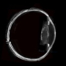
Swept-Source OCT montage of a 45-year-old male with Alports disease and posterior subcapsular cataract.
Photographer: John Golding BA, Vitreous Retina Macula Specialists of Toronto
Imaging device: Topcon DRI Triton
Condition/keywords: Alports disease, optical coherence tomography (OCT), swept source
-
 Feather like cataract
Apr 11 2023 by rodrigo torres
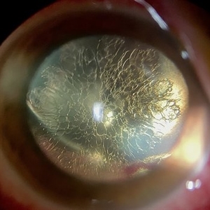
Cataract after vitrectomy and gas tamponade.
Photographer: Rodrigo Amaral Torres
Condition/keywords: cataract, pars plana vitrectomy (PPV)
-
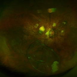
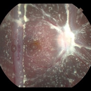
 Seedlings of Fungal Endophthalmitis
Mar 14 2025 by SHILPI H NARNAWARE, ICO ( Retina) , FAICO ( Vitreo-Retina)
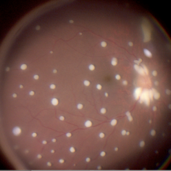
57 year diabetic female , was treated as a case of recurrent vitreous post cataract surgery. Patient was posted for vitrectomy 3 months post cataract surgery. Intra-operatively, multiple yellowish colonies were seen all over the posterior pole were seen, which were later found to be Aspergillus colonies.
Photographer: Shilpi Narnaware, Sarakshi Netralaya , Nagpur, Maharashtra , India
Imaging device: Ngenuity
Condition/keywords: endophthalmitis, fungal
-
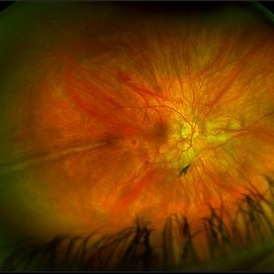
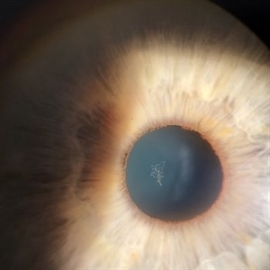
 Retinitis Pigmentosa with Macular Hole with Posterior Subcapsular Cataract
Apr 28 2025 by Malvika Singh
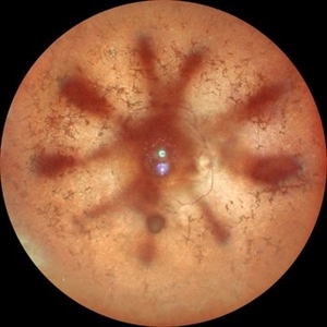
Fundus photograph of the right eye of a 31 year old with retinitis pigmentosa with a macular hole, showing the shadow of posterior subcapsular cataract over the fundus.
Photographer: Dr Malvika Singh, Retina Foundation, Ahmedabad, India
Imaging device: Mirante SLO/OCT
Condition/keywords: macular hole, posterior subcapsular cataract, retinitis pigmentosa
-
 PPV retained cataract
Apr 19 2023 by Denica Rodriguez
A 46-year-old male with hypermature dense cataract. Patient got a piece of metal in his eye when he was 5 years old and was not able to see since. Patient was having cataract surgery and phacodonesis was present. The lens dropped to the back of the eye. Patient had to have another surgery to do vitrectomy. The lens removal was done with a fragmatome handpiece.
Photographer: Denica Rodriguez COA, ST
Imaging device: Zeiss Microscope with resight
Condition/keywords: cataract, dropped nucleus, fragmatome, lens capsule, ocular trauma, pars plana vitrectomy (PPV), retained lens fragments, Retina, retina surgery, traumatic cataract
-
 Prominent Long Ciliary Nerve
Jan 25 2022 by Kachelle Brown
Ultra-wide field photograph of a 48-year-old female with a prominent long ciliary nerve. Patient presented asymptomatic, and was referred for a macula on retinal detachment. Patient was diagnosed with high myopia and a posterior vitreous detachment, and the physician discussed increased risk of floaters, myopic degeneration and retinal detachment associated with high myopia. -24.00 prior to cataract surgery OU per patient.
Photographer: Kachelle Brown
Imaging device: Optos California
Condition/keywords: fundus photograph, high myopia, long ciliary nerve, optos, right eye, ultra-widefield image
-
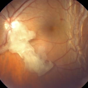 Retained Lens Fragment
Mar 2 2014 by Homayoun Tabandeh, MD, FASRS
Retained lens fragment, choroidal detachment, and serous retinal detachment post cataract surgery
Condition/keywords: retained lens fragments
-
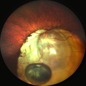
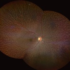
 Chorioretinal Coloboma with Retinal Detachment
Dec 5 2020 by Niloofar Piri, MD
14-year-old female with 1q21.1 microdeletion syndrome and behavioral, intellectual, and systemic abnormalities, including congenital microcornea, iris coloboma, and chorioretinal and optic nerve coloboma presented with decreased vision. Right eye fundus taken with RetCam shows coloboma with retinal detachment. (Left eye showed white cataract with funnel RD on B-scan).
Photographer: Niloofar Piri MD, Douglas Snyder MD
Condition/keywords: chorioretinal coloboma, optic nerve coloboma
-
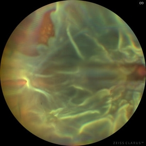 Chronic Open Funnel Retinal Detachment With Horse Shoe Tear
Feb 7 2024 by Harsh Vardhan Singh, MS
67 year old male with history of cataract surgery 1 year presented with old chronic retinal detachment with open funnel configuration with multiple breaks.
Photographer: Harsh Vardhan Singh
Imaging device: Clarus 700
Condition/keywords: chronic retinal detachment, Retinal Detachment, Retinal Detachment with multiple breaks
-
 Ciliary body melanoma
Jan 11 2013 by Alex P. Hunyor, MD
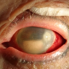
Left inferotemporal ciliary body melanoma with displacement of pupil, cataract, and large dilated episcleral vessels.
-
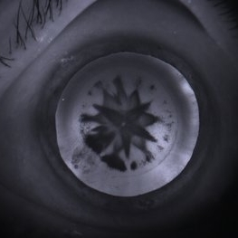 Developmental cataract
Apr 24 2015 by Mehul A Shah
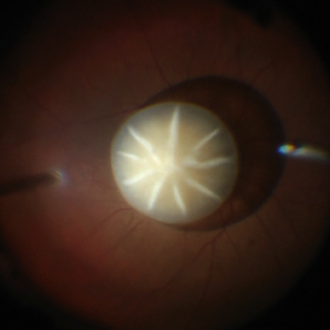
A 9-year-old girl has presented with complaint of diminished vision ou. On retro illumination this image was captured.
Photographer: Mehul Shah, Drashti Netralaya
Imaging device: FORUS
Condition/keywords: developmental cataract, pediatric cataract
-
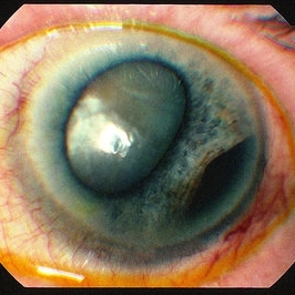
 Dislocated Brown Cataract with a Chorioretinal Coloboma
Sep 8 2021 by Ram Sudarshan
A 44 year-old male with dislocated brown cataract along with a chorioretinal coloboma.
Photographer: Dr.Sivadarshan
Condition/keywords: Brown cataract, chorioretinal coloboma, d, dislocated lens
-
 Dislocated Capsular Tension Ring in Vitreous Cavity
Dec 21 2019 by Pablo Baquero Ospina, MD
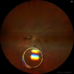
Fundus photograph of an 52-year-old woman with pseudoexfoliation glaucoma and previous cataract surgery with capsular tension ring. 5 years later she refers floaters.
Photographer: Pablo Baquero, Asociacion Para Evitar la Ceguera en Mexico, Mexico city
Imaging device: Optos/Daytona
Condition/keywords: fundus photograph, pseudoexfoliation glaucoma
-
 Dislocated Cataractous Lens
Feb 11 2024 by Anjana Mirajkar, MS Ophthalmology
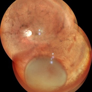
A wide field image of LE of a 40 year old male showing inferior dislocation of crystalline lens which is cataractous in vitreous cavity.
Photographer: Dr. Anjana Mirajkar -Retina Foundation, Ahmedabad
Imaging device: Mirante-Nidek
Condition/keywords: dislocated crystalline lens
-
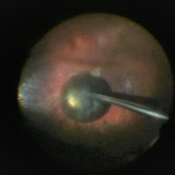
 Dislocated Cataractous Lens
Jun 19 2025 by Mrinali Gupta, MD, FASRS
Intraoperative image of a chronically dislocated cataractous lens. The patient underwent pars plana vitrectomy, lensectomy, and placement of an anterior chamber intraocular lens, with improvement in vision from Count Fingers to 20/20 without correction.
Photographer: Mrinali Gupta, MD
Imaging device: Intraoperative surgical video (Zeiss Lumera scope, Resight lens)
Condition/keywords: dislocated crystalline lens
-
 Dislocated Crystalline Lens
Mar 19 2024 by Annaka Gooding
Ultra Wide field fundus photography of a 70 year old male who presented to clinic with a sudden increase of vision due to dropped crystalline lens secondary to severely dense cataract. Patient reported seeing a full black circle in his inferior visual field. Patient's visual acuity at time of visit was 20/100 with a +5.00 diopter lens. The physician recommended surgical intervention, and discussed surgery for PPV/PPL/IOL implantation with an ACIOL.
Photographer: Annaka Gooding, CPO
Imaging device: Optos California RGB
Condition/keywords: dislocated crystalline lens, fundus photography, inferior retina, OPTOS CALIFORNIA RGB, Right Eye, Ultra-wide field retinal imaging
-
 Dislocated Intraocular Lens (IOL)
Aug 2 2019 by JEFFERSON R SOUSA, Tecg.º (Biomedical Systems Technology)
A 53-year-old male patient suffered blunt trauma 15 days after cataract surgery. Note total dislocation of the intraocular lens. No glass reaction.
Photographer: JEFFERSON R SOUSA - Study Center and Ophthalmological Research Dr. Andre M V Gomes, Institute Dr. Suel Abujamra São Paulo-Brazil
Imaging device: Topcon TRC-50 DX, Imaginet 4.0, angle de 50 graus. Flash 18w-s
Condition/keywords: dislocated intraocular lens (IOL)
-
 Dislocated IOL
May 15 2018 by Morgan Benton
Ultra-wide field pseudocolor image of a 68-year-old male with a dislocated IOL after cataract surgery in the left eye. Patient was only able to count fingers at one foot and could pinhole to 20/60.
Photographer: Morgan Benton
Imaging device: Optos
Condition/keywords: color fundus photograph, dislocated intraocular lens (IOL), left eye, Optos, ultra-wide field imaging
-
 Dislocated IOL
Sep 20 2025 by JORGE SOBERANES
Fundus photograph of a 65-year-old man with a history of cataract surgery one year ago and bad vision since that.
Photographer: Dr. Jorge Soberanes, APEC, Universidad Nacional Autónoma México
Condition/keywords: dislocated lens, intraocular lens dislocation
-
 Drusens
Sep 10 2014 by Mehul A Shah
75-year-old female patient presented with cataract having 20/20 vision post operatively.
Photographer: Drashti Netralaya,Dahod
Imaging device: Zeiss ff450
Condition/keywords: drusen
-
 Endophthalmitis
Apr 9 2014 by Aleksandra V. Rachitskaya, MD, FASRS
Slit lamp photo of a patient with endophthalmitis after cataract surgery. An infectious infiltrate is noted next to the clear corneal incision.
Photographer: Bascom Palmer Eye Institute
Condition/keywords: cataract surgery, endophthalmitis
-
 Epicapsular Stars
Jan 28 2025 by Korey Starkey
Epicapsular stars and cataract noted in natural lens of 68-year-old patient.
Photographer: Korey Starkey
Imaging device: Slit lamp camera
Condition/keywords: cataract, chicken tracks, epicapsular stars, slit lamp photography
-
 ERM
ERM
Nov 26 2020 by Priya Rasipuram Chandrasekaran, MBBS, DO, DNB, FRCS
A 58-year-old female presented with distortion of images 1 month following cataract surgery in the right eye and fundus examination showed epiretinal membrane extending from the disc to the macula and OCT macula showing epiretinal membrane with disorganization of the foveal architecture.
Condition/keywords: epiretinal membrane (ERM)
-
 Fish Hook Eye Trauma
Jun 12 2024 by Miguel Brito, MD, FASRS
Fundus photograph of a 15-year-old boy post cataract aspiration, pars plana vitrectomy, suprachoroidal drainage, and retinal reattachment surgery secondary to traumatic endophthalmitis.
Photographer: Miguel Brito
Condition/keywords: endophthalmitis, PFCL, Retinal detachment under Silicon Oil, retinal fold
-
 Fundus Albipunctatus
Aug 25 2022 by Aditya S Kelkar, MS, FRCS, FASRS,FRCOphth
65 year old female, presented for cataract evaluation. Fundus examination showed whitish-yellow flecks in the retina.
Imaging device: Clarus 500
Condition/keywords: fundus albipunctatus, fundus photograph
 Loading…