Search results (321 results)
-
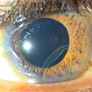
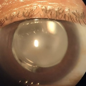
 PCO with Significant Capsular Distention
Nov 24 2025 by Virginia Gebhart
62 year old male presented with blurry vision x 3 weeks. UBM showed sulcus IOL with capsular distention, and possible aqueous misdirection into the capsule. Lens is being pushed into the iris. Pt referred back to cataract surgeon for consideration of YAG, may require PPV.
Photographer: Virginia Gebhart, Retina Consultants of Carolina
Imaging device: Ellex Eyecubed
Condition/keywords: aqueous misdirection, posterior capsule opacification
-
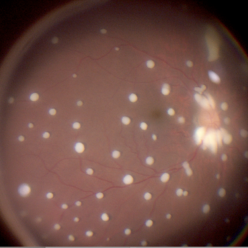
 Unilateral Pigmentary Retinopathy
Nov 9 2025 by Hrishikesh Naik, MS
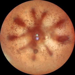
Montage fundus photographs of a 47 year old female presenting with unilateral vision loss in the left eye. Fundoscopy revealed extensive intraretinal pigment clumps, waxy disc pallor, and marked vessel attenuation in the left eye with a normal fundus in the right. Electroretinography showed unilateral reduction in rod and cone function. Unilateral pigmentary retinopathy, an uncommon variant of retinitis pigmentosa (reported incidence ˜ 5%) presents with RP-like changes in one eye, the fellow eye being completely normal. Proposed causes include lyonization and somatic mosaicism. Conditions which mimic RP should be excluded, and any diagnoses should be supported with electrodiagnostic tests and autofluorescence imaging. Management parallels RP, focusing on cataract and macular complications and long-term follow-up to monitor possible bilateral progression.
Imaging device: Zeiss Visucam 224
Condition/keywords: montage, retinitis pigmentosa, unilateral
-
 RD with GRT with Dislocated Cataractous Lens
Nov 9 2025 by SHILPI H NARNAWARE, ICO ( Retina) , FAICO ( Vitreo-Retina)
Young high myope male presented with DOV since 6 months. Examination revealed , Subluxated cataractous lens with RD with PVR with > 270 degrees GRT
Photographer: Shilpi Narnaware, Sarakshi Netralaya , Nagpur, Maharashtra , India
Imaging device: Mirante ( by Nidek)
Condition/keywords: PVR, retinal detachment
-
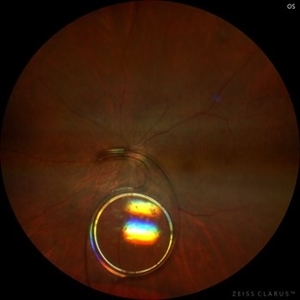
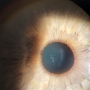
 IOL Dislocation
Sep 29 2025 by Carmen R. Negrin-Martin, MD
Male patient with a history of ocular trauma at 12 years of age. Underwent cataract surgery as an adult and now reports decreased vision of a few days’ duration. Referred to the retina service due to IOL dislocation. Visual acuity with +9.00: 20/40. Biomicroscopy: clear cornea, formed anterior chamber. The IOL haptic is observed hooked to the iris, with the optic in the posterior segment behind the iris. Posterior segment without alterations.
Photographer: Dr. Vizcaino MD
Imaging device: iPhone 16 pro Max
Condition/keywords: IOL dislocation
-
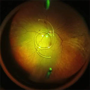
 Dislocated IOL
Sep 20 2025 by JORGE SOBERANES
Fundus photograph of a 65-year-old man with a history of cataract surgery one year ago and bad vision since that.
Photographer: Dr. Jorge Soberanes, APEC, Universidad Nacional Autónoma México
Condition/keywords: dislocated lens, intraocular lens dislocation
-
 Vitreous Cavity Migrated IOL
Sep 20 2025 by Thiago Mazzeo
Intraoperative image of a pars plana vitrectomy for the removal of migrated IOL during complicated cataract surgery.
Photographer: Thiago Mazzeo, Centro de Oftalmologia Especializade de Macaé (COEM)
Condition/keywords: scleral fixation, vitrectomy
-
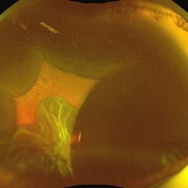 Suprachoroidal Hemorrhage
Aug 4 2025 by Anjana Mirajkar, MS Ophthalmology
A fundus photograph of a 56 year old female with a 360 degree suprachoroidal hemorrhage with a 360 degree crumpled retina during cataract surgery.
Photographer: Dr. Anjana Mirajkar- HV Desai eye hospital ,Pune
Imaging device: optos
Condition/keywords: giant retinal tear, suprachoroidal hemorrhage
-
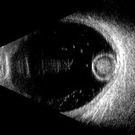 Morgagnian Ghost in the Deep
Jul 3 2025 by Gustavo Uriel Fonseca Aguirre
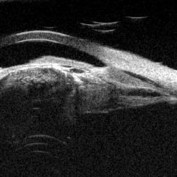
This B-mode para-axial ultrasound scan shows a posteriorly dislocated lens with cortical liquefaction, a dense nucleus, and an intact capsular bag. Vitreous bands are visible extending from the anterior to posterior segments. These findings were bilateral and not associated with trauma or prior surgery.
Photographer: Gustavo U. Fonseca Aguirre, Hospital Conde de Valenciana, Ciudad de México
Condition/keywords: ectopia lentis, morgagnian cataract
-
 Dislocated Cataractous Lens
Jun 19 2025 by Mrinali Gupta, MD, FASRS
Intraoperative image of a chronically dislocated cataractous lens. The patient underwent pars plana vitrectomy, lensectomy, and placement of an anterior chamber intraocular lens, with improvement in vision from Count Fingers to 20/20 without correction.
Photographer: Mrinali Gupta, MD
Imaging device: Intraoperative surgical video (Zeiss Lumera scope, Resight lens)
Condition/keywords: dislocated crystalline lens
-
 Snowflake Lens
May 9 2025 by BENITO VERGARA, MD
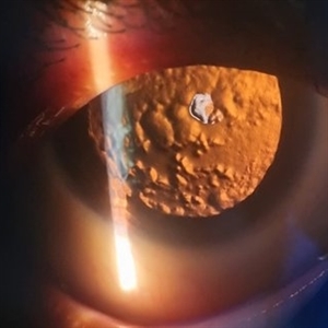
Anterior polar cataract with subcapsular component and dense posterior plaque in a 20-year-old male with congenital aniridia. The image demonstrates a well-defined anterior polar opacity with associated subcapsular changes and a prominent posterior plaque, likely reflecting long-standing progression in the setting of congenital aniridia.
Photographer: Benito Vergara, Asociación Para Evitar la Ceguera en México.
Condition/keywords: aniridia, anterior subcapsular polar cataract
-
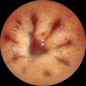 Retinitis Pigmentosa with Macular Hole with Posterior Subcapsular Cataract
Apr 28 2025 by Malvika Singh
Fundus photograph of the left eye of a 31 year old with retinitis pigmentosa, showing the shadow of posterior subcapsular cataract over the fundus.
Photographer: Dr Malvika Singh, Retina Foundation, Ahmedabad, India
Imaging device: Mirante SLO/OCT
Condition/keywords: posterior subcapsular cataract, retinitis pigmentosa
-
 Retinitis Pigmentosa with Macular Hole with Posterior Subcapsular Cataract
Apr 28 2025 by Malvika Singh
Fundus photograph of the right eye of a 31 year old with retinitis pigmentosa with a macular hole, showing the shadow of posterior subcapsular cataract over the fundus.
Photographer: Dr Malvika Singh, Retina Foundation, Ahmedabad, India
Imaging device: Mirante SLO/OCT
Condition/keywords: macular hole, posterior subcapsular cataract, retinitis pigmentosa
-
 Cyclic Membrane
Apr 23 2025 by Gustavo Uriel Fonseca Aguirre
This UBM scan reveals pars planitis with characteristic findings: an inflammatory pupillary membrane, a cataractous lens, and cyclitic membrane causing ciliary body detachment and traction. The lens demonstrates spherical deformation due to zonular laxity from ciliary body traction.
Photographer: Gustavo U. Fonseca Aguirre, Hospital Conde de Valenciana, Ciudad de México
Condition/keywords: cyclic membrane, pars planitis
-
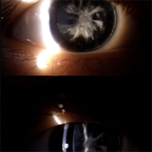
 Posterior Subcapsular Cataract
Apr 18 2025 by DR Rohit Gupta
Slit lamp image on Retroillumination showing posterior subcapsular cataract of a female patient who was on long term corticosteroids inhaler medications.
Photographer: Dr Rohit gupta
Imaging device: Samsung S21
Condition/keywords: posterior capsule opacification, posterior subcapsular cataract, posterior subcapsular changes, posterior subcapsular polar senile cataract, steroids
-
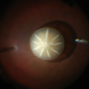
 Posterior Polar Cataract
Apr 10 2025 by DR Rohit Gupta
52 year old male presented with the gradual. Painless, diminuition of vision. On slit lamp examination an onion peel appearance opacification of lens in central part was seen.
Photographer: Dr Rohit gupta
Condition/keywords: posterior capsule opacification, Posterior polar cataract
-
 PCIOL Opacification
Mar 31 2025 by DR Rohit Gupta
A pseudophakic patient visiting after 6 months of cataract surgery. On slit lamp examination a complete hazy white PCIOL was seen, which is a rare complication after cataract surgery.
Photographer: Dr Rohit gupta
Imaging device: Samsung S21
Condition/keywords: posterior chamber intraocular lens (PCIOL)
-
 Seedlings of Fungal Endophthalmitis
Mar 14 2025 by SHILPI H NARNAWARE, ICO ( Retina) , FAICO ( Vitreo-Retina)
57 year diabetic female , was treated as a case of recurrent vitreous post cataract surgery. Patient was posted for vitrectomy 3 months post cataract surgery. Intra-operatively, multiple yellowish colonies were seen all over the posterior pole were seen, which were later found to be Aspergillus colonies.
Photographer: Shilpi Narnaware, Sarakshi Netralaya , Nagpur, Maharashtra , India
Imaging device: Ngenuity
Condition/keywords: endophthalmitis, fungal
-
 Lacteocrumenasia
Mar 11 2025 by Gustavo Uriel Fonseca Aguirre
A 75-year-old female with a history of cataract surgery with intraocular lens implantation 20 years ago presented with progressive visual loss. On slit lamp examination, opaque material was found in the capsular bag behind the intraocular lens. Ultrasound biomicroscopy revealed hyperechoic material contained in the temporal-posterior sector of the capsular bag corresponding to lacteocrumenasia.
Photographer: Gustavo U. Fonseca Aguirre, Hospital Conde de Valenciana, Ciudad de México
Condition/keywords: Lacteocrumenasia, ultrasound biomicroscopy
-
 Multimodal Imaging in CHRPE
Mar 6 2025 by Gerardo - Montante Montelongo, MD
Fundus photograph of an 83-year-old male with a history of Diabetes, smoking, cataract surgery on the right eye in 2022, and open-angle glaucoma. Asymptomatic. Indirect ophthalmoscopy revealed 80% excavation, peripapillary atrophy, and a hyperpigmented perifoveal lesion with 35% atrophy, 10% drusen, and 5.1 mm diameter, corresponding to a CHRPE. At multimodal imaging, FFA shows hypoautofluorescence of the lesion, OCT shows preservation of internal retinal layers, atrophy of external retinal layer, with an RPE disruption, and posterior shadowing. USG shows a flat hyperechoic lesion 5.1 mm in diameter and 1.32 mm in thickness, solid and with high internal reflectance.
Photographer: Gerardo Montante-Montelongo, MD, Mexican Institute of Ophthalmology
Imaging device: Clarus 700
Condition/keywords: congenital hypertrophy of the retinal pigment epithelium (CHRPE), multimodal imaging
-
 Epicapsular Stars
Jan 28 2025 by Korey Starkey
Epicapsular stars and cataract noted in natural lens of 68-year-old patient.
Photographer: Korey Starkey
Imaging device: Slit lamp camera
Condition/keywords: cataract, chicken tracks, epicapsular stars, slit lamp photography
-
 Bullous Keratopathy
Jan 4 2025 by Mosab Salah
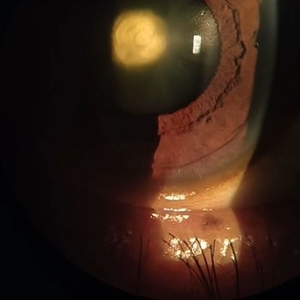
Corneal Slit photograph of an 84-year-old man underwent uneventful cataract surgery 1 year ago elsewhere, with a multiple fluid filled Bullae, not responding on conservative management and planned for KP.
Photographer: Abu-Ismail, Luai MD, The Islamic Hospital, Amman, Jordan
Imaging device: smartphone photography through SLB
Condition/keywords: bullous keratopathy, corneal edema
-
 Unilateral Coloboma Involving Disc and Macula
Dec 27 2024 by Tejaswita Verma
Fundus image of a 15 years old male presenting with unilaterally diminished vision since childhood in RE with CF3mt vision and inferior iris coloboma and retinochoroidal coloboma with nystagmus and cataract.
Photographer: DR. TEJASWITA VERMA
Imaging device: MIRANTE
Condition/keywords: chorioretinal coloboma, iridofundal coloboma
-
 Macular Degeneration
Dec 3 2024 by Sarah D Kang
Fundus photograph of an 85-year-old female patient with macular degeneration observed for retinal clearance before cataract surgery.
Condition/keywords: floaters, macular degeneration
-
 Macular Degeneration
Dec 3 2024 by Sarah D Kang
Fundus photograph of an 85-year-old female patient with macular degeneration observed for retinal clearance before cataract surgery.
Condition/keywords: floaters, macular degeneration
-
 MIDD (Maternally Inherited Diabetes and Deafness) - Left AF
Nov 30 2024 by John S. King, MD
Both right and left eyes have symmetrical ring of mottled hypo/hyper AF around the fovea and disc. The HyperAF areas correspond to RPE deposits on OCT as well as areas of blockage on FA, and drusenoid deposits seen on fundus photos 57 yo WF referred for AMD vs Pattern Dystrophy that was diagnosed 10 years ago. Reported some slow progressive vision loss in both eyes for distance and near. Denies nyctalopia or hemeralopia. Background medical history includes HTN, CVD, and DM. No family history of eye problems. Denied pentosan use. Anterior segment showed moderate cataracts (OD>OS). Posterior segment exam showed macular changes and mild NPDR. The macular appearance showed a symmetrical, paramacular ring of fleck-like drusenoid material with some faint focal areas of RPE hyperplasia. Fundus Photos, AF, OCT were performed as well as a gene test. Further questioning showed revealed that her mother and maternal grandmother had both diabetes mellitus and sensorineural hearing loss. The patient developed diabetes in her teens, and some high frequency hearing loss in her early twenties. She had not had a previous genetic test or diagnosis of MIDD. Gene testing is pending for the mitochondrial component. Invitae's retinal panel, which does not include mitochondrial disorders, only showed a variant of uncertain significance, HMCN1. I discussed this case with Dr. Freund, and it is similar to a the case report : Inoue M, Kiss S, Freund KB. MACULAR PIGMENT RINGS AS THE PRESENTING FINDING OF MITOCHONDRIAL MYOPATHY, ENCEPHALOPATHY, LACTIC ACIDOSIS, AND STROKELIKE EPISODES. Retin Cases Brief Rep. 2015 Fall;9(4):260-4. doi: 10.1097/ICB.0000000000000182. PMID: 26200388.
Photographer: Grace Melton and Carley Gunn
Imaging device: Clarus
Condition/keywords: Macular Dystrophy, Maternally Inherited Diabetes and Deafness, MIDD, Mitochondrial Disorder
 Loading…