Search results (20 results)
-
 Yellow Globular Lesion
Nov 9 2012 by Norman Byer
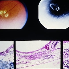
This glistening yellow globular lesion is a so-called pearl of the ora serrata in a 45-year-old man. Notice location in the tooth of the ora, which is a characteristic of this lesion. Histologically pearls are drusen-like structures which form on the inner side of Bruch’s membrane beneath the pigment epithelium. They are seen in about 20% of eyes and are often bilaterally symmetrical. They have no clinical significance but are valuable as landmarks.
Condition/keywords: Bruch's membrane, drusen-like, ora serrata
-
 Bruch’s membrane rupture
Jan 11 2013 by Hyung-Woo Kwak, MD
An area of Bruch’s membrane rupture involving the fovea is seen on indocyanine green angiography: late phase (right).
Photographer: Misook Lee, Kyung Hee Univsersity Hospital, Seoul
Imaging device: Zeiss f 450 plus
Condition/keywords: Bruch's membrane, myopic choroidal neovascularization (CNV)
-
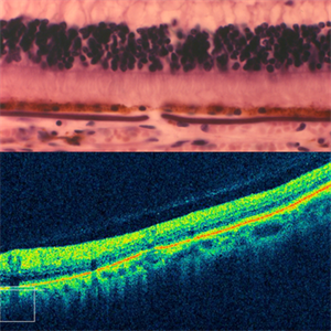
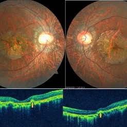
 Autosomal Dominant Drusen of Bruch's Membrane, OCT
May 3 2018 by Alexandr Stepanov
Autosomal dominant drusen of Bruch's membrane, OCT.
Photographer: Alexandr Stepanov MD, PhD, FEBO, Faculty Hospital Hradec Kralove, Czech Republic
Condition/keywords: Bruch's membrane, drusen, optical coherence tomography (OCT)
-
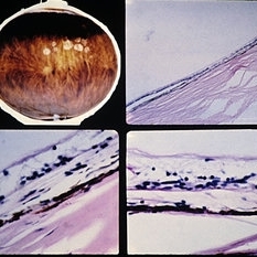
 Angioid streaks - Light microscopy and OCT
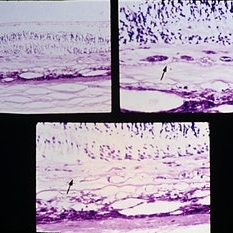
Jan 11 2013 by Gerardo Garcia-Aguirre, MD
Top: Light microscopy of an angioid streak. Note: Bruch's membrane below the RPE, which is thickened and fractured. Bottom: OCT scan of an angioid streak (not of the same patient) showing fracture of the RPE.
Photographer: Alfredo Gomez-Leal, MD (top) and Gerardo Garcia-Aguirre, MD (bottom)
Condition/keywords: angioid streaks
-
 Angioid streaks - Light microscopy and OCT
Jan 11 2013 by Gerardo Garcia-Aguirre, MD
Top: Light microscopy of an angioid streak. Note: Bruch's membrane below the RPE, which is thickened, elevated and fractured. Bottom: OCT scan of an angioid streak (not of the same patient) showing elevation and fracture of the RPE.
Photographer: Alfredo Gomez-Leal, MD (top) and Gerardo Garcia-Aguirre, MD (bottom)
Condition/keywords: angioid streaks
-
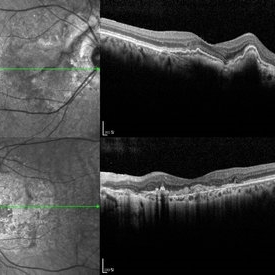 Angioid Streak-Associated Choroidal Neovasclar Membranes
Dec 27 2016 by Young Hee Yoon, MD, PhD
Optical coherence tomogaphs of an 74-year-old woman who received several anti-VEGF injections due to CNV associated with angioid streak in both eyes. There are diffuse CNVM in her right eye and subretinal scar in her left eye. Note the irregular crack in IR image of right eye and the focal Bruch's membrane dehiscence in corresponding B-scan image.
Photographer: Young Hee Yoon, University of Ulsan, Asan Medical Center, Seoul, Korea
Imaging device: Spectralis
Condition/keywords: angioid streaks, choroidal neovascularization (CNV)
-
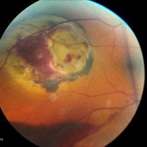 OD Choroidal Tumor CF2
Oct 14 2015 by Darin R. Goldman, MD
55-year-old female with multiple prior primary systemic cancers. She presented with a choroidal tumor, felt to be a primary choroidal melanoma, that had ruptured Bruch's membrane, causing subretinal hemorrhage.
-
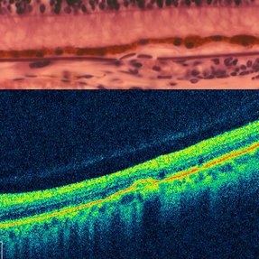
 Autosomal Dominant Drusen of Bruch's Membrane
May 3 2018 by Alexandr Stepanov
Autosomal dominant drusen of Bruch's membrane.
Photographer: Alexandr Stepanov MD, PhD, FEBO, Faculty Hospital Hradec Kralove, Czech Republic
Condition/keywords: Bruch's membrane, drusen
-
 Central Areolar Choroidal Dystrophy
May 4 2021 by Priya Rasipuram Chandrasekaran, MBBS, DO, DNB, FRCS
Fundus photo of a 34-year-old male showing bilaterally symmetrical atrophy of retinal pigment epithelium (RPE) and choriocapillaris involving the fovea and highlighting the underlying large choroidal vessels. OCT macula shows atrophy of the outer retinal layers up to the external limiting membrane along with thinning of RPE and Bruch's membrane complex. Rosette - like hyperreflective structures causing retinal elevation at the border of atrophic area (yellow arrows) are seen categorizing this into stage 4 disease.
Condition/keywords: central areolar choroidal dystrophy (CACD)
-
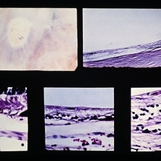
 Slide 9-63
Feb 26 2019 by Lancaster Course in Ophthalmology
Paving-stone degeneration. There is loss of the choriocapillaris, RPE, and outer retinal layers. The thinned inner nuclear layer of the retina rests against Bruch's membrane, and there is no reparative proliferation. Adjacent RPE is hypertrophic.
Condition/keywords: Bruch's membrane, choriocapillaris, retinal pigment epithelium
-
 Slide 9-65
Feb 26 2019 by Lancaster Course in Ophthalmology
Elschnig spot. Localized choroidal infarction with loss of choriocapillaris, RPE, and outer layers of the retina. The thinned inner nuclear layer of the retina rests against Bruch's membrane. At the anterior margin (lower left view) there is an abrupt transition (arrow) between the normal area (left) where the choriocapillaris and RPE are intact and the area of post-ischemic atrophy of the structures (right). A similar but reversed configuration is observed at the posterior margin (lower right view).
Condition/keywords: Bruch's membrane, choroidal infarction, Elschnig's spots
-
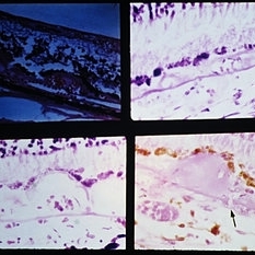 Slide 9-88
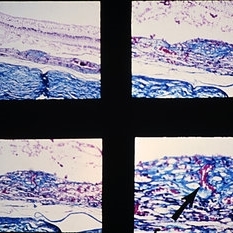
Feb 26 2019 by Lancaster Course in Ophthalmology
Vascularized drusen in periphery (upper left) and macular area (upper right and lower left). Note the tiny break in Bruch's membrane (arrow) with a choroidal vessel with an erythrocyte extending into a peripapillary druse.
Condition/keywords: Bruch's membrane, drusen, macular
-
 Slide 9-84
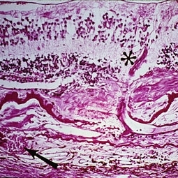
Feb 26 2019 by Lancaster Course in Ophthalmology
Senile macular degeneration with disciform scar. A retinal arteriole (asterisk) extends into the subretinal component of the scar, through a break in the thickened and detached inner layer of Bruch's membrane, and then into the vascularized intra-Bruch's-membrane component of the scar. Study of serial sections disclosed this retinal vessel to anastomose with the choroidal vessel (arrow) which extends through a branch in Bruch's membrane.
Condition/keywords: Bruch's membrane, disciform scar, macular degeneration, retinal arteriole
-
 Slide 9-83
Feb 26 2019 by Lancaster Course in Ophthalmology
Senile macular degeneration. A disciform scar is shown in a typical two component configuration. Arrows indicate the thickened and detached inner aspect of Bruch's membrane. There is one component of the disciform lesion that is located between the retina and the detached inner aspect of Bruch's membrane. RPE hyperplasia is evident in this component. The second component is located between the two layers of Bruch's membrane, and this component has choroidal neovascular tissue in the lower view.
Condition/keywords: Bruch's membrane, choroidal neovascular tissue, disciform scar, macular degeneration, retinal pigment epithelium
-
 Slide 9-87
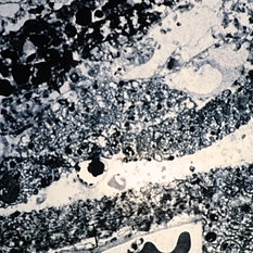
Feb 26 2019 by Lancaster Course in Ophthalmology
Ultrastructural appearance of Bruch's membrane and drusen material in eye with serous detachment of RPE. The RPE basement membrane is intact and normal (arrows). The inner collagenous zone of Bruch's membrane is greatly thickened (between larger arrows) by the accumulation of small vesicles, electron-dense particles, fibrils, and clusters of widely spaced collagen (large circle and inset). The middle-elastic layer of Bruch's membrane (small circle) is essentially normal. The outer collagenous zone (bracket) is mildly thickened with accumulation of material similar to that seen in the inner zone. Splitting of the thickened inner collagenous zone (asterisk) has occurred with the accumulation of a finely granular material, membranous structures, and electron-dense particles (CC =choriocapillaris).
Condition/keywords: Bruch's membrane, drusen, retinal pigment epithelium, serous retinal detachment
-
 Slide 9-94
Feb 26 2019 by Lancaster Course in Ophthalmology
Macular disciform lesion in the ocular histoplasmosis syndrome. Note choroidal scar with vessels (arrow) extending through a break in Bruch's membrane.
Condition/keywords: Bruch's membrane, disciform macular lesion, ocular histoplasmosis syndrome (OHS)
-
 Slide 9-66
Feb 26 2019 by Lancaster Course in Ophthalmology
Midperipheral punched-out lesions in the presumed ocular histoplasmosis syndrome. There is scarring in the choroid and retina, discontinuity in Bruch's membrane, and loss of the RPE. An infiltrate of lymphocytes is present in the subjacent choroid (lower middle and right).
Condition/keywords: Bruch's membrane, ocular histoplasmosis syndrome (OHS), retinal pigment epithelium, scar
-
 Slide 9-80
Feb 26 2019 by Lancaster Course in Ophthalmology
Senile macular degeneration. Note presence of drusen and diffuse thickening of the inner aspect of Bruch's membrane, areolar RPE and photoreceptor atrophy, and choroidal neovascularization (arrows).
Condition/keywords: Bruch's membrane, choroidal neovascularization (CNV), drusen, macular degeneration, retinal pigment epithelium
-
 Slide 9-96
Feb 26 2019 by Lancaster Course in Ophthalmology
Hypertrophy of peripapillary RPE associated with a vascularized druse with the vessel extending from the choroid through a very small break in Bruch's membrane (arrow).
Condition/keywords: Bruch's membrane, retinal pigment epithelium
-
 Slide 9-82
Feb 26 2019 by Lancaster Course in Ophthalmology
Senile macular degeneration. Hemorrhagic detachment of the RPE is contiguous with sub-RPE neovascularization (arrows, lower middle and lower right). The thickened inner aspect of Bruch's membrane and drusen (upper right, arrows) are detached along with the RPE.
Condition/keywords: Bruch's membrane, hemorrhagic detachment, macular degeneration, retinal pigment epithelium
 Loading…