Search results (65 results)
-
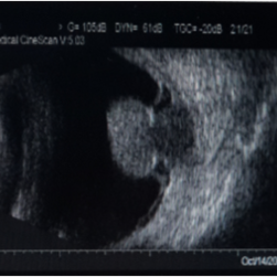
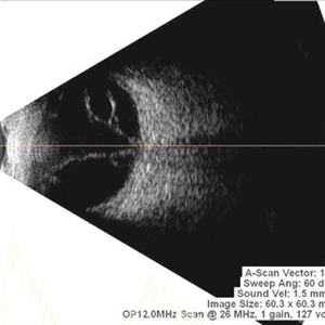
 Open Funnel Retinal Detachment
Oct 13 2012 by Geoffrey G. Emerson, MD, PhD, FASRS
Open funnel retinal detachment
Condition/keywords: B scan ultrasound, open funnel RD
-
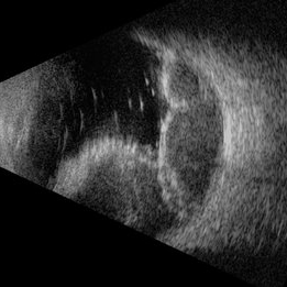
 Chronic Retinal Detachment
Oct 12 2012 by Jeffrey G. Gross, MD, FASRS
Chronic RD with multiple retinal cysts, B scan ultrasound.
Condition/keywords: B scan ultrasound, chronic retinal detachment, retinal cyst
-
 Retinoblastoma Ultrasound
Oct 2 2015 by Aparna Ramasubramanian
Ultrasonography of a retinoblastoma tumor shows hyperreflective echoes suggestive of calcification. It is seen in 90% of retinoblastoma patients and is an important diagnostic sign.
Photographer: Aparna Ramasubramanian
Condition/keywords: A-scan ultrasound, B scan ultrasound, calcification, retinoblastoma
-
 Retinoblastoma
Oct 5 2012 by Ronald C. Gentile, MD
B scan ultrasonography of the large endophytic retinoblastoma revealing internal hyper-reflective spots consistent with internal calcification.
Photographer: The New York Eye & Ear Infirmary Department of Medical Imaging
Condition/keywords: B scan ultrasound, retinoblastoma
-
---thumb.jpg/image-square;max$300,300.ImageHandler) Sturge-Weber Diffuse Hemangioma and Retinal Detachment on B-scan
Sturge-Weber Diffuse Hemangioma and Retinal Detachment on B-scan
Apr 18 2014 by Susanna S. Park, MD, PhD
B-scan ultrasonogram of the right eye of an 8 year old Hispanic boy with Sturge -Weber Syndrome showing diffuse choroidal thickening from diffuse choroidal hemangioma and associated total exudative retinal detachment.
Photographer: Ellen Redenbo, University of California Davis Eye Center
Condition/keywords: B scan ultrasound, diffuse choroidal hemangioma, Sturge-Weber syndrome
-
---thumb.jpg/image-square;max$300,300.ImageHandler) Choroidal Metastasis B-Scan
Choroidal Metastasis B-Scan
Jan 10 2014 by Susanna S. Park, MD, PhD
B-scan ultrasound image showing choroidal thickening and exudative retinal detachment in a patient with diffuse choroidal metastasis from breast carcinoma.
Photographer: Ellen Redenbo, University of California Davis Eye Center
Condition/keywords: B scan ultrasound, choroidal metastasis
-
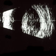
 Encircling Scleral Buckle Axial View
Dec 10 2012 by Yale L. Fisher, MD
The buckle is an encircling element. the anterior posterior view shows a cross section above and below on the screen. Rotation vertically demonstrates a superior and inferior cross-section of a highly elevated scleral buckle. There is movement of the separated formed vitreous and far anterior (visible at the left of the screen). Optic nerve shadow is visible as the nerve moves vertically.
Condition/keywords: B scan ultrasound, scleral buckle, video
-
---thumb.jpg/image-square;max$300,300.ImageHandler) Asteroids-B Scan
Asteroids-B Scan
Apr 18 2014 by Susanna S. Park, MD, PhD
B-scan ultrasound image of the left eye of a 95-year-old Hispanic diabetic man with dense media opacity from asteroids hyalosis. Visual acuity is 20/60.
Photographer: Ellen Redenbo, UC Davis Eye Center
Condition/keywords: asteroid hyalosis, B scan ultrasound, vitreous opacity
-
 Melanoma-B-Mode Ultrasonography
Nov 12 2013 by Xuefeng Feng, MD, PhD
B-mode ultrasonography of a 52-year-old man with a choriod melanoma.
Photographer: Xing Wang, Ophthalmology Department, Peking University Third Hospital
Condition/keywords: B scan ultrasound
-
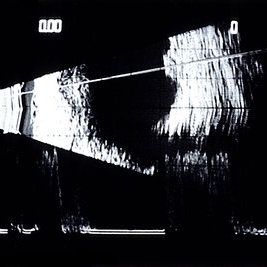
 Choroidal Osteoma 5
Oct 5 2012 by Ronald C. Gentile, MD
B scan ultrasonography with representative A scan of the macular choroidal osteoma. The B scan reveals a characteristic highly reflective plaque consistent with its bone-like calcium composition that persists with low gain. The A scan reveals a large spike.
Photographer: The New York Eye & Ear Infirmary Department of Medical Imaging
Condition/keywords: B scan ultrasound, choroidal tumor, macular choroidal osteoma
-
 Discrete Choroidal Hemangioma B-Scan
Jul 14 2014 by Susanna S. Park, MD, PhD
B-scan ultrasonography of a discrete choroidal hemangioma.
Photographer: Ellen Redenbo
Condition/keywords: B scan ultrasound, choroidal hemangioma
-
 Choroidal melanoma; B-scan
May 2 2013 by Henry J. Kaplan, MD
B-scan of a choroidal melanoma shows dome shaped lesion #1.
Condition/keywords: B scan ultrasound
-
 Ciliary Body Melanoma B-Scan Ultrasound
Feb 27 2014 by Susanna S. Park, MD, PhD
Large ciliary body melanoma in a 57-year-old man.
Photographer: Ellen Redenbo, University of California Davis Eye Center
Condition/keywords: B scan ultrasound, ciliary body melanoma
-
 Choroidal Hemangioma
Mar 29 2013 by Henry J. Kaplan, MD
B-scan of the same patient showing the lesion with high internal reflectivity #2.
Condition/keywords: B scan ultrasound
-
 Optic nerve melanocytoma case 3 - Bscan
Jan 11 2013 by Alex P. Hunyor, MD
Optic nerve melanocytoma, left eye. B scan ultrasound demonstrating the lesion with high internal reflectivity.
Condition/keywords: optic disc melanocytoma
-
 Diffuse Choroid hemangioma
Nov 7 2012 by Rajiv Anand, MD, FRCS, FASRS
B scan ultrasound shows the thickened choroid
Condition/keywords: choroidal thickening, Sturge-Weber syndrome
-
---thumb.jpg/image-square;max$300,300.ImageHandler) Osteoma B Scan
Osteoma B Scan
Apr 18 2014 by Susanna S. Park, MD, PhD
72-year-old woman noted with a mid-peripheral amelanotic choroidal lesion with minimal elevation which shows marked shadowing on ultrasonography consistent with a choroidal osteoma. Optic nerve is shown in the bottom of the image.
Photographer: Ellen Redenbo, University of California Davis Eye Center
Condition/keywords: B scan ultrasound
-
 Tube Shunt Plate
Jul 14 2013 by Jason S. Calhoun
BSCAN ultrasound of tube shunt plate.
Photographer: Jason S. Calhoun, Department of Ophthalmology, Mayo Clinic Jacksonville, Florida
Imaging device: BSCAN
Condition/keywords: B scan ultrasound
-
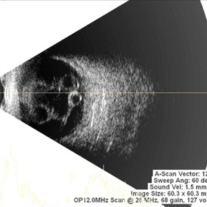
 Multiple Retinal Cysts Associated With Chronic Retinal Detachment
Sep 24 2018 by samarth mishra
Patient presented with a diminution of vision in left eye since few months. On B-scan ultrasonography multiple retinal cysts with a total retinal detachment were noted.
Photographer: Aditya Birla Sankara Nethralaya, West Bengal , Kolkata , India
Condition/keywords: B scan ultrasound, chronic retinal detachment, intraretinal cyst, retinal cyst
-
---thumb.jpg/image-square;max$300,300.ImageHandler) Sturge-Weber B Scan Post-Radiation
Sturge-Weber B Scan Post-Radiation
Apr 18 2014 by Susanna S. Park, MD, PhD
B-scan ultrasound image of the right eye of an 8-year-old Hispanic boy with Sturge -Weber Syndrome and diffuse choroidal hemangioma one year after proton beam irradiation showing complete resolution of retinal detachment with some thinning of the choroidal hemangioma.
Photographer: Ellen Redenbo, University of California Davis Eye Center
Condition/keywords: after proton beam irradiation, B scan ultrasound, diffuse choroidal hemangioma, Sturge-Weber syndrome
-
 Multiple Retinal Cysts Associated With Chronic Retinal Detachment
Sep 24 2018 by samarth mishra
Patient presented with a diminution of vision in left eye since few months. On B-scan ultrasonography multiple retinal cysts with a total retinal detachment were noted.
Photographer: Aditya Birla Sankara Nethralaya, West Bengal , Kolkata , India
Condition/keywords: B scan ultrasound, chronic retinal detachment, intraretinal cyst, retinal cyst
-
 Choroidal Hemangioma
Sep 7 2014 by Thomas A. Ciulla, MD, MBA, FASRS
B-scan shows shallow echodense lesion nasal to the optic nerve with shallow overlying subretinal fluid.
Condition/keywords: B scan ultrasound, choroidal hemangioma
-
 Multiple Retinal Cysts Associated With Chronic Retinal Detachment
Sep 24 2018 by samarth mishra
Patient presented with a diminution of vision in left eye since few months. On B-scan ultrasonography multiple retinal cysts with a total retinal detachment were noted.
Photographer: Aditya Birla Sankara Nethralaya, West Bengal , Kolkata , India
Condition/keywords: B scan ultrasound, chronic retinal detachment, intraretinal cyst, retinal cyst
-
 Suprachoroidal Hemorrhage
Nov 16 2019 by Sophia El Hamichi, MD
Ultrasound of the right eye of 43-year-old male presenting with suprachoroidal hemorrhage, note the multilobulated heterogenous echogenic mass aspect of the choroid
Photographer: Fiona J Ehlies, Murray Ocular Oncology and Retina, Miami
Condition/keywords: B scan ultrasound, suprachoroidal hemorrhage
 Loading…