Search results (253 results)
-
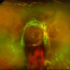
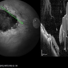
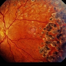
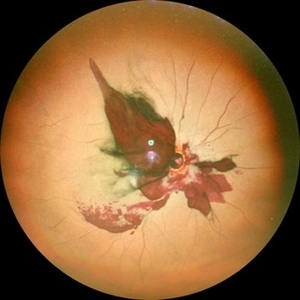 Optic Nerve Avulsion with Vitreous Hemorrhage and Pale Retina
Jan 25 2021 by Sham Talati, DOMS
A 30-year-old male presented with history of trauma to RE with NO Perception of light in the affected eye.
Photographer: Dr. Sham Talati,Retina Foundation,Ahmedabad
Imaging device: Nidek Mirante
Condition/keywords: optic nerve, pale retina
-
 Candy Stripe Sign
Mar 30 2023 by pedro fernandes souza neto
Candy Stripe Sign, patient with proliferative diabetic retinopathy progressing to vitreous hemorrhage and subsequently to ghost cell glaucoma.
Photographer: Marlos Henrique Oliveira Junior, Federal University of Bahia.
Condition/keywords: dehemoglobinized hemorrhage, diabetes, diabetic glaucoma
-
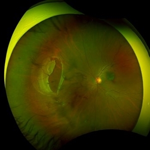
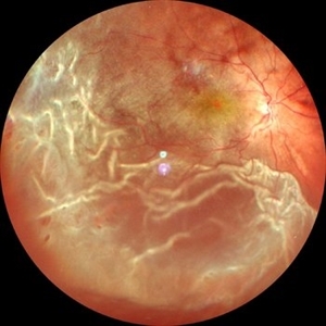
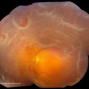 Retinal Detachment With Multiple Retinal Tears
May 18 2017 by Kamal Kishore, MD, MBBS
77-year-old female presented with a report of gradual decreased vision over the span of one week. Vision finger count. Examination showed retinal detachment with multiple retinal tears and vitreous hemorrhage present.
Photographer: Lindsay Shepard, Illinois Retina and Eye Associates, Peru, IL
Imaging device: Topcon TRC- 50 EX
Condition/keywords: retinal tear
-
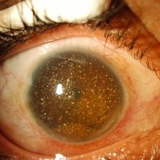
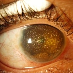 Synchysis Scintillans
Sep 17 2015 by Jessica G Lee, MD
24-year-old male with history of chronic retinal detachment.
Photographer: Bob Masini
Condition/keywords: cholesterol crystals, refractile bodies, synchysis scintillans, trauma, vitreous hemorrhage
-
 Choroidal Melanoma Masquerading as Subretinal Hemorrhage With Breakthrough VH
Jan 23 2025 by Tejaswita Verma
A 65 year old diabetic male presented with large nasal retinal mass giving the appearance of organized dehaemoglobinized subretinal hemorrhage with breakthrough vitreous hemorrhage , with 6/6P vision. Enucleation specimen showed histopathology confirmed choroidal melanoma.
Photographer: DR. TEJASWITA VERMA
Imaging device: MIRANTE
-
 Chronical Submacular Hemorrhage in the Setting of Neovascular AMD
Mar 23 2015 by Rita Couceiro, MD, MS
An 80-year-old male, with a history of hypertension and high cholesterol, complained of acute and painless vision loss in his left eye (OS) in the previous 5 months. On observation best corrected visual acuity in OS was hand motion. A dense vitreous opacity in OS precluded fundus examination. Ocular ultrasound revealed vitreous hemorrhage and thickening of the macular area. The patient was submitted to pars plana vitrectomy, which disclosed a large submacular hemorrhage with chronical features and disciform scarring in the setting of neovascular AMD.
Imaging device: Intraoperative fundus photograph
Condition/keywords: neovascular age-related macular degeneration (AMD), submacular hemorrhage, wet age-related macular degeneration (wet AMD)
-
 Ciliary Body Melanoma
Feb 12 2025 by Virginia Gebhart
91 year old female with large collar button tumor emanating from the ciliary body with resolving vitreous hemorrhage. Melanoma cells in the AV as well as studded on the entire retina surface. Pt scheduled for enucleation. CT scans of chest and abdomen showed no evidence of metastatic disease.
Photographer: Virginia Gebhart, Retina Consultants of Carolina
Imaging device: Optos California
Condition/keywords: asteroid hyalosis, ciliary body mass, ciliary body melanoma, vitreous hemorrhage
-
 Diabetic Tractional Retinal Detachment
Jan 23 2019 by Olivia Rainey
Ultra-wide field pseudocolor image of an 43-year-old female with a diabetic tractional retinal detachment and a vitreous hemorrhage affecting her right eye.
Photographer: Olivia Rainey
Imaging device: Optos
Condition/keywords: diabetes, diabetic traction detachment, Optos, pan-retinal photocoagulation (PRP), proliferative diabetic retinopathy (PDR), pseudocolor, ultra-wide field imaging, vitreous hemorrhage
-
 Large Retinal Tear from a Shuttlecock Injury
Oct 11 2024 by Ahmad B. Tarabishy, MD
27 year old woman presenting with floaters and a shadow in her temporal visual field OS. Approximately one week earlier, she was injured in her left eye by a shuttlecock while playing badminton. Fundus exam reveals mild vitreous hemorrhage and a large retinal tear with a small cuff of surrounding SRF.
Photographer: Angela Rico, M.D.
Imaging device: Optos
Condition/keywords: blunt trauma, ocular trauma, retinal tear
-
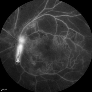 Optic Nerve Head Cannonball
Dec 15 2019 by Veer Singh, MS, FVRS, FMRF, FICO (Retina)
This is the fundus fluorescein angiography (FFA) of the left eye of a 62-year-old diabetic patient with proliferative diabetic retinopathy and neovascularization of disc who bled from the disc while he was undergoing an FFA procedure. The bleed from the disc gives the appearance of a cannonball fired from a cannon hence the caption "Optic Nerve Head Cannonball".
Photographer: Dr. Veer Singh
Imaging device: Heidelberg Spectralis HRA
Condition/keywords: fluorescein angiogram (FA), neovascularization of the disc (NVD), optic nerve head, proliferative diabetic retinopathy (PDR), vitreous hemorrhage
-
 Pan-Retinal Photocoagulation
Apr 5 2018 by Mohamed Tawfik, MD
Wide field FFA post phaco vitrectomy of a case of vitreous hemorrhage show PRP with regression of diabetic retinopathy.
Photographer: Mohamed A,Tawfik MD,FRCSed
Condition/keywords: pan-retinal photocoagulation (PRP)
-
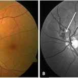 Peri-papillary Vascular Loop
Jun 2 2020 by Dhaivat Shah
Peri-papillary vascular loops (PVL) are rare congenital vascular malformations, which are usually detected as accidental finding during routine fundus examination. They can often be confused with tributary vein occlusion or racemose hemangioma. Although benign and asymptomatic, they can be rarely associated with vitreous hemorrhage and arterial occlusion. We herein present a case of a 60-year-old hypertensive male, who was diagnosed elsewhere to have a tributary vein occlusion and was referred to us. FFA was advised to rule out neovascularization, surrounding capillary non perfusion and mass lesion (hemangioma). On FFA, the arterial loop showed a slightly delayed filling (3-5 seconds) as compared to the other arterial vessels and the original vessel appeared to be a branch arising from central retinal artery. The choroidal filling was delayed in the area supplied by the loop. A cilioretinal artery was also noted. The patient was diagnosed to have a Peri-papillary vascular arterial loop (PVL), likely to be congenital in origin. The patient was reassured and was advised yearly follow up. These loops are usually accidental findings discovered during routine fundus examination. Since these vessels are looped and tortuous, they exhibit a slower and laminar blood flow, which make them more prone for arterial occlusions. The vitreous in this area tends to be adherently attached, so during PVD induction, it is likely to cause a tear and hemorrhage leading to vitreous hemorrhage. Until and unless there is a break, this hemorrhage tends to resolve on its own and does not warrant treatment. If there is an evident break, it can be dealt with laser barrage.
Photographer: Choithram Netralaya
Condition/keywords: congenital prepapillary vascular loop
-
 Retinal Cavernous Hemangioma
Oct 22 2020 by Olivia Rainey
Widefield OCT of a 31-year-old male presenting with a retinal cavernous hemangioma affecting his left eye. Patient was 18-years-old when he was diagnosed with a retinal cavernous hemangioma. He has had a few episodes of vitreous hemorrhages since then. His vision was 20/20-1 in both eyes.
Photographer: Becca Harris
Imaging device: Heidelberg Spectralis
Condition/keywords: 50 degrees, cavernous hemangioma of the retina, Heidelburg Spectralis, left eye, optical coherence tomography (OCT), wide angle imaging
-
 ROP
ROP
Mar 26 2025 by Korey Starkey
9 month old patient presents today with Retinopathy of Prematurity in both eyes. Patient was born at gestational age of 25 weeks 2 days, 940g. Left eye presents with vitreoretinal traction and peripheral VH with regressed stage 3 and persistent stage 2 disease.
Photographer: Korey Starkey
Imaging device: Optos
Condition/keywords: retinopathy of prematurity stage 2, rop, stage 3, vitreoretinal traction, vitreous hemorrhage
-
 Synchisis Scintillans
Sep 17 2015 by Jessica G Lee, MD
24-year-old male with history of chronic retinal detachment.
Condition/keywords: cholesterol crystals, refractile bodies, synchysis scintillans, trauma, vitreous hemorrhage
-
 Vitreous Hemorrhage
Jul 10 2018 by Karen Panzegrau
SD-OCT of a 35-year-old female presenting with a vitreous hemorrhage of her left eye. Patient has active proliferative diabetic retinopathy, as well as a completed posterior vitreous detachment in the left eye.
Photographer: Karen Panzegrau
Condition/keywords: diabetes, Heidelburg Spectralis, left eye, optical coherence tomography (OCT), posterior vitreous detachment, proliferative diabetic retinopathy (PDR), vitreous hemorrhage
-
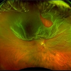
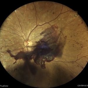
 Von Hippel Lindau Syndrome
Jun 9 2024 by Anjana Mirajkar, MS Ophthalmology
A widefield montage of a 23 year old female of LE case of VHL syndrome showing some hemorrhages with traction superiorly in a silicon oil filled eye with central settled retina. Cryo and laser marks are noted in periphery.
Photographer: Dr. Anjana Mirajkar -Retina Foundation, Ahmedabad
Imaging device: Mirante-Nidek
Condition/keywords: cryotherapy, exudative detachment, laser photocoagulation, vitreous hemorrhage, Von Hippel-Lindau
-
 Optos Giant Tear within Retinal Detachment
Apr 30 2019 by Lauren Whaley
Noticed an inferior visual field defect on a patient with history of vitreous hemorrhage. Decided to take an Optos image and this is what we found. Doctor performed pneumatic retinopexy in office and patient recovering well.
Photographer: Lauren R. Whaley
Imaging device: Optos
Condition/keywords: Optos, retinal tear, subretinal fluid
-
 Sickle Cell Neovascularization and Vitreous Hemorrhage
Oct 30 2015 by David Callanan, MD
Female patient, sickle cell neovascularization and vitreous hemorrhage; pre and post laser.
Condition/keywords: neovascularization (NV), sickle cell, vitreous hemorrhage
-
 4 Point Scleral Fixation Akreos AO60 With Gore Tex Suture
May 20 2021 by Jesus Lozano, MD
Optos Silverstone fundus image of a 54-year-old man after 4 point scleral fixation Akreos AO60 with Gore Tex suture plus PPV who had a severe traumatic iris defect and was aphakic after ocular trauma.
Photographer: Yair Bet Yosef, Hadassah Medical Center. Israel
Imaging device: Optos Silverstone
Condition/keywords: aphakia, globe perforation, lens, pars plana vitrectomy (PPV), penetrating trauma, vitreous hemorrhage
-
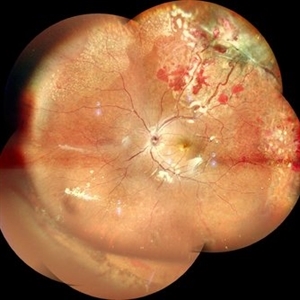
 Blunt Ocular Trauma Due to Firework Injury
Jun 9 2020 by Brittany Rota
Ultra- widefield pseudocolor image of an 18-year-old male with blunt ocular trauma in the right eye due to a firework injury. The patient presented with commotio retinae (sclopteria), an acute vitreous hemorrhage, choroidal rupture, and a subretinal hemorrhage. The referring physician performed surgery on the lateral rectus muscle which was macerated but not severed, and several orbital fibrous foreign bodies were removed from the posterior orbit. The globe was intact. There is no evidence of retinal tear in the region of sclopetaria; however, there is complete necrosis of the temporal peripheral choroid and retina. The vitreous hemorrhage was slowly clearing on his exam 6-9-2020. The patient is developing subretinal fibrosis. The physician is concerned about the choroidal rupture that is visible through the submacular hemorrhage. There is one rupture that appears to course directly under the fovea. The physician states that if this is the case, his vision most likely will be 20/200 or worse. His vision was hand motion in all fields except nasally, which he was unable to see hand motion at his visit on 6-9-2020.
Photographer: Brittany Rota
Imaging device: Optos California
Condition/keywords: blunt trauma, choroidal rupture, commotio retinae, fibrosis, firework injury, fundus photograph, hand motion, necrotizing retina, Optos, pseudocolor, subretinal hemorrhage, vitreous hemorrhage
-
---thumb.jpg/image-square;max$300,300.ImageHandler) Lupus Anticoagulant Disorder
Lupus Anticoagulant Disorder
Feb 26 2013 by Henry J. Kaplan, MD
Occluded retinal vessels and vitreous hemorrhage apparent.
Condition/keywords: lupus anticoagulate factor
-
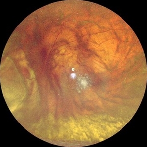
 Retinal Detachment with Vitreous Hemorrhage
Jun 10 2020 by Manish Nagpal, MD, FRCS (UK), FASRS
Retinal detachment with folds and minimal vitreous hemorrhage.
Photographer: Gayathri Mohan, Retina Foundation
Imaging device: NIDEK SLO MIRANTE
Condition/keywords: vitreous hemorrhage
-
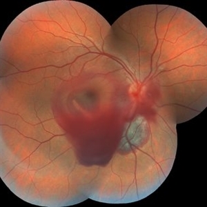
 Subhyaloid Hemorrhage With Vitreous Hemorrhage in a Case of Proliferative Diabetic Retinopathy
May 7 2024 by Akansha Sharma
Color fundus photograph of a 39 year old female with vitreous hemorrhage and subhyaloid hemorrhage in proliferative diabetic retinopathy.
Photographer: Dr. Akansha Sharma, Bharati Eye Hospital
Condition/keywords: PDR, proliferative diabetic retinopathy (PDR), SHH, subhyaloid hemorrhage, vitreous hemorrhage
-
 Trio of Retinal Hemorrhages
Dec 8 2020 by Priya Rasipuram Chandrasekaran, MBBS, DO, DNB, FRCS
This is the fundus photo of a 29-year-old following blunt trauma showing hemorrhages in all the three layers of the retina (vitreous hemorrhage, subhyaloid hemorrhage and subretinal hemorrhage)
Condition/keywords: blunt trauma, retinal hemorrhage
 Loading…