Search results (47 results)
-
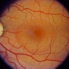
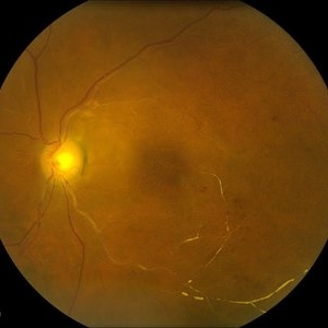 Calcium Deposits of Central Retinal Vein
Jul 14 2013 by Jason S. Calhoun
Vein occlusion with severe blockage inferiorly. Calcium deposits seen through the veins branching out with thinning of the retina.
Photographer: Jason S. Calhoun, Department of Ophthalmology, Mayo Clinic Jacksonville, Florida
Imaging device: TOPCON TRC 50-EX
Condition/keywords: branch retinal vein occlusion (BRVO)
-
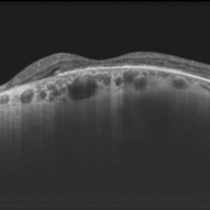
 Dome-Shaped Macula With Subretinal Fluid
Jun 14 2018 by Gerardo Garcia-Aguirre, MD
EDI OCT of the right eye of a 17-year-old highly myopic girl. Subfoveal fluid is present. There is choroidal thinning, and scleral thickening in the foveal area.
Photographer: Gerardo Garcia-Aguirre, MD
Imaging device: Heidelberg Spectralis
Condition/keywords: dome shaped macula, high myopia
-
 Choroidal Rupture, Subretinal and Vitreous Hemorrhage Secondary to Blunt Trauma
Dec 29 2012 by Humberto Ruiz-Garcia, MD
SD-OCT obtained at 72 hours which shows neurosensory macular detachment and severe thinning (impending macular hole).
Photographer: Humberto Ruiz-Garcia
Imaging device: Cirrus HD OCT
Condition/keywords: traumatic macular hole
-
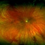 High Myopia with Cobblestone Degeneration
Nov 5 2019 by Nichole Lewis
50-year-old female with high myopia, diffuse myopic thinning and cobblestone degeneration.
Photographer: Nichole Lewis
Imaging device: Optos
Condition/keywords: high myopia, myopia, paving stone degeneration
-
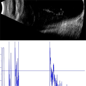 AL 39.16 mm
Sep 10 2025 by Gustavo Uriel Fonseca Aguirre
This axial B-scan reveals an elongated globe with an axial length of 39.16 mm, consistent with high axial myopia. Posterior staphyloma and scleral thinning are observed, though the retina remains attached.
Photographer: Gustavo U. Fonseca Aguirre, Hospital Conde de Valenciana, Ciudad de México
Condition/keywords: high myopia
-
 Alport's Syndrome
Aug 29 2018 by Abhishek Das, MBBS, MS
OCT of a 54-year-old woman diagnosed to have Alport's syndrome. OCT shows temporal thinning of retina with nasal retina preserved.
Photographer: Abhishek Das, The Eye Foundation,Coimbatore,India
Imaging device: Optovue
Condition/keywords: Alports disease
-
 Biclonal Gammopathy
Jan 29 2014 by Mallika Goyal, MD
OCT of the left eye of a 17-year-old girl with monoclonal gammopathy shows extreme thinning of the foveal centre, likely from ischaemia associated atrophy.
Photographer: Mallika Goyal, MD, Apollo Health City, Hyderabad, India
Condition/keywords: biclonal gammopathy, optical coherence tomography (OCT)
-
 Central Areolar Choroidal Dystrophy
May 4 2021 by Priya Rasipuram Chandrasekaran, MBBS, DO, DNB, FRCS
Fundus photo of a 34-year-old male showing bilaterally symmetrical atrophy of retinal pigment epithelium (RPE) and choriocapillaris involving the fovea and highlighting the underlying large choroidal vessels. OCT macula shows atrophy of the outer retinal layers up to the external limiting membrane along with thinning of RPE and Bruch's membrane complex. Rosette - like hyperreflective structures causing retinal elevation at the border of atrophic area (yellow arrows) are seen categorizing this into stage 4 disease.
Condition/keywords: central areolar choroidal dystrophy (CACD)
-
 Central Serous Chorioretinopathy (CSR)
Sep 21 2023 by Ben Serar
Fundus photograph showing increased cup-disc ratio with nasalisation of vessels , with thinning of Neuroretinal rim and bayonetting of vessels in a case of Glaucomatous Optic Atrophy (GOA) Fundus photograph of LE showing serous macular detachment in a case of Central Serous Chorioretinopathy (CSR).
Condition/keywords: Central Serous Chorioretinopathy (CSR)
-
 Colorized BR Disc Heme Wedge Defect
Oct 5 2015 by Jared Watson
+FHx, HVF defects corresponding to RNFL thinning and ONH appearance.
Photographer: Jared Watson COT
Imaging device: Heidelberg Spectralis
Condition/keywords: primary angle-closure glaucoma
-
 Commotio-Retinae
Sep 22 2021 by Luiz Guilherme Freitas, MD, MsC, PhD
Fundus photograph of a 30-year-old male patient with blunt injury to the globe. Commotio retinae is retinal whitening/opacification that results from a blunt injury. The ocular findings will often resolve in a matter of days to weeks. Vision loss can result from commotio involving the posterior pole (historically referred to as Berlin’s edema). Clinical findings of commotio include the characteristic retinal whitening. Commotio may result in significant vision loss that can be transient. Healing can result in pigmentary changes and retinal thinning which may be associated with poor visual recovery if the area of involvement is macular.
Photographer: Diogo Melo, Santa Luzia Eye Hospital Recife - PE – Brazil
Condition/keywords: Berlin's edema, blunt trauma, commotio retinae, retinal whitening
-
 cRORA
cRORA
Aug 5 2020 by Dhaivat Shah
A 54-year-old healthy male presented to us with a decreased vision in right eye since past 8 years. The patient gave a history of bleed in right eye before 8 years for which some intravitreal injection was given; post which there no major visual improvement. No details or documentation was available regarding the same. His BCVA in the right eye was 5/60. Fundus examination revealed a sharply demarcated hypopigmented patch over the macula with mild posterior excavation suggestive of macular scar. OCT image shows foveal thinning with loss of Retinal pigment epithelium and outer retinal layers (RORA). There are 2 types of RORAs, complete and incomplete. Complete RORA and incomplete RORA are entities defined by various imaging modalities describing atrophy of the retinal pigment epithelial and the outer retinal layers. OCT imaging defines incomplete RORA (iRORA) as a region of signal hyper transmission into the choroid and a corresponding zone of attenuation ordisruption of the RPE (<250um) and evidence of overlying photoreceptor degeneration (<250um). There should not be any RPE tear associated with it. OCT imaging describes complete RORA (cRORA) based on 4 inclusion criteria. These include, area of hypertransmission of more than 250um, zone of attenuation or disruption of the RPE of more than 250um in diameter, evidence of overlying photoreceptor degeneration and absence of scrolled RPE or other signs of an RPE tear. Other modalities used to define these include fundus autoflourescence(FAF), near infrared reflectance(NIR) and color fundus photograph(CFP). On CFP, it shows a sharply demarcated hypopigmented of >250um size with better visibility of choroidal vessels. FAF shows a hypo autoflourescent patch with sharply demarcated borders of size >250um, the colour of which is similar to that of the optic nerve head or retinal blood vessels excluding any pigmentation or artefact. On NIR, it shows a hyperreflective area with sharply demarcated borders of >250um size excluding any artefact. RORA can be seen in conditions like geographical atrophy in ARMD, central areolar choroidal dystrophy, atrophy secondary to anti-VEGF treatment. References: 1. Sadda SR, Guymer R, Holz FG, et al. Consensus Definition for Atrophy Associated with Age-Related Macular Degeneration on OCT: Classification of Atrophy Report 3 [published correction appears in Ophthalmology. 2019 Jan;126(1):177]. Ophthalmology. 2018;125(4):537-548. 2. Guymer RH, Rosenfeld PJ, Curcio CA, et al. Incomplete Retinal Pigment Epithelial and Outer Retinal Atrophy in Age-Related Macular Degeneration: Classification of Atrophy Meeting Report 4. Ophthalmology. 2020;127(3):394-409. 3. Eng VA, Rayess N, Nguyen HV, Leng T. Complete RPE and outer retinal atrophy in patients receiving anti-VEGF treatment for neovascular age-related macular degeneration. PLoS One. 2020;15(5):e0232353.
Photographer: Miss Anjum Zafar Khan
Imaging device: Choithram Netralaya
Condition/keywords: macular scar, outer retina, retinal pigment epithelium
-
 Disc Heme Wedge Defect Blue Reflectance
Oct 5 2015 by Jared Watson
+FHx, HVF defects corresponding to RNFL thinning and ONH appearance.
Photographer: Jared Watson COT
Imaging device: Heidelberg Spectralis
Condition/keywords: glaucoma
-
 Disc Pallor With Retinal Atrophy Status Post Ischaemic Vascular Event
Apr 8 2024 by Akansha Sharma
Color fundus photograph of a 22 year old female with disc pallor with retinal atrophy status post ischaemic vascular event.
Photographer: Dr. Akansha Sharma, Bharati Eye Hospital
Condition/keywords: inner retinal thinning, optic disc pallor
-
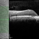
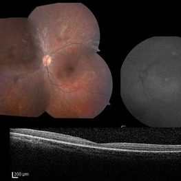
 Dome-Shaped Macula With Pachyvessels
May 3 2024 by Sonia Lee, MD
A 66 year old woman with high miopia with 20/60 vision in her left eye shows an OCT with dome-shaped macula and pachyvessels. A nasal focal thinning of the retina is seen.
Photographer: Sonia Lee, University of Sao Paulo, Brazil.
Imaging device: DRI OCT-1 Triton Topcon
Condition/keywords: dome shaped macula, pachyvessels
-
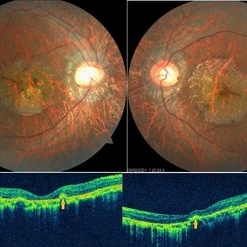
 Extensive Macular Atrophy with Pseudodrusen-Like Appearance
Dec 10 2020 by Cláudia Farinha
71-year-old male presented with progressive vision loss OD, now reduced to CF, without nyctalopia. The SD-OCT scans are one year apart and show extensive and progressive macular atrophy with marked disruption of the outer retinal layers and slightly larger vertical diameter, plus choroidal thinning. Reticular pseudodrusen-like deposits are heavily present in the posterior pole and are better depicted in the infra-red. The widefield imaging shows extensive paving stone peripheral degeneration. The patient denies any systemic medication or known disease. No family history of similar findings.
Photographer: Claudia Farinha
Imaging device: Optomap ultra-widefield imaging, Optos
Condition/keywords: macular atrophy
-
 Extensive Macular Atrophy with Pseudodrusen-Like Appearance
Dec 10 2020 by Cláudia Farinha
71-year-old male presented with progressive vision loss OD, now reduced to CF, without nyctalopia. The SD-OCT scans are one year apart and show extensive and progressive macular atrophy with marked disruption of the outer retinal layers and slightly larger vertical diameter, plus choroidal thinning. Reticular pseudodrusen-like deposits are heavily present in the posterior pole and are better depicted in the infra-red. The widefield imaging shows extensive paving stone peripheral degeneration. The patient denies any systemic medication or known disease. No family history of similar findings.
Photographer: Claudia Farinha
Imaging device: Heidelberg Spectralis SD-OCT
Condition/keywords: macular atrophy
-
 Foveal atrophy post Chronic CSCR
Oct 20 2022 by T. P . VIGNESH, MBBS,MS
SD- OCT image of a 45 year old man revealing severe foveal thinning and RPE atrophy post Chronic CSCR.
Photographer: Shivanath
Imaging device: Heidelberg Spectralis
Condition/keywords: chronic central serous chorioretinopathy (CSCR)
-
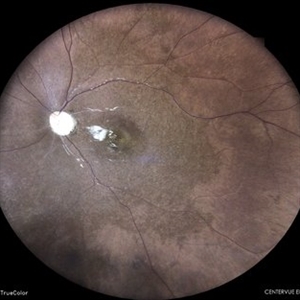
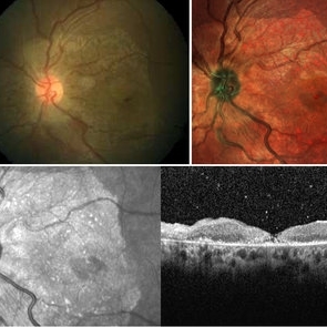 Foveal Thinning Post Blunt Trauma
Aug 25 2018 by Dhaivat Shah
28-year-old male. Post blunt trauma with tennis ball. Fundus color photo shows large area of retinal thinning. Multi color image shows dull red color over fovea, depicting thinning. SD-OCT shows inner retinal ischemia and foveal thinning with early macular hole formation.
Imaging device: Spectralis
Condition/keywords: blunt trauma
-
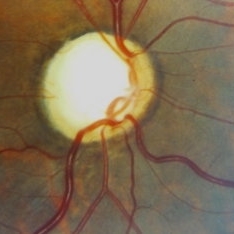 Glaucomatous Optic Atrophy (GOA)
Sep 21 2023 by Ben Serar
Fundus photograph showing increased cup-disc ratio with nasalisation of vessels , with thinning of Neuroretinal rim and bayonetting of vessels in a case of Glaucomatous Optic Atrophy (GOA).
Condition/keywords: Glaucomatous Optic Atrophy (GOA)
-
 Heredomacular Degeneration (HMD)
Sep 12 2023 by Ben Serar
Fundus photograph of RE showing Foveal thinning with scarring in a case of Heredomacular degeneration.
Condition/keywords: foveal scarring, foveal thinning, Heredomacular Degeneration (HMD)
-
 Keratoconus
Oct 2 2013 by Jerald A. Bovino, MD
This is a profile picture of a patient with keratoconus.
Condition/keywords: cornea, degeneration, ectasia, thinning
-
 LCA Type 2
Apr 10 2025 by Joshua Friedman
LCA Type 2 (RPE65) showing characteristic hypoautofluorescence and retinal thinning. 8F with best corrected visual acuity of 20/400 (OD) and 20/150 (OS). Small white intraretinal spots and RPE mottling are visible on color fundus photography. Blue light autofluorescence reveals near-complete loss of signal, while OCT demonstrates widespread outer retinal thinning.
Photographer: Stephen Tsang, MD, PhD
Condition/keywords: Leber Congenital Amaurosis
-
 Mooren Ulcer
May 18 2020 by McGill University Health Centre
A Mooren ulcer is a rapidly progressive ulcerative keratitis that first affects the periphery of the cornea before spreading circumferentially toward its center. Mooren ulcer is a diagnosis of exclusion: it can only be diagnosed after ruling out infectious and systemic causes. In this enucleation specimen, the periphery of the cornea is circumferentially ulcerated (arrow) and the cornea is thinning. There is also scarring of the corneoscleral junction.
Condition/keywords: Mooren's ulcer, ulcerative keratitis
-
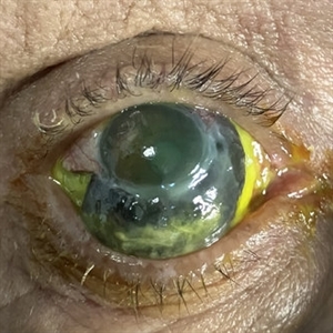 Necrotizing Scleritis
Apr 17 2025 by Gustavo Uriel Fonseca Aguirre
The clinical photograph shows necrotizing scleritis with perilimbal involvement, featuring marked scleral thinning and violaceous episcleral injection in the inferior quadrant. Focal uveal prolapse is visible at the area of maximal scleral necrosis, accompanied by peripheral ulcerative keratitis. Fluorescein staining residue is observed on the ocular surface. Associated findings include mild conjunctival chemosis and dilated episcleral vessels.
Photographer: Gustavo U. Fonseca Aguirre, Hospital Conde de Valenciana, Ciudad de México
Condition/keywords: necrotizing scleritis
 Loading…