Search results (111 results)
-
 Wyburn-Mason Syndrome (Racemose Angioma)
Mar 23 2024 by Pushkar Mahale
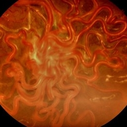
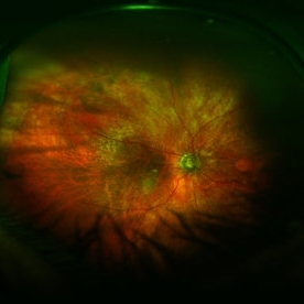
Fundus photograph of a 10 year old child presenting with no perception of light in right eye. Fundus examination revealed dilated and tortuous retinal vessels suggestive of Racemose Hemangioma.
Photographer: Dr Pushkar Mahale
Condition/keywords: racemose hemangioma, Wyburn -Mason Syndrome
-
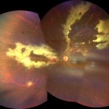
 Amelanotic Melanoma
Sep 19 2025 by Aditya S Kelkar, MS, FRCS, FASRS,FRCOphth
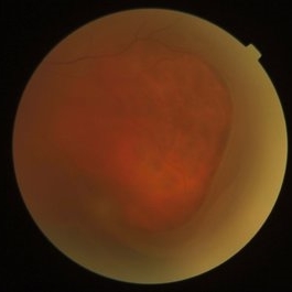
Widefield fundus photograph of a 37 year old showing a large, dome-shaped, intraocular mass involving the temporal retina. The lesion appears elevated and lacks surface pigmentation. Overlying retinal vessels are displaced and draped across the tumor surface, with surrounding retinal elevation noted. The appearance is suggestive of amelanotic variant of choroidal melanoma.
Photographer: Dr. Muskan Mangal
Imaging device: Optos Daytona
Condition/keywords: choroidal melanoma, intraocular tumor
-
 CMV Retinitis
Feb 17 2024 by Eloy Mata-Cortes, MD
Fundus photograph of left eye showing Cytomegalovirus retinitis of a 40-year-old male with positive HIV history. He presented with CD4 cell count of 50 cells/mm3 and decreased vision of left eye. In the photograph we can see the three typical patterns in this retinitis: a hemorrhagic appearance in superior temporal arcade and between nasal arcades, granular pattern in superior temporal retina, and a “frosted branch” angiitis surrounding the retinal vessels in nasal and superior retina.
Photographer: Eloy Mata-Cortes, Instituto Mexicano de Oftalmologia, Queretaro, Mexico
Imaging device: Clarus 700
Condition/keywords: CMV retinitis, cytomegalovirus (CMV), frosted branch angiitis, Frosted Branch Angitis
-
 Macular Pucker
Mar 29 2013 by Henry J. Kaplan, MD
Large epiretinal membrane with straightened vessels in the papillomacular bundle and distorsion of retinal vessels.
Condition/keywords: epiretinal membrane (ERM), macular pucker
-
 Moyamoya: FA 2 Min OD of an Acute CRAO with CRA Sparing
Nov 17 2019 by John S. King, MD
60-year-old white female presented with five days of acute vision loss in the right eye. She was seen initially by referring doctor after hours five days ago and diagnosed with a CRAO and sent to ED to be evaluated stroke team. Right ICA was 100% closed but completely bypassed. She called four days later c/o redness and eye pain; at this point prominent iris vessels were seen, and she was sent to us. Her background history includes a diagnosis of moyamoya (underwent bilateral cerebral artery bypass 2015); atorvastatin for hypercholesterolemia; ASA; no hx of HTN or heart disease. She had a scleral buckle repair OD in 2017 and later developed a thick ERM, which was repaired in 2018; on her previous visit her acuity was noted at 20/40. On presentation her visual acuity was HM OD and 20/15 OS. IOP was 8 OD and 10 OS. There were prominent iris vessels in the right eye, no cell or flare, and an IOL. The posterior segment exam showed diffuse retinal whitening with attenuated vessels and boxcarring; there was sparing retinal whitening in a central area of the macula that appeared to be supplied by a cilio-retina artery. The FA showed very slow filling of the retinal vessels; there was some early perfusion secondary to the cilio-retinal artery. At 7 minutes there was still significant areas of non-perfusion, as well as macular ischemia. Avastin was administered, and one week later, PRP was performed. On the day PRP was performed, the irregular iris vessels had regressed completely. She said that she had a "sliver" of vision centrally in that eye; her acuity was CF 2' and IOP 12.
Photographer: Gretchen Harper
Imaging device: Topcon
Condition/keywords: central retinal artery occlusion (CRAO), cilioretinal sparing, moyamoya, neovascularization of iris (NVI)
-
 Myelinated Nerve Fibers
Apr 18 2025 by DR Rohit Gupta
The **myelinated nerve fibers of the optic disc** (also known as **medullated nerve fibers**) are retinal nerve fibers that retain their myelin sheath as they pass through the optic nerve head. Normally, retinal nerve fibers are unmyelinated to allow for light transparency, but in some cases, myelination extends anteriorly into the retina, appearing as a striking white, feathery patch on the optic disc or peripapillary retina. ### **Key Features:** 1. **Appearance:** - Dense, white, striated patches with feathery edges. - Typically located at the superior or inferior pole of the optic disc. - May obscure retinal vessels underneath. 2. **Clinical Significance:** - Usually **benign** and asymptomatic. - **Congenital** (present at birth or early childhood). - Rarely associated with **visual field defects** (e.g., scotomas corresponding to the area of myelination). - Occasionally linked with **high myopia** or **amblyopia** if extensive. 3. **Pathophysiology:** - Failure of oligodendrocytes or Schwann cells to stop myelination at the lamina cribrosa. - Normally, myelination stops at the optic nerve head, but in this condition, it extends into the retina. 4. **Diagnosis:** - **Fundoscopy:** Classic white, feathery appearance. - **Optical Coherence Tomography (OCT):** Shows thickened retinal nerve fiber layer (RNFL). - **Visual Field Testing:** May detect defects if large. 5. **Differential Diagnosis:** - Optic disc edema - Cotton wool spots - Retinoblastoma (rarely, but must be ruled out in children) 6. **Management:** - No treatment required if asymptomatic. - Monitor for amblyopia in children. - Rare cases with significant visual impairment may need further evaluation. ### **Fun Fact:** Myelinated nerve fibers are seen in **~0.5-1%** of the population and are usually an incidental finding.
Photographer: Dr Rohit gupta
Imaging device: Samsung S21
Condition/keywords: Medulated Nerve fibre, Medullated Nerve fibres, myelinated nerve fibers, Myelinated Nerve Fibres, optic disc drusen
-
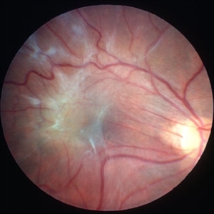
 Optic Atrophy and Attenuated Retinal Vessels Following Endophthalmitis
Jul 12 2014 by Philip J. Polkinghorne, MD
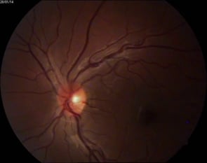
This elderly lady underwent a vitrectomy for post-surgical endophthalmitis. The infection was successfully treated but the functional outcome was poor because of optic atrophy and attenuated retinal vessels.
Photographer: Alex Fraser
Imaging device: Optos Camera
Condition/keywords: attenuated vessels, endophthalmitis, optic atrophy, post-vitrectomy
-
 Outer Retinal Tear in Schisis-Detachment
Mar 25 2016 by Gregory R. Blaha, MD, PhD
Large outer retinal tear in combined retinoschisis-detachment. The retinal vessels are visible going over the retinal break.
Photographer: Janice Neal, Gurley Eye Care Associates
Imaging device: Topcon Mark II
Condition/keywords: retinal tear, retinoschisis
-
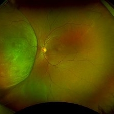
 Remnant of Hyaloidal Artery
Feb 5 2014 by Gerardo Garcia-Aguirre, MD
Video of the fundus of the left eye of a 14-year-old asymptomatic female, where a prepapillary vitreous opacity is observed. The opacity is attached to the origin of the retinal vessels in the optic nerve head, and is considered to be a remnant of the hyaloidal artery.
Photographer: Gerardo Garcia-Aguirre, MD
Condition/keywords: persistence of the hyaloid artery
-
 Retina
May 31 2014 by ruth pav
A 32-year-old woman with a history of drug abuse was admitted due to acute manifestation of multiple infarcts, including acute stroke, splenic and renal infarcts, and multiple cutaneous hematomas. Due to decreased vision in her left eye the patient was referred for ophthalmic evaluation. On exam, visual acuity was 6/10 in the right eye and no light perception in her left eye. Ophthalmoscopic examination was normal in the right eye but showed pallor of the optic nerve head with attenuated retinal vessels in the left eye. Fluorescein angiography showed an oval area of hyperfluorescence from from non-perfusion involving the macular center with staining of overlying retinal capillaries.
Photographer: Ruth Pav, Rambam medical center,Hifa Israel.
Imaging device: Zeiss FF4
Condition/keywords: retina
-
 Intraocular Multiple Cysticercus
Oct 10 2018 by Vishal Agrawal, MD, FRCS,FACS,FASRS
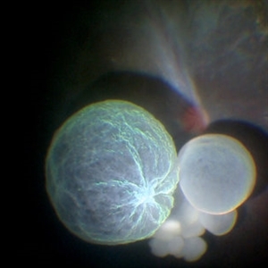
Intraoperative fundus picture of right eye of a 18-year-old boy with complaints of DOV for the past 2 months. There were 12 intravitreal cysts in total with vitritis sclerosis retinal vessels and TRD. To note here, the largest cyst has a flimsy wall and no scolex (possibly ruptured) and the rest of the smaller cysts have a scolex and a taut wall.
Photographer: Vishal Agrawal MD,FRCS
Imaging device: SONY PMW-10 MD HD
Condition/keywords: cysticercosis, scolex
-
 Choroidal Osteoma
Jun 2 2018 by awaneesh m upadhyay, MBBS, DNB
23-year-old patient's fundus photograph having complaints of defective vision, metamorphosia over 6 months shows yellow orange elevated well defined submacular lesion with normal overlying retinal vessels and normal disc . Vision left eye is 20/80.
Photographer: Hiteshwar Saikia
Condition/keywords: macular choroidal osteoma
-
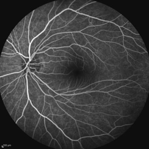
 Tortuous Retinal Vessels
Apr 29 2021 by Giselle DeOliveira
Infared photograph of 30-year-old male with tortuous retinal vessels .
Photographer: Giselle DeOliveira
Imaging device: Heidelberg Spectralis
Condition/keywords: tortuous vessels
-
 Combined Hamartoma of the Retinal Pigment Epithelium Case 1
Oct 5 2012 by Ronald C. Gentile, MD
A peripapilary combined hamartoma of the retinal pigment epithelium involving the nasal disc margin. This tumor is slightly elevated, charcoal grey in color with grey-white tissue on it surface. The underlying retinal vessels are obscured.
Photographer: The New York Eye & Ear Infirmary Department of Medical Imaging
Condition/keywords: hamartoma, retinal pigment epithelium
-
---thumb.jpg/image-square;max$300,300.ImageHandler) Lupus Anticoagulant Disorder
Lupus Anticoagulant Disorder
Feb 26 2013 by Henry J. Kaplan, MD
Occluded retinal vessels and vitreous hemorrhage apparent.
Condition/keywords: lupus anticoagulate factor
-
 Asymptomatic Eye in FEVR
Jul 7 2015 by Hamid Ahmadieh, MD
FA image of the asymptomatic left eye of a 28-year-old man with total RD secondary to advanced FEVR in his right eye. Notice straightening of the retinal vessels.
Photographer: Soulmaz Shahmohammad, Negah Eye Center, Tehran, Iran
Imaging device: Specteralis
Condition/keywords: asymptomatic, familial exudative vitreoretinopathy (FEVR)
-
 Asymptomatic Eye in FEVR
Jul 7 2015 by Hamid Ahmadieh, MD
Color fundus photograph of the asymptomatic eye of a patient with FEVR. Notice straightening of the retinal vessels.
Photographer: Soulmaz Shahmohammad, Negah Eye Center, Tehran, Iran
Condition/keywords: color fundus photograph, familial exudative vitreoretinopathy (FEVR)
-
 BRVO With Non-Perfusion
May 3 2014 by Mallika Goyal, MD
Early phase fluorescein angiogram in an eye with superotemporal BRVO shows delayed filling of retinal vessels in the affected quadrant.
Photographer: Mallika Goyal, MD, Apollo Health City, Jubilee Hills, Hyderabad, India
Condition/keywords: non-perfused branch retinal vein occlusion (BRVO)
-
 BRVO With Non-Perfusion
May 3 2014 by Mallika Goyal, MD
Mid-phase fluorescein angiogram in an eye with superotemporal BRVO shows delayed filling of retinal vessels and non-perfusion in the affected quadrant.
Photographer: Mallika Goyal, MD, Apollo Health City, Jubilee Hills, Hyderabad, India
Condition/keywords: non-perfused branch retinal vein occlusion (BRVO)
-
 BRVO With Non-Perfusion
May 3 2014 by Mallika Goyal, MD
Fluorescein angiogram in an eye with superotemporal BRVO shows delayed filling of retinal vessels and non-perfusion in the affected quadrant.
Photographer: Mallika Goyal, MD, Apollo Health City, Jubilee Hills, Hyderabad, India
Condition/keywords: non-perfused branch retinal vein occlusion (BRVO)
-
 BRVO With Non-perfusion
May 3 2014 by Mallika Goyal, MD
Mid-phase fluorescein angiogram in an eye with superotemporal BRVO shows delayed filling of retinal vessels and non-perfusion in the affected quadrant.
Photographer: Mallika Goyal, MD, Apollo Health City, Jubilee Hills, Hyderabad, India
Condition/keywords: non-perfused branch retinal vein occlusion (BRVO)
-
 BRVO With Non-perfusion
May 3 2014 by Mallika Goyal, MD
Fluorescein angiogram in an eye with superotemporal BRVO shows delayed filling of retinal vessels and non-perfusion in the affected quadrant.
Photographer: Mallika Goyal, MD, Apollo Health City, Jubilee Hills, Hyderabad, India
Condition/keywords: non-perfused branch retinal vein occlusion (BRVO)
-
 BRVO With Non-perfusion
May 3 2014 by Mallika Goyal, MD
Late phase fluorescein angiogram in an eye with superotemporal BRVO shows delayed filling of retinal vessels and non-perfusion in the affected quadrant.
Photographer: Mallika Goyal, MD, Apollo Health City, Jubilee Hills, Hyderabad, India
Condition/keywords: non-perfused branch retinal vein occlusion (BRVO)
-
 BRVO With Non-perfusion
May 3 2014 by Mallika Goyal, MD
Late phase fluorescein angiogram in an eye with superotemporal BRVO shows delayed filling of retinal vessels and non-perfusion in the affected quadrant .
Photographer: Mallika Goyal, MD, Apollo Health City, Jubilee Hills, Hyderabad, India
Condition/keywords: non-perfused branch retinal vein occlusion (BRVO)
-
 BRVO With Non-perfusion
May 3 2014 by Mallika Goyal, MD
Late phase fluorescein angiogram of an eye with superotemporal BRVO shows delayed filling of retinal vessels, dilation and tortuosity of the affected veins, and non-perfusion in the affected quadrant .
Photographer: Mallika Goyal, MD, Apollo Health City, Jubilee Hills, Hyderabad, India
Condition/keywords: non-perfused branch retinal vein occlusion (BRVO)
 Loading…