Search results (648 results)
-
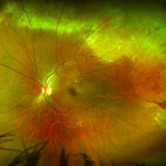
 Massive Subretinal Hemorrhage With Near Total Retina Detachment
Nov 27 2013 by David W. Faber, MD
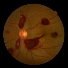
Fundus photo of an 71-year-old male with massive subretinal hemorrhage. Had been given 6 week Avastin treatments. Was put on coumadin for 6 weeks following knee surgery.
Photographer: Donna Knight, Rocky Mountain Retina Consultants, Salt Lake City, Utah
-
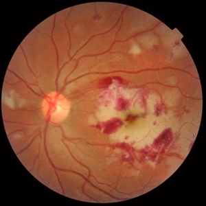
 Ocular Manifestation of Acute Leukemia
Sep 8 2012 by Hamid Ahmadieh, MD
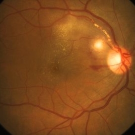
Color fundus photograph of a 26-year-old man with acute leukemia.
Photographer: Hamid Ahmadieh, MD, Ophthalmic Research Center, Labbafinejad Medical Center, Shahid Beheshti University of Medical Sciences , Tehran
Imaging device: Topcon Fundus Camera
Condition/keywords: acute leukemia, white centered retinal hemorrhage (Roth Spot)
-
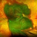
 Choroidal Melanoma
Nov 3 2022 by pedro fernandes souza neto
Transillumination of Enucleation specimen of Choroidal Melanoma: anterior chamber is closed. Total secondary retinal detachment with subretinal serous fluid and some subretinal hemorrhages are present.
Photographer: Eduardo Marback, Federal University of Bahia, Brazil
Condition/keywords: enucleation, melanoma
-
 CMV Retinitis with Frosted Branch Angiitis
Sep 23 2020 by Nimesh A. Patel, MD, FASRS
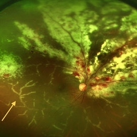
Fundus photo showing peri-vascular inflammation of both arteries and veins with translucent exudation (yellow arrow). Superior nasally, there is classic retinal whitening with retinal hemorrhages superior. This patient was found to have a low CD4 count and a diagnosis of AIDS was made.
Condition/keywords: cytomegalovirus (CMV), HIV, uveitis
-
 Choroidal Fracture
Oct 27 2024 by César Adrián Gómez Valdivia, MD
Fundus photograph of a traumatic choroidal fracture & extra-macular sub-retinal hemorrhage.
Photographer: @eyemissu2
Imaging device: TOPCON TRC-50DX
Condition/keywords: Choroidal Fracture
-
 Lady in a dress
Feb 9 2023 by Shelby Helton
Fluorescein Angiography on a 67-year-old male with significant RPE changes secondary to a severe subretinal hemorrhage that required a vitrectomy with subretinal TPA in 2013.
Photographer: Shelby Helton
Imaging device: Heidelberg Spectralis
Condition/keywords: wet age-related macular degeneration (wet AMD)
-
 Pancytopenia
Mar 24 2016 by Mitzy E Torres Soriano, MD
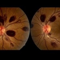
Fundus photographs of a 35-year-old woman with pancytopenia.
Photographer: Mitzy E. Torres Soriano, Hospital Central de Maracay, Venezuela
Condition/keywords: pancytopenia, retinal hemorrhage
-
 Subretinal BSS and air
Apr 12 2022 by Nassim Alejandro Abreu Arbaje, MD
67 year old female who presented with complaints of 5 days of decreased vision of her left eye. She underwent PPV + BSS and Air injection in the subretinal space
Photographer: Nassim Abreu, Dr. Elias Santana Hospital
Imaging device: Ngenuity 3D system screenshot
Condition/keywords: subretinal hemorrhage
-
 Acute Idiopathic Occlusive Retinal Vasculitis
May 31 2014 by Hamid Ahmadieh, MD
Color fundus photograph of the right eye of a 28-year-old woman with sudden drop of vision due to acute occlusive retinal vasculitis leading to extensive nerve fiber layer infarction and retinal hemorrhages.
Photographer: Naghmeh Nozhat, Negah Eye Center, Tehran
Condition/keywords: color fundus photograph, cotton wool spots, retinal hemorrhage, retinal ischemia
-
 Aggressive Posterior Retinopathy of Prematurity with Macular Hemorrhage
Oct 9 2012 by Audina M. Berrocal, MD FASRS
APROP with multiple pre-retinal hemorrhages
Photographer: Ditte Hess CRA, BPEI
Imaging device: RETCAM
Condition/keywords: macular hemorrhage, retinopathy of prematurity (ROP)
-
 Benign Idiopathic Hemorrhagic Retiopathy - RF Image
Feb 6 2013 by Roy Schwartz, MD
A healthy 31-year-old female presented with visual deterioration in her left eye. On examination thick macular retinal hemorrhages were seen, as well as some small minopr hemorrhages in the periphery. A workup for hypercoagulability was negative, and sources of emboli were not found. A diagnosis of a unilateral idiopathic hemorrhagic retinopathy was made.
Photographer: Galit Yair-Pur
Condition/keywords: benign idiopathic hemorrhagic retinopathy, red-free
-
 Cat Scratch Disease
Mar 29 2021 by Gabriel Costa Andrade, PhD
Fundus photograph of an 36-year-old woman with a macular vasculitis, pre retinal hemorrhage and exudation due to Bartonella henselae infection.
Photographer: Gabriel Andrade
Condition/keywords: cat scratch retinitis
-
 Commotio Retinae with Retinal Hemorrhages
Mar 27 2018 by Nichole Lewis
14-year-old male hit in the right eye with a stick. Commotio Retinae with retinal hemorrhages and peripapillary hemorrhage.
Photographer: Nichole Lewis
Condition/keywords: commotio retinae, peripapillary hemorrhage, retinal hemorrhage
-
 Leukemic Retinopathy
Oct 9 2012 by Sharon Fekrat, MD FACS FASRS
22-year-old female with new diagnosis of acute myelogenous leukemia. White blood cell count was 35,000,000,000 cells/L. Note Roth Spots.
Photographer: Tiffanie Keaton, Duke Eye Imaging, Durham, NC
Condition/keywords: acute leukemia, white centered retinal hemorrhage (Roth Spot)
-
 Massive Commotio Retinae
Oct 20 2020 by Veronika Yehezkeli
Fundus photograph of a 24-year-old male, made after blunt trauma with a plastic bottle. Note massive commotio retinae and preretinal hemorrhages in the contralateral to trauma area.
Photographer: Veronika Yehezkeli, Meir medical center, Israel
Condition/keywords: blunt trauma, commotio retinae, preretinal hemorrhage, trauma
-
 Massive SRH in Patient on Coumadin Being Treated for Exudative AMD
Sep 30 2019 by John S. King, MD
78-year-old white female using 1mg of warfarin for atrial fibrillation, who had a large PED, Type 1 lesion from AMD. Noticed acute darkening of vision one week after anti-VEGF injection. Has very large SRH, subRPE heme, and corrugated retinal appearance post RPE tear. Vision HM (from 20/100). 20/25 in the fellow eye that has dry AMD.
Photographer: Shelly Blair
Imaging device: Optos CA
Condition/keywords: subretinal hemorrhage, wet age-related macular degeneration (wet AMD)
-
 PEHCR (Peripheral Exudative Hemorrhagic Chorioretinopathy)
May 12 2023 by Niloofar Piri, MD
Ultrawide fundus photograph of the left eye demonstrating extensive peripheral hemorrhagic exudative detachment in a 79 yo Caucasian female with prior history of non-exudative AMD. Recent diagnosis of Acute myeloid leukemia with low platelet count which might have contributed to the above presentatuon. Please note the temporal subretinal hemorrhage as well as RPE atrophy and hyperplasia in the macula.
Photographer: Rocio Bentivegna, MD, Saint Louis University; Jessica Maddox, COA, Saint Louis University
Condition/keywords: peripheral exudative hemorrhagic chorioretinopathy (PEHCR)
-
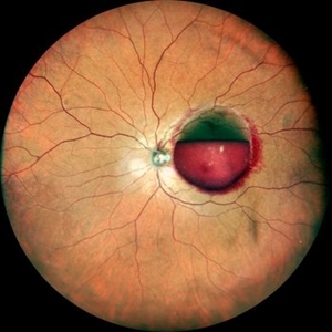 Premacular Subhyaloid Hemorrhage
Jan 20 2021 by Nivesh Gupta
A 41-year-old male patient complaining of diminution of vision in left eye since 6 days. His best corrected visual acuity finger counting at 2 meters.
Photographer: Nivesh Gupta, Retina Fellow, Retina Foundation, Ahmedabad, India
Condition/keywords: hypertensive retinopathy, preretinal hemorrhage, subhyaloid hemorrhage
-
 Preretinal Hemorrhage
May 6 2017 by Mitzy E Torres Soriano, MD
Fundus photograph of a 36-year-old-woman with a preretinal subhyaloid hemorrhage (valsalva retinopathy).
Photographer: Mitzy Torres Soriano
Condition/keywords: macular hemorrhage, premacular hemorrhage, preretinal hemorrhage, subhyaloid hemorrhage, valsalva retinopathy
-
 Proliferative Diabetic Retinopathy with Pre-retinal Hemorrhage
Jan 16 2018 by Olivia Rainey
Ultra-wide field pseudo-color image of an 57-year-old male with a large pre-retinal hemorrhage secondary to proliferative diabetic retinopathy affecting his left eye.
Photographer: Olivia Rainey
Imaging device: Optos California
Condition/keywords: color fundus photograph, diabetic mellitus, hemorrhage, left eye, neovascularization (NV), Optos, proliferative diabetic retinopathy (PDR), pseudocolor, ultra-wide field imaging
-
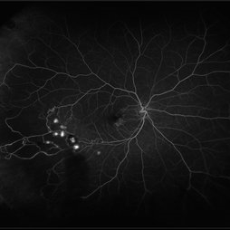 Retinal Hemorrhages
Mar 10 2021 by Kachelle Brown
Ultra widefield Fluorescein Angiography of a 48-year-old female with retinal hemorrhages affecting her right eye. Physician suspect sickle cell due to family history, and has ordered labs to rule out.
Photographer: Kachelle Brown
Imaging device: Optos California
Condition/keywords: fluorescein angiogram (FA), fluorescein leakage, Optos, retinal hemorrhage, sickle cell retinopathy, ultra-wide field imaging
-
---thumb.jpg/image-square;max$300,300.ImageHandler) Active Choroidal Neovascularization With Subretinal Hemorrhage
Active Choroidal Neovascularization With Subretinal Hemorrhage
Nov 25 2013 by Maurice F. Rabb
Active choroidal neovascularization with subretinal hemorrhage.
Condition/keywords: choroidal neovascularization (CNV), subretinal hemorrhage
-
 Acute Idiopathic Occlusive Retinal Vasculitis
May 31 2014 by Hamid Ahmadieh, MD
Color fundus photograph of the left eye of a 28-year-old woman with acute drop of vision due to occlusive retinal vasculitis leading to extensive nerve fiber layer infarction and retinal hemorrhages.
Photographer: Naghmeh Nozhat, Negah Eye Center, Tehran
Condition/keywords: color fundus photograph, cotton wool spots, retinal hemorrhage, retinal ischemia
-
---thumb.JPG/image-square;max$300,300.ImageHandler) Acute myeloid leukemia
Acute myeloid leukemia
Dec 9 2012 by Mallika Goyal, MD
Right eye of a 21-year-old gentleman with acute myeloid leukemia who is undergoing chemotherapy and has low platelet counts (17,000) shows multiple pre-retinal haemorrhages. Other eye has similar picture. There is no vascular occlusion or inflammation. Visual prognosis remains good with spontaneous resolution expected over few weeks.
Photographer: Mallika Goyal, MD, Apollo Health City, Hyderabad, India
Condition/keywords: preretinal hemorrhage
-
 Branch Retinal Vein Occlusion
Oct 17 2012 by Sharon Fekrat, MD FACS FASRS
Fluorescein angiography of an inferior perfused branch retinal vein occlusion in the left eye of a middle aged male with hypertension. The foveal avascular zone is irregular. Subretinal hemorrhage is present.
Photographer: John Reaves, Ophthalmic Photographer, Durham VA Medical Center Eye Clinic Imaging Suite, Durham, NC
Imaging device: Fluorescein Angiography
Condition/keywords: branch retinal vein occlusion (BRVO), subretinal hemorrhage
 Loading…