Search results (119 results)
-
 PRP laser
Mar 29 2013 by Henry J. Kaplan, MD
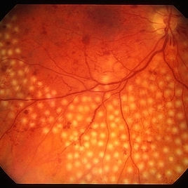
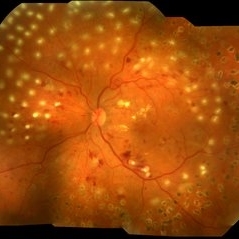
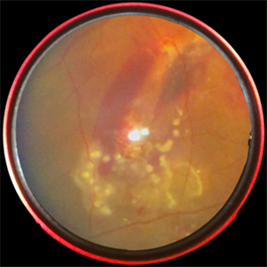
Right after PRP laser in PDR.
Condition/keywords: laser photocoagulation, pan-retinal photocoagulation (PRP)
-
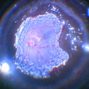
 Aggressive Posterior Retinopathy of Prematurity (APROP)
May 16 2025 by KANWALJEET HARJOT MADAN, M.S. (Ophthalmology); FAICO (Vitreous - Retina)
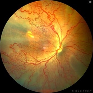
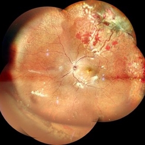
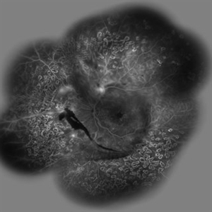
This is the fundus picture of right eye of a premature neonate depicting Aggressive Posterior Retinopathy of Prematurity (APROP). It is a severe rapidly progressing form of retinopathy that can lead to vision loss and blindness. It requires prompt diagnosis and treatment in the form of anti-VEGF agents and laser photocoagulation.
Photographer: Dr. Kanwaljeet Harjot Madan, Thind Eye Hospital, Jalandhar City (Punjab) INDIA.
Imaging device: Zeiss Clarus
Condition/keywords: Oxygen Exposure, retinopathy of prematurity (ROP)
-
 Coloboma involving the Optic nerve, Retina, and Choroid
Dec 6 2021 by Jesus Lozano, MD
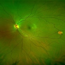
78-year-old woman after prophylactic laser photocoagulation (PLP) for her RE Coloboma involving the optic nerve, retina, and choroid. At 6 month follow up, patient preserved her FC vision as it was before the procedure. Retina attached.
Photographer: Yair Bet Yosef, Hadassah Medical Center. Israel
Imaging device: Optos Silverstone fundus image
Condition/keywords: coloboma, coloboma of choroid, coloboma of macula, coloboma of optic disc, PLP, prophylactic photocoagulation
-
 CSCR Mushroom Cloud
Feb 25 2015 by James J. Bedrick, MD
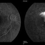
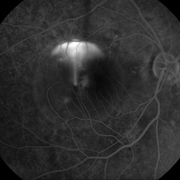
Late transit FA of a large active subfoveal CSCR leak. Focus is on peri-foveal vessels to give sense of height of large serous RD of macula. This patient presented with a BCVA of 20/200 and fluorescein and historic evidence of prior episodes of leakage. After discussion of known treatment options including observation, he was initially treated with rifampin and had partial resolution to 20/70 BCVA but this was short-lived with reaccumulation of the large serous detachment within 3 months. He then received sub-threshold micro-pulse laser photocoagulation with an 810 nm diode laser which resulted 1 month later in complete drying of the serous detachment and BCVA of 20/25.
Photographer: Diana Bodnar, COT
Imaging device: Topcon 50X with OIS capture station
Condition/keywords: CSCR subfoveal leak
-
 CSCR Mushroom Cloud
Feb 23 2015 by James J. Bedrick, MD
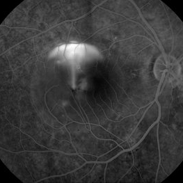
Late transit FA of a large active sub-foveal CSCR leak. You may view this pair in stereo to appreciate the plume of leakage within this large serous RD of the macula. This patient presented with a BCVA of 20/200 and fluorescein and historic evidence of prior episodes of leakage. After discussion of known treatment options including observation, he elected to be treated initially with oral rifampin and BCVA improved to 20/40 with persistent metamorphosis and a shallower persistent macular detachment over several visits. Rifampin was discontinued and he then received sub-threshold micro-pulse laser photocoagulation with an 810 diode which resulted in the patient reporting full restoration of his vision subjectively within a month. He failed to keep his follow-up appointment.
Photographer: Diana Bodnar, COT
Imaging device: Topcon 50X with Merge capture station
Condition/keywords: CSCR subfoveal leak
-
 Diabetic Retinopathy With Laser Photocoagulation.
Sep 10 2017 by JEFFERSON R SOUSA, Tecg.º (Biomedical Systems Technology)
Patient atient 55-years-old, female, attended the clinic with complaint of low visual acuity. It was subjected to laser photocoagulation.
Photographer: JEFFERSON R SOUSA - Study Center and Ophthalmological Research Dr. Andre M V Gomes, Dr. Suel Abujamra Institute São Paulo-Brazil
Imaging device: Topcon TRC-50 DX, Imaginet, 50 degree field. Flash 75, mosaic with eleven images.
Condition/keywords: background diabetic retinopathy (BDR)
-
 Exudative Macular Detachment After Intensive Laser Photocoagulation
Mar 12 2016 by Sjakon G Tahija, MD
Fundus photograph of 44-year-old man with exudative detachment of the macula after vitrectomy and ILM peeling for proliferative diabetic retinopathy combined with intensive endolaser photocagulation.
Photographer: Avris Siahaan, Klinik Mata Nusantara
Condition/keywords: exudative detachment, pan-retinal photocoagulation (PRP)
-
 Familial Exudative Vitreoretinopathy
Nov 25 2022 by Aditya S Kelkar, MS, FRCS, FASRS,FRCOphth
Colour fundus photograph of the right eye of a 56-year-old lady showing lasered FEVR with epiretinal membrane and vitreous band.
Photographer: Dr. Pranali Surawase. National Institute of Ophthalmology, Pune, Maharashtra, India
Imaging device: Zeiss Clarus 500
Condition/keywords: ERM, familial exudative vitreoretinopathy (FEVR), laser photocoagulation
-
 Isolated Retinal Capillary Hemangioblastoma
Mar 11 2022 by Bryon R McKay, MD, PhD, FRCSC, DRCPSC - Retina
Optos widefield fundus photograph and IVFA of a 23-year-old female with asymptomatic isolated retinal capillary hemangioblastoma without exudation. IVFA demonstrates some mild late leakage. The tumor measures 1.5mm and was effectively ablated with laser photocoagulation.
Imaging device: Optos
Condition/keywords: retina capillary hemangioblastoma
-
 Laser Induced BRAO in IRVAN Syndrome
May 3 2019 by Deependra Vikram Singh, MD FASRS
Fundus photograph of a 26-year-old man with IRVAN syndrome referred for vitreous surgery in OS for secondary rhegmatogenous retinal detachment. OD has received laser photocoagulation for capillary nonperfusion areas and retinal artery macroaneurysm associated with retinal vasculitis. Fundus photograph of OD shows laser induced nasal BRAO. Case re-emphasizes why laser for macroaneurysm should be avoided in cases with IRVAN.
Photographer: Deependra V Singh, Eye-Q Superspecialty Eye Hospitals. Gurugram, India
Imaging device: Zeiss Visucam 500
Condition/keywords: arteriolar macroaneurysm, branch retinal artery occlusion (BRAO), laser photocoagulation
-
 Laser Photocoagulation
Nov 9 2012 by Norman Byer
This shows the same lesion four days after laser photo coagulation. The new hemorrhages seen in this photograph did not occur during the photocoagulation but developed within the next four days.
Condition/keywords: argon photocoagulation, laser photocoagulation, preretinal hemorrhage
-
 Lasered Proliferative Diabetic Retinopathy
May 31 2018 by awaneesh m upadhyay, MBBS, DNB
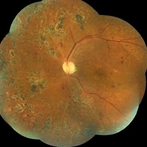
Left eye fundus photography of a 63-year-old male having proliferative diabetic retinopathy after laser photocoagulation.
Photographer: Dr Awaneesh Upadhyay
Condition/keywords: laser photocoagulation, proliferative diabetic retinopathy (PDR)
-
 Retinal Detachment Following Scleral Buckling, Retinectomy, Laser, and Oil
Jan 31 2022 by Ahmad B. Tarabishy, MD
Ultra wide-field fundus photograph of a 55-year-old gentleman who is 4 days after surgery with scleral buckling, pars plana vitrectomy, perfluoron tamponade, membrane peeling, direct fluid-PFO-oil exchange, nasal and temporal retinectomies, and endolaser photocoagulation. Visual acuity was 20/150 under oil.
Photographer: Megan McLandsborough, Lakeland Eye Clinic
Imaging device: Optos California UWF Camera
Condition/keywords: endolaser, Membrane Peel, PPV, proliferative retinopathy, proliferative vitreoretinopathy (PVR), Retinal Detachment, retinal detachment with retinal defect, scleral buckle, submacular perfluorocarbon liquid (PFO)
-
 Subretinal Worm with Laser Marks - Smartphone Fundus Photograph
Jun 13 2019 by Prithvi Chandrakanth
42-year-old, male came with chief complaints of diminished vision and floaters in right eye for past one week. On fundus examination noted to have subretinal haemorrhage and edema at the posterior pole and a subretinal live mobile worm at the periphery. Laser photocoagulation done followed by pars plana vitrectomy.
Photographer: Dr.PRITHVI CHANDRAKANTH, Dr.CHANDRAKANTH MALABAR NETHRALAYA, KOZHIKODE
Imaging device: TRASH TO TREASURE RETCAM - SMARTPHONE FUNDUS CAMERA DEVICE
Condition/keywords: laser photocoagulation, smartphone fundus photography, subretinal hemorrhage, uveitis, worm
-
 Von Hippel Lindau Syndrome
Jun 9 2024 by Anjana Mirajkar, MS Ophthalmology
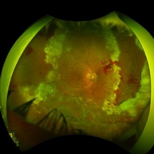
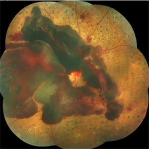
A widefield montage of a 23 year old female of LE case of VHL syndrome showing some hemorrhages with traction superiorly in a silicon oil filled eye with central settled retina. Cryo and laser marks are noted in periphery.
Photographer: Dr. Anjana Mirajkar -Retina Foundation, Ahmedabad
Imaging device: Mirante-Nidek
Condition/keywords: cryotherapy, exudative detachment, laser photocoagulation, vitreous hemorrhage, Von Hippel-Lindau
-
 Retinoblastoma Stage 5 After One Cycle of Systemic Chemotherapy and Laser Ablation
Sep 17 2019 by Sophia El Hamichi, MD
A 1-year-old patient with stage 5B retinoblastoma, fundus after one cycle of systemic chemotherapy and laser ablation.
Photographer: Abby Orcutt-Hayes, Murray Ocular Oncology and Retina
Condition/keywords: chemoreduction, laser photocoagulation, montage, retinoblastoma, stage 5
-
 Subhyaloid Hemorrhage, Proliferative Diabetic Retinopathy
May 31 2018 by awaneesh m upadhyay, MBBS, DNB
Right eye fundus photography of a 63-year-old male came with sudden onset defective vision with history of laser photocoagulation done for proliferative diabetic retinopathy.
Photographer: Dr Awaneesh Upadhyay
Condition/keywords: laser photocoagulation, proliferative diabetic retinopathy (PDR), subhyaloid hemorrhage
-
 CSCR Mushroom Cloud
Feb 25 2015 by James J. Bedrick, MD
Late transit FA of a large active subfoveal CSCR leak. Focus is on peri-foveal vessels to give sense of height of large serous RD of macula. This patient presented with a BCVA of 20/200 and fluorescein and historic evidence of prior episodes of leakage. After discussion of known treatment options including observation, he was initially treated with rifampin and had partial resolution to 20/70 BCVA but this was short-lived with reaccumulation of the large serous detachment within 3 months. He then received sub-threshold micro-pulse laser photocoagulation with an 810 nm diode laser which resulted 1 month later in complete drying of the serous detachment and BCVA of 20/25.
Photographer: Diana Bodnar, COT
Imaging device: Topcon 50X with OIS capture station
Condition/keywords: CSCR subfoveal leak
-
 Proliferative Diabetic Retinopathy (PDR)
Sep 11 2012 by Hamid Ahmadieh, MD
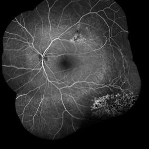
Wide- field FA image of a 55-year-old woman with active PDR and the history of scatter laser photocoagulation.
Photographer: Hamid Ahmadieh, MD, Ophthalmic Research Center, Labbafinejad Medical Center, Shahid Beheshti University of Medical Sciences
Imaging device: Heidelberg HRA
Condition/keywords: preretinal hemorrhage, retinal neovascularization, scatter laser photocoagulation
-
 Coats' Disease
Aug 24 2018 by Kim Barrett
Montage fluorescein angiography of 14-year-old male with Coats' Disease of the left eye. Multiple focal laser treatments. Current uncorrected visual acuity is 20/15-1 OU.
Photographer: Kim Barrett, C.O.A. Retina Specialist of Michigan
Imaging device: Heidelberg Spectralis
Condition/keywords: adolescent, Coats' disease, fluorescein angiogram (FA), Heidelburg Spectralis, laser photocoagulation, left eye, macroaneurysm, montage
-
 Proliferative Diabetic Retinopathy
Sep 15 2012 by Hamid Ahmadieh, MD
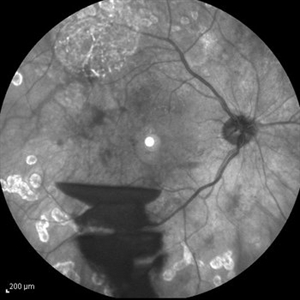
Infrared image of a 30-year-old woman with the history of scatter laser photocoagulation and a preretinal hemorrhage due to active PDR .
Photographer: Hamid Ahmadieh, MD, Ophthalmic Research Center, Labbafinejad Medical Center, Shahid Beheshti University of Medical Sciences
Imaging device: Heidelberg HRA
Condition/keywords: infrared image, preretinal hemorrhage
-
 Regressed Proliferative Diabetic Retinopathy following PRP
Sep 6 2012 by Sharon Fekrat, MD FACS FASRS
58-year-old man with regressed proliferative diabetic retinopathy in the left eye following panretinal laser photocoagulation. Note attenuated retinal vasculature.
Photographer: Sarah Enfiedjian, Ophthalmic Photographer, Durham VA Medical Center, Durham, NC
Imaging device: Zeiss
Condition/keywords: attenuated vessels, pan-retinal photocoagulation (PRP)
-
 Marked Retinal Ischemia in Patient with Mixed Connective Tissue Disease
Feb 26 2013 by Sharon Fekrat, MD FACS FASRS
Fluorescein angiogram of the right eye of a 27-year-old female with mixed connective tissue disease and marked retinal ischemia. Panretinal laser photocoagulation (PRP) has been performed for neovascularization elsewhere (NVE).
Condition/keywords: mixed connective tissue disease, retinal ischemia
-
 Proliferative Diabetic Retinopathy
Sep 15 2012 by Hamid Ahmadieh, MD
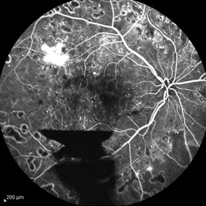
FA image of a 30-year-old woman with the history of scatter laser photocoagulation, NVE and a preretinal hemorrhage due to active PDR .
Photographer: Hamid Ahmadieh, MD, Ophthalmic Research Center, Labbafinejad Medical Center, Shahid Beheshti University of Medical Sciences
Imaging device: Heidelberg HRA
Condition/keywords: preretinal hemorrhage
-
 360 Degree Retinectomy
Feb 2 2022 by Manish Nagpal, MD, FRCS (UK), FASRS
Intraoperative photo of a case of retinal detachment with extensive PVR, which underwent 360 degree relaxing retinectomy followed by 360 laser barrage just prior to silicone oil injection.
Photographer: Manish Nagpal, Retina Foundation, Ahmedabad, India
Imaging device: Sony PMW -10 MD surgical camera
Condition/keywords: laser, laser photocoagulation, proliferative vitreoretinopathy (PVR), relaxing retinectomy, retinectomy, silicone oil
 Loading…