Search results (31 results)
-
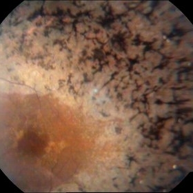
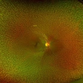 Benign Familial Fleck Retina
Feb 2 2023 by Hemanth Murthy, MBBS, MD, FASRS
12 year boy first born of consanguineous marriage, came for routine eye check up with BCVA 20/40 OU. He has no night blindness. His OCT showed thickening of the RPE with dome like elevations involving the ellipsoid layer. Dark adapted ERG showed normal 'b' wavesPhotopic ERG showed reduced 'a' and b waves.
Photographer: Veda Vyas
Imaging device: Optos Daytona
Condition/keywords: Benign familial fleck retina
-
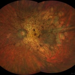 Rod Cone dystrophy
Nov 29 2022 by Niloofar Piri, MD
Fundus photograph of the left eye in a 58 yo male with rod cone dystrophy. He presented with night blindness and peripheral vision loss since youth and recent decrease in central vision for the past 10 years. Notice waxy pallor of the nerve, severe arterial narrowing and chorioretinal atrophy mainly around the arcades as well as posterior pole along with RPE hyperplastic changes and atrophy. RPE atrophy in midperiphery has coin shaped appearance. FAF has characteristic appearance (uploaded separately) He has one pathogenic variants of both CEP290 and PRPH2 genes.
Photographer: Sean Kelso, Saint Louis University
Condition/keywords: hereditary retinal deg, hereditary retinal dystrophy, Rod cone dystrophy
-
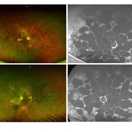
 Rod Cone dystrophy
Nov 29 2022 by Niloofar Piri, MD
Fundus autofluorescence of the left eye in a 58 yo male with rod cone dystrophy. He presented with night blindness and peripheral vision loss since youth and recent decrease in central vision for the past 10 years. Notice multiple coin shaped hypoautofluorescent pacthes within central 20 degrees which are coalescing centrally. (fundus photo uploaded separately) He has one pathogenic variants of both CEP290 and PRPH2 genes.
Photographer: Sean Kelso, Saint Louis University
Condition/keywords: hereditary retinal degeneration, hereditary retinal dystrophy, rod cone dystrophy
-
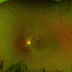
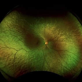
 Mizuo Nakamura phenomenon
Apr 16 2022 by Hemanth Murthy, MBBS, MD, FASRS
Oguchi's disease showing the Mizo Nakamura phenomenon in wide field Fundus photo
Photographer: Mr Veda Vyas
Imaging device: Optos Daytona
Condition/keywords: congenital stationary night blindness (CSNB)
-
 Gyrate Atrophy
Oct 31 2018 by Dhaivat Shah
50-year-old male came in with complaint of daytime vision loss for a year and nighttime vision loss for more than 20 years, gradually increasing day by day. Fundus showed paving-stone like areas of atrophy of the RPE involving the macula which coalesces to form a characteristic scalloped border at the junction of normal and abnormal RPE. Gyrate atrophy is an autosomal recessive dystrophy caused by tenfold elevations of plasma ornithine, which is toxic to the RPE and choroid. Patients with gyrate atrophy have hyperpigmented fundi, with lobular loss of the RPE and choroid, normally sparing the fovea. The finding of generalized hyperpigmentation of the remaining RPE helps to clinically distinguish gyrate atrophy from choroideremia. Affected patients usually develop night blindness during the first decade of life and experience progressive loss of visual field and visual acuity later in the disease course. Early diagnosis is crucial because treatment in form of Arginine free diet and oral pyridoxine helps in slowing the progression of disease.
Imaging device: Optos
Condition/keywords: fundus autofluorescence (FAF), gyrate atrophy
-
 Fundus Albipunctatus
Mar 29 2013 by Henry J. Kaplan, MD
Fundus albipunctatus (one of the stationary night blindness syndromes with multiple white dots in the periphery and normal optic disc and vessels).
Condition/keywords: fundus albipunctatus
-
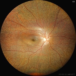 Benign Familial Fleck Retina
Dec 2 2024 by KANWALJEET HARJOT MADAN, M.S. (Ophthalmology); FAICO (Vitreous - Retina)
This is fundus picture of a 21 year old female patient who had come for refractive surgery consultation. Her best corrected vision in both eyes was 20/20. She had myopic astigmatism in both eyes. Fundus exam revealed presence of multiple yellowish white flecks spread throughout retina sparing macular area in both eyes. Her color vision was normal. Electroretinogram and electrooculogram were normal. She gave no history of night blindness. A diagnosis of Benign Familial Fleck Retina was made. She was also advised ocular exam of her parents and elder brother which was normal.
Photographer: Dr. Kanwaljeet Harjot Madan, M.S. (Ophthalmologist) Fellow in Vitrous & Retina. Thind Eye Hospital, Jalandhar City. Punjab. India
Imaging device: Zeiss Clarus
Condition/keywords: Benign familial fleck retina, Night Blindness
-
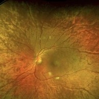
 Benign familial Fleck Retina-left eye
Feb 2 2023 by Hemanth Murthy, MBBS, MD, FASRS
12 year boy first born of consanguineous marriage, came for routine eye check up with BCVA 20/40 OU. He has no night blindness. His OCT showed thickening of the RPE with dome like elevations involving the ellipsoid layer. Dark adapted ERG showed normal 'b' wavesPhotopic ERG showed reduced 'a' and b waves.
Photographer: Veda Vyas
Imaging device: Optos Daytona
Condition/keywords: Benign familial Fleck Retina
-
 Choroideremia
May 8 2024 by KANWALJEET HARJOT MADAN, M.S. (Ophthalmology); FAICO (Vitreous - Retina)
These are the fundus pics of a 28 year young male who presented with history of night blindness. Fundus examintaion revealed presence of Choroideremia. There is diffuse pigment clumping followed by atrophy of retinal pigment epithelium, photoreceptors and choriocapillaris with visible sclera and choroidal vessels in this condition. Atrophy progresses centripetally and the fovea is the last to become affected.
Photographer: Dr. Kanwaljeet Harjot Madan
Imaging device: Zeiss Clarus
Condition/keywords: choriocapillaris, choroideremia, nightblindness
-
 CSNB - Oguchi's Disease
Feb 9 2021 by Dinesh Rungta, MBBS, DNB
• Golden tapetal reflex suggestive of CSNB - Oguchi disease. • MFERG – shows grossly reduced scotopic responses with normal photopic responses in both eyes
Photographer: Dr Shivam Madan , Giridhar Eye Institute, Kerala, India
Imaging device: CARL ZEISS FF450 FUNDUS CAMERA
Condition/keywords: congenital stationary night blindness (CSNB), multifocal ERG (MFERG), Oguchi's disease
-
CSNB-ERG-crop
Aug 17 2021 by Christine Kay, MD
This is a full-field ERG of a patient with X-linked incomplete congenital stationary night blindness with proven mutation in CACNA1F, showing a "negative B wave" pattern.
Photographer: Christine Kay, MD
Condition/keywords: X-linked CSNB
-
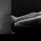
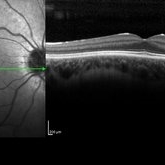 CSNB-OCT-OD
Aug 23 2021 by Jennifer Carstens
OCT/infrared image showing myopic fundus with normal retinal structure in patient with CACNA1F associated X-linked CSNB (OD).
Photographer: Jing Zhang, Ophthalmic Photographer
Condition/keywords: congenital stationary night blindness (CSNB), infrared image, optical coherence tomography (OCT)
-
 CSNB-OCT-OS
Aug 23 2021 by Jennifer Carstens
OCT/infrared image showing myopic fundus with normal retinal structure in patient with CACNA1F associated X-linked CSNB (OS).
Photographer: Jing Zhang, Ophthalmic Photographer
Condition/keywords: congenital stationary night blindness (CSNB), infrared image, optical coherence tomography (OCT)
-
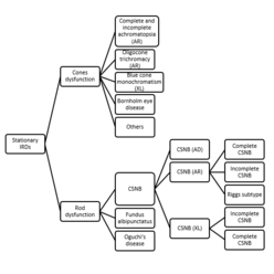 Figure 1: Classification of stationary inherited retinal disease
Dec 15 2023 by Joshua Friedman
Abbreviations: AD, autosomal dominant; AR, autosomal recessive; CSNB, congenital stationary night blindness; IRD, inherited retinal disease; XL, X-linked.
Condition/keywords: stationary IRD
-
 Fundus Albipunctata
Dec 27 2016 by Elad Moisseiev, MD
A 53-year-old female patient with high myopia and complaints of stationary night blindness since childhood. Fundus: myopic fundus with yellow dots in the posterior pole. Genetics: Homozygous mutations in RDH5 gene - c.160C>T (p.R54X), confirming the diagnosis of fundus albipunctata.
Photographer: Galit Yair-Pur
Condition/keywords: fundus albipunctatus
-
 Fundus Albipunctatus
Mar 29 2013 by Henry J. Kaplan, MD
Typical fundus albipunctatus a kind of stationary night blindness; notice the normal disc and vessels #1.
Condition/keywords: fundus albipunctatus
-
 Fundus Albipunctatus
Mar 29 2013 by Henry J. Kaplan, MD
Typical fundus albipunctatus a kind of stationary night blindness ; notice the normal disc and vessels #2.
Condition/keywords: fundus albipunctatus
-
 Fundus Albipunctatus
Apr 27 2021 by Priya Rasipuram Chandrasekaran, MBBS, DO, DNB, FRCS
This is the fundus photo montage of a 23-year-old male demonstrating whitish-yellow spots all over the fundus sparing the fovea at the level of retinal pigment epithelium. This belongs to the group of congenital stationary night blindness with flecks in the retina.
Condition/keywords: fleck retinopathy
-
 Macular Dystrophy
Sep 20 2014 by Mehul A Shah
A 28-year-old female presented with complaint of exotropia and night blindness, on examination she was found to have this picture.
Photographer: Drashti Netralaya,Dahod
Imaging device: Zeiss ff450
Condition/keywords: macular dystrophy
-
 Mizuo Nakamura phenomenon
Apr 16 2022 by Hemanth Murthy, MBBS, MD, FASRS
Oguchi's disease showing the Mizo Nakamura phenomenon in wide field Fundus image
Photographer: Mr Veda Vyas
Imaging device: Optos Daytona
Condition/keywords: congenital stationary night blindness (CSNB)
-
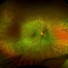
 Mizuo Nakamura phenomenon
Apr 16 2022 by Hemanth Murthy, MBBS, MD, FASRS
Oguchi's disease showing the Mizo Nakamura phenomenon with autofluorescence image showing normal Fundus
Photographer: Mr Veda Vyas
Imaging device: Optos Daytona
Condition/keywords: congenital stationary night blindness (CSNB)
-
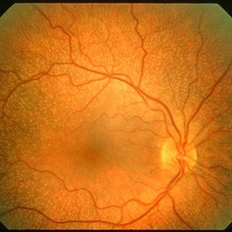
 Oguchi Disease
Aug 12 2025 by Debarun Sharma
A 21 year old female presented with a history of night blindness for the past 16 years when she had difficulty in doing work and navigating places at night. BCVA OU was 6/6. Fundus examination showed the Mizuo-Nakamura phenomenon. ERG was done which showed extinguished rod responses with slightly diminished cone responses. A of Oguchi’s disease was made. The patient was advised for genetic testing and sibling screening. Oguchi’s disease is a rare cause of congenital stationary night blindness with characteristic fundus appearance.
Photographer: Debarun Sharma
Imaging device: Optos
Condition/keywords: Oguchi disease
-
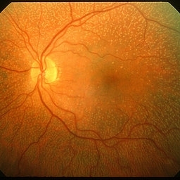
 Oguchi Disease
Sep 27 2024 by juhy cherian
Right eye fundus photograph of a 16 year old girl with golden sheen after light exposure.
Imaging device: Optos image
Condition/keywords: congenital stationary night blindness (CSNB)
-
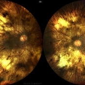
 Oguchi's Disease
Feb 5 2021 by Dinesh Rungta, MBBS, DNB
• Montage image of a 19-year-old male with history of night blindness since childhood showing Bilateral Golden Tapetal Reflex suggestive of CSNB - Oguchi disease. • MFERG – shows grossly reduced scotopic responses with normal photopic responses in both eyes.
Photographer: Dr Shivam Madan, Giridhar Eye Institute, Kerala, India
Imaging device: CARL ZEISS FUNDUS CAMERA
Condition/keywords: multifocal ERG (MFERG), Oguchi's disease
 Loading…