Search results (42 results)
-
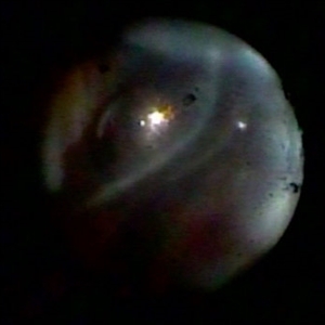
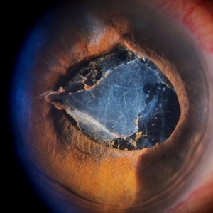 Inflammatory pupillary membrane in patient with endophthalmitis
Jan 28 2023 by Kingston Rodolfo Ureña-Wong, MD, Opht, MSc
Anterior segment photography of a 54-year-old woman with post phacoemulsification endophthalmitis. She did not improve after first intravitreal antibiotics injection and develop an inflammatory pupillary membrane. After two vitrectomies, and a complete three intravitreal injections scheme, we decided to remove the intraocular lens and capsules.
Photographer: Marco Antonio Rubio-Atonal,UNAM, Asociación para evitar la ceguera en México
Imaging device: Zeiss Clarus 700
Condition/keywords: endophthalmitis, pupillary membranes
-
 Proliferative Diabetic Retinopathy
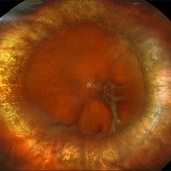
Mar 1 2025 by Neeket R. Patel, MD
A fundus photograph of a 28-year-old male monocular patient diagnosed with proliferative diabetic retinopathy, who is highly motivated to pursue retinal surgery. This case presents a unique challenge in both management and communication, given the patient's strong desire for intervention and the guarded prognosis for visual improvement.
Condition/keywords: Diabetes, membranes, retinopathy, Traction retinal detachment
-
 Repaired Retinal Detachment with Scleral Buckle
Mar 25 2025 by Kimberly Wakester
Optomap RGB montage of an 64-year-old woman with a repaired retinal detachment with scleral buckle in the right eye. There is nasal and inferior pre-retinal membranes with traction. PPV was recommended but patient defers to proceed with sx at this time. Will continue to follow patient closely for worsening traction. Patient was educated on how to monitor their peripheral vision and was advised to report any changes immediately.
Photographer: Kimberly Wakester, COA, OCT-C
Imaging device: Optos California
Condition/keywords: pre-retinal membrane with traction, repaired RD, scleral buckle
-
10 steps to follow in PDR
Sep 5 2024 by DAVID PÉREZ GONZÁLEZ, MD
Cases of proliferative diabetic retinopathy with fibrovascular membranes are among the most complex in retinal surgery. Here are 10 steps I follow in each case to achieve the best possible outcome, particularly when performing one-handed dissection with the vitrector.
Condition/keywords: diabetic retinopathy, Fibrovascular, Membranes, Retinal Surgery
-
25 Gauge Vitrectomy Membrane Shaving
Jan 31 2015 by Thomas A. Ciulla, MD, MBA, FASRS
Membrane shaving of dense membranes in diabetic traction detachment using 25 gauge vitrectomy.
Condition/keywords: diabetes, pars plana vitrectomy (PPV), retina surgery, tractional retinal detachment, vitreoretinal surgery
-
 360 Retinotomy in a closed Funnel combined Tractional and rhegmatogenous retinal detachment
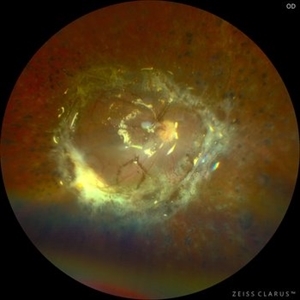
Jan 1 2023 by Malek Yassine, MD
This is the results at 6 months of a Bimanual 23 G-PPV with a very extensive and posterior 360 retinotomy for the management of a combined longstanding closed funnel RD, with submacular membranes, intraretinal PVR. Preop VA was a doubtful light perception. Borders of the retinotomy are stable at 6 months under 1300 Cs Silicon oil with some pigmented PVR developping the edges. Macula appears spared. Silicon oil emulsification droplets are well visualized beneath the superior temporal arcade.
Imaging device: Zeiss Clarus 700
Condition/keywords: combined retinal detachment, retinotomy, silicone oil
-
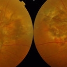
 Advanced Angioid Streak-Associated Choroidal Neovasclar Membranes
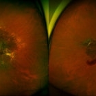
Dec 27 2016 by Young Hee Yoon, MD, PhD
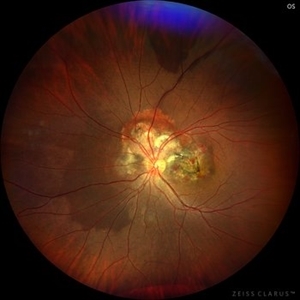
UWF fundus photographs of an 74-year-old woman who received several anti-VEGF injections due to CNV associated with angioid streak in both eyes. Note diffuse scar change and pigmentary degenerations aroud disc in both eyes. There is some retinal hemorrhage due to CNV in her left eye.
Photographer: Young Hee Yoon, University of Ulsan, Asan Medical Center, Seoul, Korea
Imaging device: Optomap
Condition/keywords: angioid streaks, choroidal neovascularization (CNV)
-
 Alports Disease
Jul 29 2013 by H. Michael Lambert, MD
Alports disease, macular diseases not associated with FA changes. Spotty window defects with mid peripheral lesions. Disease may represent ABNL in basement membranes. Basal laminar drusen.
Condition/keywords: Alports disease
-
 Alports Disease
Jul 29 2013 by H. Michael Lambert, MD
Alports disease, macular diseases not associated with FA changes. Spotty window defects with mid peripheral lesions. Disease may represent ABNL in basement membranes. Basal laminar drusen.
Condition/keywords: Alports disease
-
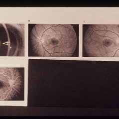
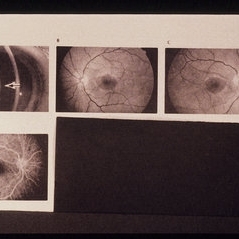
 Angioid Streak-Associated Choroidal Neovasclar Membranes
Dec 27 2016 by Young Hee Yoon, MD, PhD
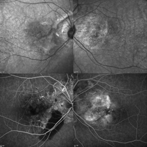
Optical coherence tomogaphs of an 74-year-old woman who received several anti-VEGF injections due to CNV associated with angioid streak in both eyes. There are diffuse CNVM in her right eye and subretinal scar in her left eye. Note the irregular crack in IR image of right eye and the focal Bruch's membrane dehiscence in corresponding B-scan image.
Photographer: Young Hee Yoon, University of Ulsan, Asan Medical Center, Seoul, Korea
Imaging device: Spectralis
Condition/keywords: angioid streaks, choroidal neovascularization (CNV)
-
 Angioid streak-associated choroidal neovasclar membranes
Dec 27 2016 by Young Hee Yoon, MD, PhD
Fundus photographs of an 67-year-old woman with CNV associated with angioid streak in both eyes.
Photographer: Young Hee Yoon, University of Ulsan, Asan Medical Center, Seoul, Korea
Condition/keywords: angioid streaks, choroidal neovascularization (CNV)
-
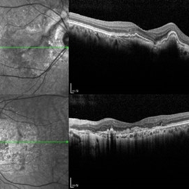
 Angioid streak-associated choroidal neovasclar membranes
Dec 27 2016 by Young Hee Yoon, MD, PhD
Optical coherence tomogaphs of an 67-year-old woman with CNVM associated with angioid streak in both eyes. (upper row : IR image) Irregular crak-like streaks (lower row : FAG image) Block fluorescence due to subretinal hemorrhage in her right eye and classic CNV in her left eye.
Photographer: Young Hee Yoon, University of Ulsan, Asan Medical Center, Seoul, Korea
Imaging device: Spectralis
Condition/keywords: angioid streaks, choroidal neovascularization (CNV)
-
 Aniridic Fibrosis Syndrome - #7 of 7
Jan 24 2013 by Christopher D. Riemann, MD
6-year-old pseudophakic girl with aniridic fibrosis syndrome. Superonasal view with HD endoscope. Note: 20 gauge probe applying endocautery to ciliary body bleed resulting from stripping of epiciliary membranes
Photographer: Christopher Riemann MD, Cincinnati Eye Institute, University of Cincinnati
Imaging device: Endoscope
Condition/keywords: aniridia, epiciliary membrane
-
Bimanual Tractional Retinal Detachment Repair with Viscous Subretinal Fluid Removal
Nov 28 2022 by Nimesh A. Patel, MD, FASRS
Using a chandelier endoillumination, a 2-handed approach with forceps and a vitrector can be used to remove preretinal membranes in a TRD from PDR. The subretinal fluid had a high viscosity. It was removed mechanically by rotating the vitrector.
Condition/keywords: subretinal fluid, video
-
 blue2
blue2
Jun 30 2012 by Stanislao Rizzo, MD
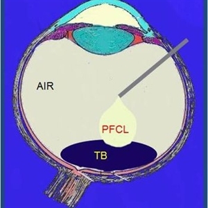
PFO associated with blue staining facilitates the PVR membranes removal
-
 Choroidal Osteoma
Oct 14 2022 by JORGE SOBERANES
A 32-year-old woman with diagnosis of choroidal osteoma and history of multiple choroidal neovascular membranes.
Photographer: }Dr. Jorge I. Soberanes, Asociación para Evitar la Ceguera en México.
Imaging device: Zeiss Clarus 700
Condition/keywords: choroidal osteoma, tumor
-
 Combined Hamartoma of the Retinal Pigment Epithelium Case 2
Oct 5 2012 by Ronald C. Gentile, MD
Magnified view of the peripapilary combined hamartoma of the retinal pigment epithelium involving the inferior disc margin. This tumor and slightly elevated, charcoal grey to light grey in color with grey-white tissue on it surface. The underlying retinal vessels are obscured with some epiretinal vitreous membranes.
Photographer: The New York Eye & Ear Infirmary Department of Medical Imaging
Condition/keywords: hamartoma, retinal pigment epithelium
-
 Diagnosed Case of Sjogren Larsson
Aug 12 2020 by Thirumalesh Mochi Basavaraj, MD
Hyperreflective deposits seen in inner plexiform layers. Sjogren Larsson is inborn error of lipid metabolism caused by mutation in FADH gene which leads to MCFA, LCFA build up specifically in the membranes of skin and brain. This picture shows shows deposits in IPL.
Photographer: Ravikrishna, Puttaswamy
Imaging device: DRI OCT triton SSOCT-Topcon
Condition/keywords: Sjogren's syndrome
-
 Diffuse Uveitis
May 18 2020 by McGill University Health Centre
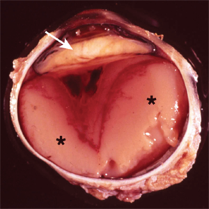
In this enucleation specimen, the uveal tract is completely replaced by purulent material (*). There is a subretinal hemorrhage underlying the detached retina and fibrous material in the vitreous cavity (arrowhead). The lens is cataractous and is surrounded by fibrinoid membranes (arrow).
Condition/keywords: uveitis
-
 Endophtalmitis
Apr 3 2025 by Gustavo Uriel Fonseca Aguirre
B-mode ultrasound imaging of a vitrectomized eye showing vitreous cavity membranes secondary to endophthalmitis.
Photographer: Gustavo U. Fonseca Aguirre, Hospital Conde de Valenciana, Ciudad de México
Condition/keywords: endophthalmitis
-
Endophthalmitis
Apr 9 2025 by Gustavo Uriel Fonseca Aguirre
B-mode ultrasound video of a vitrectomized eye reveals characteristic vitreous cavity membranes secondary to endophthalmitis. The real-time imaging demonstrates these inflammatory membranes exhibit semi-rigid dynamics, displaying viscoelastic behavior with limited displacement during ocular movements while maintaining structural integrity. This restricted mobility pattern, showing both resistance to kinetic forces and slow recoil, represents a pathognomonic feature of advanced fibrotic organization in endophthalmitis.
Condition/keywords: endophthalmitis
-
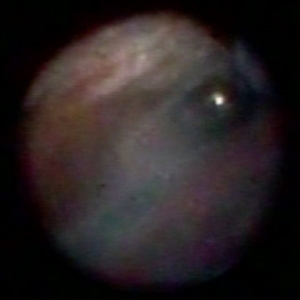
 Endoscopic View - The Start of Anterior Hyaloid Membrane Pneumatic Dissection
Oct 2 2019 by Radwan S. Ajlan, MBBCh, FRCS(C)
Endoscopic view - the start of anterior hyaloid membrane pneumatic dissection.
Condition/keywords: endoscopy, hyaloid, membranes
-
 Endoscopic View: the Beginning of Anterior Hyaloid Membrane Pneumatic Dissection
Oct 2 2019 by Radwan S. Ajlan, MBBCh, FRCS(C)
Endoscopic view: the beginning of anterior hyaloid membrane pneumatic dissection.
Condition/keywords: endoscopy, hyaloid, membranes
-
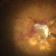 Endstage Diabetic Traction Retinal Detachment
Nov 1 2014 by Maria Stephanie R. Jardeleza, MD
Surgical photograph of optic disc avulsion secondary to extensive diabetic traction retinal membranes over the optic nerve head.
Photographer: Maria Stephanie R. Jardeleza, M.D., UTHSCSA-Texas Diabetes Institute
Imaging device: Zeiss Lumera Operating Microscope
Condition/keywords: proliferative diabetic retinopathy (PDR), tractional retinal detachment
-
 Funnel Retinal Detachment
Jun 11 2023 by Ethan K Sobol, MD
Intraoperative view of a funnel retinal detachment with proliferative vitreoretinoapthy in an eye with previous open globe injury. PVR membranes were peeled, and the retina was flattened and re-attached with an inferior relaxing retinotomy and silicone oil tamponade
Condition/keywords: intraoperative, open funnel RD, open globe injury, proliferative vitreoretinopathy (PVR)
 Loading…