Search results (42 results)
-
Endophthalmitis
Apr 9 2025 by Gustavo Uriel Fonseca Aguirre
B-mode ultrasound video of a vitrectomized eye reveals characteristic vitreous cavity membranes secondary to endophthalmitis. The real-time imaging demonstrates these inflammatory membranes exhibit semi-rigid dynamics, displaying viscoelastic behavior with limited displacement during ocular movements while maintaining structural integrity. This restricted mobility pattern, showing both resistance to kinetic forces and slow recoil, represents a pathognomonic feature of advanced fibrotic organization in endophthalmitis.
Condition/keywords: endophthalmitis
-
 Endophtalmitis
Apr 3 2025 by Gustavo Uriel Fonseca Aguirre
B-mode ultrasound imaging of a vitrectomized eye showing vitreous cavity membranes secondary to endophthalmitis.
Photographer: Gustavo U. Fonseca Aguirre, Hospital Conde de Valenciana, Ciudad de México
Condition/keywords: endophthalmitis
-
 Repaired Retinal Detachment with Scleral Buckle
Mar 25 2025 by Kimberly Wakester
Optomap RGB montage of an 64-year-old woman with a repaired retinal detachment with scleral buckle in the right eye. There is nasal and inferior pre-retinal membranes with traction. PPV was recommended but patient defers to proceed with sx at this time. Will continue to follow patient closely for worsening traction. Patient was educated on how to monitor their peripheral vision and was advised to report any changes immediately.
Photographer: Kimberly Wakester, COA, OCT-C
Imaging device: Optos California
Condition/keywords: pre-retinal membrane with traction, repaired RD, scleral buckle
-
 Proliferative Diabetic Retinopathy
Mar 1 2025 by Neeket R. Patel, MD
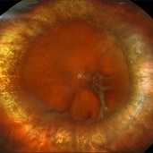
A fundus photograph of a 28-year-old male monocular patient diagnosed with proliferative diabetic retinopathy, who is highly motivated to pursue retinal surgery. This case presents a unique challenge in both management and communication, given the patient's strong desire for intervention and the guarded prognosis for visual improvement.
Condition/keywords: Diabetes, membranes, retinopathy, Traction retinal detachment
-
10 steps to follow in PDR
Sep 5 2024 by DAVID PÉREZ GONZÁLEZ, MD
Cases of proliferative diabetic retinopathy with fibrovascular membranes are among the most complex in retinal surgery. Here are 10 steps I follow in each case to achieve the best possible outcome, particularly when performing one-handed dissection with the vitrector.
Condition/keywords: diabetic retinopathy, Fibrovascular, Membranes, Retinal Surgery
-
 Membranes Formed Under Silicon Oil and Retina
Jul 18 2024 by Anjana Mirajkar, MS Ophthalmology
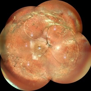
A montage color photo of RE of a 7 year old male with membranes formed between silicon and the retina injected for retinal detachment.
Photographer: Dr. Anjana Mirajkar -Retina Foundation, Ahmedabad
Imaging device: Mirante-Nidek
Condition/keywords: sub-silicon membranes
-
 Proliferative Vitreoretinopathy
Jun 9 2024 by Marcelo Zas, MD PhD
We present a case of a 20-year-old patient who underwent surgery for congenital cataract when he was born and 20 years after he developed a retinal detachment with proliferative vitreoretinopathy. Proliferative vitreoretinopathy (PVR), a major complication of rhegmatogenous retinal detachment (RRD), is an abnormal process whereby proliferative, contractile cellular membranes form in the vitreous and on both sides of the retina, resulting in tractional retinal detachment with fixed retinal folds. PVR arises in an estimated 5-10% of RRD cases, and therefore represents a major complication of retinal detachment. The best treatment of PVR is its prevention. Clinical factors associated with increased risk of PVR include: • Chronic RRD • 2 o more horseshoe retinal tears and RRD exposing three-disc diameters or more of RPE • RD associated with giant retinal • RD associated with choroidal detachment • Ocular Trauma • RRD associated with vitreous hemorrhage • Aphakia and RRD • Failure of previous surgery or multiple retinal surgeries • Aggressive retinitis, etc.
Photographer: Luciano Scorsetti MD
Condition/keywords: proliferative vitreoretinopathy (PVR)
-
 Macular Telangiectasia Type 2
Mar 29 2024 by Lucy V Cobbs, M.D.
Color fundus photograph of the left eye of a 70-year-old male with a disciform scar resulting from a neovascular membrane. A minority of MacTel type 2 patients develop neovascular disease, and the gold standard treatment is anti-VEGF intravitreal therapy. Without treatment, membranes may progress to severe central macular scarring. Late stages of proliferative MacTel type 2 may be confused with AMD, and a differentiating aspect is that MacTel type 2 typically lacks pigment epithelial detachments and drusen.
Condition/keywords: Mac Tel type 2
-
 Proliferative vitreoretinopathy in complex retinal redetachment
Dec 6 2023 by Ma. Guadalupe Perez Guevara
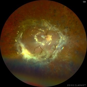
A 58-year-old female underwent phacovitrectomy + scleral buckling by retinal detachment. Silicon oil was used as endo tamponade. In the post-surgical period, the formation of a subretinal PRV is observed with a significant circumferential contraction that generates an inferior redetachment. It was decided to undergo a vitreous cavity lavage to remove membranes + 180° retinectomy.
Photographer: Ma. Guadalupe Pérez-Guevara. Fundación Hospital Nuestra Señora de la Luz I.A.P
Imaging device: Optos
Condition/keywords: PVR, retina surgery complications
-
 Funnel Retinal Detachment
Jun 11 2023 by Ethan K Sobol, MD
Intraoperative view of a funnel retinal detachment with proliferative vitreoretinoapthy in an eye with previous open globe injury. PVR membranes were peeled, and the retina was flattened and re-attached with an inferior relaxing retinotomy and silicone oil tamponade
Condition/keywords: intraoperative, open funnel RD, open globe injury, proliferative vitreoretinopathy (PVR)
-
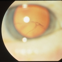
 Inflammatory pupillary membrane in patient with endophthalmitis
Jan 28 2023 by Kingston Rodolfo Ureña-Wong, MD, Opht, MSc
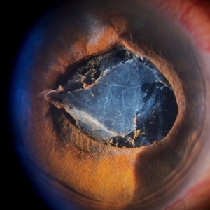
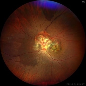
Anterior segment photography of a 54-year-old woman with post phacoemulsification endophthalmitis. She did not improve after first intravitreal antibiotics injection and develop an inflammatory pupillary membrane. After two vitrectomies, and a complete three intravitreal injections scheme, we decided to remove the intraocular lens and capsules.
Photographer: Marco Antonio Rubio-Atonal,UNAM, Asociación para evitar la ceguera en México
Imaging device: Zeiss Clarus 700
Condition/keywords: endophthalmitis, pupillary membranes
-
Vitrectomy TRD in Proliferative diabetic retinopathy
Jan 2 2023 by Manish Nagpal, MD, FRCS (UK), FASRS
Vitrectomy for PDR and TRD using Cutter based dissection| This is a case of subhyaloid hemorrhage and Tractional retinal detachment in a diabetic patient. The subhyaloid hemorrhage is aspirated using the cutter . 25 gauge bevelled cutter is used to dissect all the epiretinal proliferations and tractional components. The ports of these cutters can reach very close to the retinal surface and cut flush without causing any iatrogenic damage to the retinal surface. Forceps are used to peel adherent membranes Bleeders are stopped raising pressure and applying diathermy. Once the retina is flattened endolaser is done 360 degree to achieve long term regression.
Condition/keywords: cutter, diabetic retinopathy, endolaser, forceps, PDR, peeling, proliferative diabetic retinopathy (PDR), PRP, tractional retinal detachment, TRD, video, vitrectomy
-
 360 Retinotomy in a closed Funnel combined Tractional and rhegmatogenous retinal detachment
Jan 1 2023 by Malek Yassine, MD
This is the results at 6 months of a Bimanual 23 G-PPV with a very extensive and posterior 360 retinotomy for the management of a combined longstanding closed funnel RD, with submacular membranes, intraretinal PVR. Preop VA was a doubtful light perception. Borders of the retinotomy are stable at 6 months under 1300 Cs Silicon oil with some pigmented PVR developping the edges. Macula appears spared. Silicon oil emulsification droplets are well visualized beneath the superior temporal arcade.
Imaging device: Zeiss Clarus 700
Condition/keywords: combined retinal detachment, retinotomy, silicone oil
-
Bimanual Tractional Retinal Detachment Repair with Viscous Subretinal Fluid Removal
Nov 28 2022 by Nimesh A. Patel, MD, FASRS
Using a chandelier endoillumination, a 2-handed approach with forceps and a vitrector can be used to remove preretinal membranes in a TRD from PDR. The subretinal fluid had a high viscosity. It was removed mechanically by rotating the vitrector.
Condition/keywords: subretinal fluid, video
-
Surgery for retinal detachment with starfolds
Oct 24 2022 by Manish Nagpal, MD, FRCS (UK), FASRS
This video show s vitrectomy being done for retinal detachment with starfolds, membranes are removed following which the sequential steps of settling a retinal detachment are carried out
Photographer: Manish Nagpal
Condition/keywords: PVR, RD, starfolds, tears, video, vitrectomy
-
 Choroidal Osteoma
Oct 14 2022 by JORGE SOBERANES
A 32-year-old woman with diagnosis of choroidal osteoma and history of multiple choroidal neovascular membranes.
Photographer: }Dr. Jorge I. Soberanes, Asociación para Evitar la Ceguera en México.
Imaging device: Zeiss Clarus 700
Condition/keywords: choroidal osteoma, tumor
-
 Human Embryonic Eye
Sep 1 2020 by J. Sebag, MD, FACS, FRCOphth, FARVO
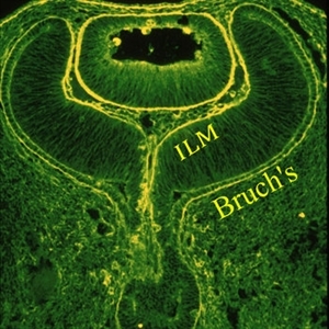
Immunohistochemistry of a human fetal eye after invagination of the optic vesicle demonstrates basement membrane staining with fluorescein conjugated ABA lectin staining. Continuity of the membranes destined to be Bruch’s membrane and the Inner Limiting Membrane (ILM) of the retina is evident (see loops at upper aspects of this image), suggesting a common embryologic origin and similar composition. Thus, there may be important similarities during pathologies (especially neovascularization) at these two interfaces, which may provide insights into the process of pathologic cell migration and proliferation. Anterior to the ILM and behind the lens is the vascular primary vitreous. [Cover Photo - Sebag J and Hageman G: Interfaces. Farina Publishers, Rome, 2000; reprinted in Sebag J: Vitreous – in Health & Disease (J. Sebag, ed.) Springer, New York, 2014; image © Springer Nature, reprinted with permission]
Condition/keywords: embryonic eye
-
 Diagnosed Case of Sjogren Larsson
Aug 12 2020 by Thirumalesh Mochi Basavaraj, MD
Hyperreflective deposits seen in inner plexiform layers. Sjogren Larsson is inborn error of lipid metabolism caused by mutation in FADH gene which leads to MCFA, LCFA build up specifically in the membranes of skin and brain. This picture shows shows deposits in IPL.
Photographer: Ravikrishna, Puttaswamy
Imaging device: DRI OCT triton SSOCT-Topcon
Condition/keywords: Sjogren's syndrome
-
 Toxoplasmic Uveitis
May 18 2020 by McGill University Health Centre
This enucleation specimen shows multiple irregular chorioretinal scars, surrounded by hyperplastic retinal pigment epithelium (arrowheads). Some adherent membranes are present in the vitreous cavity (arrow). The lens has a total cataract and the anterior chamber is filled with whitish material that corresponds to hypopyon.
Condition/keywords: toxoplasmosis uveitis, uveitis
-
 Diffuse Uveitis
May 18 2020 by McGill University Health Centre
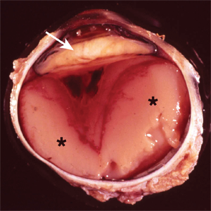
In this enucleation specimen, the uveal tract is completely replaced by purulent material (*). There is a subretinal hemorrhage underlying the detached retina and fibrous material in the vitreous cavity (arrowhead). The lens is cataractous and is surrounded by fibrinoid membranes (arrow).
Condition/keywords: uveitis
-
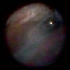
 Endoscopic View - The Start of Anterior Hyaloid Membrane Pneumatic Dissection
Oct 2 2019 by Radwan S. Ajlan, MBBCh, FRCS(C)
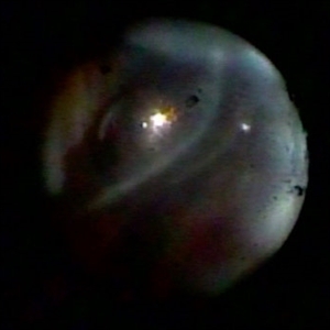
Endoscopic view - the start of anterior hyaloid membrane pneumatic dissection.
Condition/keywords: endoscopy, hyaloid, membranes
-
 Endoscopic View: the Beginning of Anterior Hyaloid Membrane Pneumatic Dissection
Oct 2 2019 by Radwan S. Ajlan, MBBCh, FRCS(C)
Endoscopic view: the beginning of anterior hyaloid membrane pneumatic dissection.
Condition/keywords: endoscopy, hyaloid, membranes
-
 Vitreous Membranes
Apr 8 2019 by Gary R. Cook, MD, FACS
60-year-old white female with vitreous syneresis and membranes
Imaging device: Topcon VT-50
Condition/keywords: vitreous membranes and strands
-
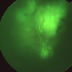 Vitreous Membranes
Apr 8 2019 by Gary R. Cook, MD, FACS
Red-free photograph of the vitreous membrane ahead of the optic disc in the patient with retinal vasculitis.
Imaging device: Topcon VT-50
Condition/keywords: retinal vasculitis, vitreous membranes and strands
-
 Vitreous Membranes
Apr 8 2019 by Gary R. Cook, MD, FACS
Vitreous membrane ahead of the optic disc in a patient with retinal vasculitis.
Condition/keywords: retinal vasculitis, vitreous membranes and strands
 Loading…