Search results (42 results)
-
 Subretinal Bands in CTRRD
Mar 1 2014 by Homayoun Tabandeh, MD, FASRS
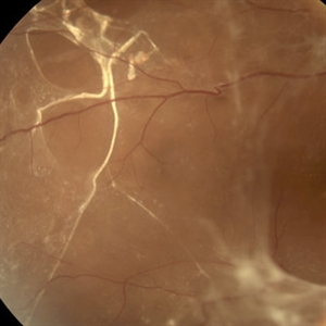
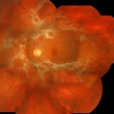
Fundus photograph of a diabetic man with subretinal and pre-retinal bands and membranes associated with combined tractional rhegmatogenous retinal detachment due to proliferative diabetic retinopathy.
Photographer: Daniel Rivas
Condition/keywords: proliferative diabetic retinopathy (PDR), subretinal bands, tractional retinal detachment
-
 Combined Hamartoma of the Retinal Pigment Epithelium Case 2
Oct 5 2012 by Ronald C. Gentile, MD
Magnified view of the peripapilary combined hamartoma of the retinal pigment epithelium involving the inferior disc margin. This tumor and slightly elevated, charcoal grey to light grey in color with grey-white tissue on it surface. The underlying retinal vessels are obscured with some epiretinal vitreous membranes.
Photographer: The New York Eye & Ear Infirmary Department of Medical Imaging
Condition/keywords: hamartoma, retinal pigment epithelium
-
 Neurofibromatosis 2
Oct 9 2012 by Audina M. Berrocal, MD FASRS
Typical vertical membranes seen in cases of NF2
Photographer: BPEI
Condition/keywords: neurofibromatosis
-
 Angioid Streak-Associated Choroidal Neovasclar Membranes
Dec 27 2016 by Young Hee Yoon, MD, PhD
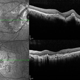
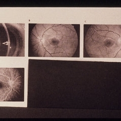
Optical coherence tomogaphs of an 74-year-old woman who received several anti-VEGF injections due to CNV associated with angioid streak in both eyes. There are diffuse CNVM in her right eye and subretinal scar in her left eye. Note the irregular crack in IR image of right eye and the focal Bruch's membrane dehiscence in corresponding B-scan image.
Photographer: Young Hee Yoon, University of Ulsan, Asan Medical Center, Seoul, Korea
Imaging device: Spectralis
Condition/keywords: angioid streaks, choroidal neovascularization (CNV)
-
 Retinal Detachment With PVR and Macular Hole
Aug 1 2017 by Manish Nagpal, MD, FRCS (UK), FASRS
65 year old woman had come to us post macular hole surgery with further drop in vision due to the onset of a retinal detachment leading to PVR and enlargement of the macular hole
Photographer: pooja barot
Condition/keywords: macular hole, membranes, proliferative vitreoretinopathy (PVR), star folds
-
25 Gauge Vitrectomy Membrane Shaving
Jan 31 2015 by Thomas A. Ciulla, MD, MBA, FASRS
Membrane shaving of dense membranes in diabetic traction detachment using 25 gauge vitrectomy.
Condition/keywords: diabetes, pars plana vitrectomy (PPV), retina surgery, tractional retinal detachment, vitreoretinal surgery
-
 Aniridic Fibrosis Syndrome - #7 of 7
Jan 24 2013 by Christopher D. Riemann, MD
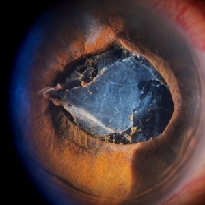
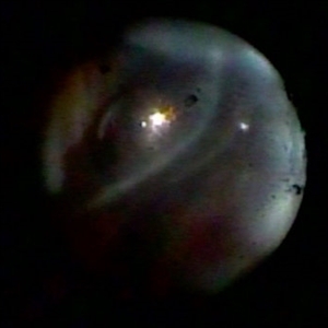
6-year-old pseudophakic girl with aniridic fibrosis syndrome. Superonasal view with HD endoscope. Note: 20 gauge probe applying endocautery to ciliary body bleed resulting from stripping of epiciliary membranes
Photographer: Christopher Riemann MD, Cincinnati Eye Institute, University of Cincinnati
Imaging device: Endoscope
Condition/keywords: aniridia, epiciliary membrane
-
 Endstage Diabetic Traction Retinal Detachment
Nov 1 2014 by Maria Stephanie R. Jardeleza, MD
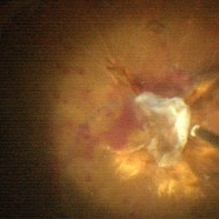
Surgical photograph of optic disc avulsion secondary to extensive diabetic traction retinal membranes over the optic nerve head.
Photographer: Maria Stephanie R. Jardeleza, M.D., UTHSCSA-Texas Diabetes Institute
Imaging device: Zeiss Lumera Operating Microscope
Condition/keywords: proliferative diabetic retinopathy (PDR), tractional retinal detachment
-
 Angioid streak-associated choroidal neovasclar membranes
Dec 27 2016 by Young Hee Yoon, MD, PhD
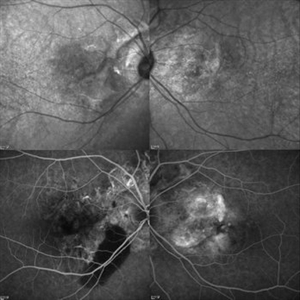
Optical coherence tomogaphs of an 67-year-old woman with CNVM associated with angioid streak in both eyes. (upper row : IR image) Irregular crak-like streaks (lower row : FAG image) Block fluorescence due to subretinal hemorrhage in her right eye and classic CNV in her left eye.
Photographer: Young Hee Yoon, University of Ulsan, Asan Medical Center, Seoul, Korea
Imaging device: Spectralis
Condition/keywords: angioid streaks, choroidal neovascularization (CNV)
-
 Post Endophthalmitis Sub-Silicon Membranes
Jul 11 2016 by Manish Nagpal, MD, FRCS (UK), FASRS
Post endophthalmitis sub silicon membranes.
Condition/keywords: endophthalmitis
-
 Inflammatory pupillary membrane in patient with endophthalmitis
Jan 28 2023 by Kingston Rodolfo Ureña-Wong, MD, Opht, MSc
Anterior segment photography of a 54-year-old woman with post phacoemulsification endophthalmitis. She did not improve after first intravitreal antibiotics injection and develop an inflammatory pupillary membrane. After two vitrectomies, and a complete three intravitreal injections scheme, we decided to remove the intraocular lens and capsules.
Photographer: Marco Antonio Rubio-Atonal,UNAM, Asociación para evitar la ceguera en México
Imaging device: Zeiss Clarus 700
Condition/keywords: endophthalmitis, pupillary membranes
-
 Advanced Angioid Streak-Associated Choroidal Neovasclar Membranes
Dec 27 2016 by Young Hee Yoon, MD, PhD
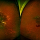
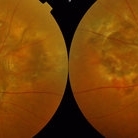
UWF fundus photographs of an 74-year-old woman who received several anti-VEGF injections due to CNV associated with angioid streak in both eyes. Note diffuse scar change and pigmentary degenerations aroud disc in both eyes. There is some retinal hemorrhage due to CNV in her left eye.
Photographer: Young Hee Yoon, University of Ulsan, Asan Medical Center, Seoul, Korea
Imaging device: Optomap
Condition/keywords: angioid streaks, choroidal neovascularization (CNV)
-
 Alports Disease
Jul 29 2013 by H. Michael Lambert, MD
Alports disease, macular diseases not associated with FA changes. Spotty window defects with mid peripheral lesions. Disease may represent ABNL in basement membranes. Basal laminar drusen.
Condition/keywords: Alports disease
-
 Funnel Retinal Detachment
Jun 11 2023 by Ethan K Sobol, MD
Intraoperative view of a funnel retinal detachment with proliferative vitreoretinoapthy in an eye with previous open globe injury. PVR membranes were peeled, and the retina was flattened and re-attached with an inferior relaxing retinotomy and silicone oil tamponade
Condition/keywords: intraoperative, open funnel RD, open globe injury, proliferative vitreoretinopathy (PVR)
-
 Diagnosed Case of Sjogren Larsson
Aug 12 2020 by Thirumalesh Mochi Basavaraj, MD
Hyperreflective deposits seen in inner plexiform layers. Sjogren Larsson is inborn error of lipid metabolism caused by mutation in FADH gene which leads to MCFA, LCFA build up specifically in the membranes of skin and brain. This picture shows shows deposits in IPL.
Photographer: Ravikrishna, Puttaswamy
Imaging device: DRI OCT triton SSOCT-Topcon
Condition/keywords: Sjogren's syndrome
-
 blue2
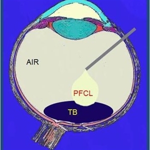
blue2
Jun 30 2012 by Stanislao Rizzo, MD
PFO associated with blue staining facilitates the PVR membranes removal
-
 Alports Disease
Jul 29 2013 by H. Michael Lambert, MD
Alports disease, macular diseases not associated with FA changes. Spotty window defects with mid peripheral lesions. Disease may represent ABNL in basement membranes. Basal laminar drusen.
Condition/keywords: Alports disease
-
 Angioid streak-associated choroidal neovasclar membranes
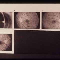
Dec 27 2016 by Young Hee Yoon, MD, PhD
Fundus photographs of an 67-year-old woman with CNV associated with angioid streak in both eyes.
Photographer: Young Hee Yoon, University of Ulsan, Asan Medical Center, Seoul, Korea
Condition/keywords: angioid streaks, choroidal neovascularization (CNV)
-
 Slide 2-16
Feb 19 2019 by Lancaster Course in Ophthalmology
Organization of fibrinous exudate from iris, causing posterior synechiae and pupillary membrane.
Condition/keywords: fibrinous exudate, membranes, posterior synechiae
-
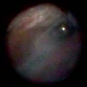 Endoscopic View: the Beginning of Anterior Hyaloid Membrane Pneumatic Dissection
Oct 2 2019 by Radwan S. Ajlan, MBBCh, FRCS(C)
Endoscopic view: the beginning of anterior hyaloid membrane pneumatic dissection.
Condition/keywords: endoscopy, hyaloid, membranes
-
 Toxoplasmic Uveitis
May 18 2020 by McGill University Health Centre
This enucleation specimen shows multiple irregular chorioretinal scars, surrounded by hyperplastic retinal pigment epithelium (arrowheads). Some adherent membranes are present in the vitreous cavity (arrow). The lens has a total cataract and the anterior chamber is filled with whitish material that corresponds to hypopyon.
Condition/keywords: toxoplasmosis uveitis, uveitis
-
 Endoscopic View - The Start of Anterior Hyaloid Membrane Pneumatic Dissection
Oct 2 2019 by Radwan S. Ajlan, MBBCh, FRCS(C)
Endoscopic view - the start of anterior hyaloid membrane pneumatic dissection.
Condition/keywords: endoscopy, hyaloid, membranes
-
 Diffuse Uveitis
May 18 2020 by McGill University Health Centre
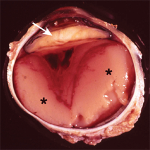
In this enucleation specimen, the uveal tract is completely replaced by purulent material (*). There is a subretinal hemorrhage underlying the detached retina and fibrous material in the vitreous cavity (arrowhead). The lens is cataractous and is surrounded by fibrinoid membranes (arrow).
Condition/keywords: uveitis
-
 Slide 10-12
Feb 26 2019 by Lancaster Course in Ophthalmology
Anterior subcapsular cataract associated with severe inflammation of the anterior segment and an inflammatory pupillary membrane ( x40). (Scheie Eye Institute, No. 3952.)
Condition/keywords: anterior subcapsular polar cataract, pupillary membranes
-
 Human Embryonic Eye
Sep 1 2020 by J. Sebag, MD, FACS, FRCOphth, FARVO
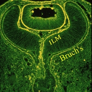
Immunohistochemistry of a human fetal eye after invagination of the optic vesicle demonstrates basement membrane staining with fluorescein conjugated ABA lectin staining. Continuity of the membranes destined to be Bruch’s membrane and the Inner Limiting Membrane (ILM) of the retina is evident (see loops at upper aspects of this image), suggesting a common embryologic origin and similar composition. Thus, there may be important similarities during pathologies (especially neovascularization) at these two interfaces, which may provide insights into the process of pathologic cell migration and proliferation. Anterior to the ILM and behind the lens is the vascular primary vitreous. [Cover Photo - Sebag J and Hageman G: Interfaces. Farina Publishers, Rome, 2000; reprinted in Sebag J: Vitreous – in Health & Disease (J. Sebag, ed.) Springer, New York, 2014; image © Springer Nature, reprinted with permission]
Condition/keywords: embryonic eye
 Loading…