Search results (2800 results)
-
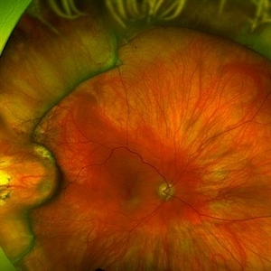
 Wide-Field-OCT-montage
Jan 8 2018 by Netan Choudhry, MD, FRCS(C) FASRS
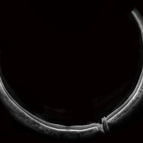
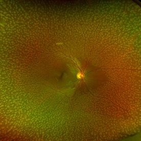
This is an SD-OCT montage image of a 55 year old male with optic neuropathy representing a wide-field OCT spanning 130 degrees.
Photographer: John Golding, Vitreous Retina Macula Specialists of Toronto
Imaging device: Heidelberg Spectralis OCT system
Condition/keywords: wide angle imaging
-
 Branch Retinal Artery Occlusion With Calcium Embolus at the Disc - Fundus Autofluorescence Imaging (FAF)
Apr 7 2018 by Rameez N Hussain, MD
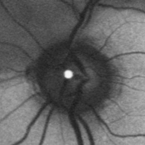
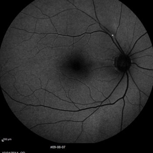
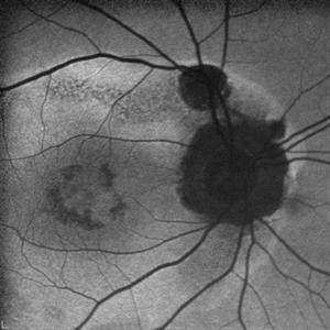
Acute branch retinal artery occlusion with a calcium embolus at the disc which is hyper autofluorescent in fundus autofluorescence imaging (FAF) -resembles an LED light source ('LED sign').
Photographer: DR RAMEEZ N HUSSAIN
Imaging device: Heidelberg Spectralis
Condition/keywords: branch retinal artery occlusion (BRAO), embolus, fundus autofluorescence (FAF), retinal edema
-
 Central Retinal Artery Occlusion & Cilioretinal Artery Sparing
Dec 22 2012 by Hamid Ahmadieh, MD
Early phase FA image of the right eye of a 34-year-old man with sudden drop of vision due to CRAO. The macula is involved despite cilioretinal artery sparing .
Photographer: Zohre Salimi; Labbafinejad Medical Center, Shahid Beheshti University of Medical Sciences , Tehran
Imaging device: Heidelberg HRA
Condition/keywords: central retinal artery occlusion (CRAO), cilioretinal sparing
-
 Oil Bubbles
Apr 27 2018 by Mark Lazcano
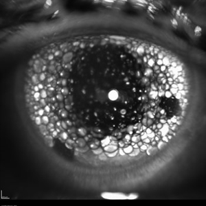
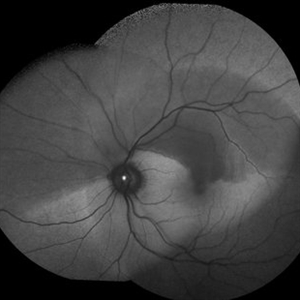
Infrared photograph of 56-year-old male with retinal detachment oil on lens.
Photographer: Mark Lazcano, University of Miami, Bascom Palmer Eye Institute
Imaging device: Heidelberg Spectralis
Condition/keywords: silicone oil
-
 Relentless Placoid Chorioretinitis
Jan 9 2019 by Janet Brazil
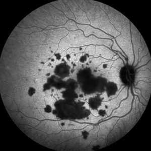
ICG photo of a 24-year-old male with Relentless placoid chorioretinitis
Photographer: Janet Atkinson, Eye Associates of New Mexico
Imaging device: Heidelberg HRA Spectralis
Condition/keywords: placoid retinal lesions
-
 Benign Familial Fleck Retina
Feb 2 2023 by Hemanth Murthy, MBBS, MD, FASRS
12 year boy first born of consanguineous marriage, came for routine eye check up with BCVA 20/40 OU. He has no night blindness. His OCT showed thickening of the RPE with dome like elevations involving the ellipsoid layer. Dark adapted ERG showed normal 'b' wavesPhotopic ERG showed reduced 'a' and b waves.
Photographer: Veda Vyas
Imaging device: Optos Daytona
Condition/keywords: Benign familial fleck retina
-
 BRAO Rianto AF
Apr 12 2014 by Sjakon G Tahija, MD
Auto fluorescence fundus image of a 70-year-old man with a superior temporal branch retinal artery occlusion. The emboli can be very clearly seen as the white dot of AF blockage.
Photographer: Avris Siahaan
Imaging device: Heidelberg Spectralis
Condition/keywords: branch retinal artery occlusion (BRAO)
-
 Choroidal Excavation
Jun 2 2019 by Nelson Chamma Capelanes, MD
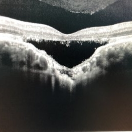
SD-OCT of a 32-year-old woman showing a subfoveal choroidal excavation associated with chronic central serous chorioretinopathy.
Photographer: Nelson Chamma Capelanes, Promacula, Brazil
Imaging device: Heidelberg Spectralis SD-OCT
Condition/keywords: choroidal excavation, chronic central serous chorioretinopathy (CSCR), pachychoroid
-
 Seedlings of Fungal Endophthalmitis
Mar 14 2025 by SHILPI H NARNAWARE, ICO ( Retina) , FAICO ( Vitreo-Retina)
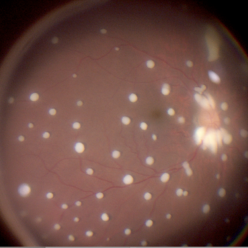
57 year diabetic female , was treated as a case of recurrent vitreous post cataract surgery. Patient was posted for vitrectomy 3 months post cataract surgery. Intra-operatively, multiple yellowish colonies were seen all over the posterior pole were seen, which were later found to be Aspergillus colonies.
Photographer: Shilpi Narnaware, Sarakshi Netralaya , Nagpur, Maharashtra , India
Imaging device: Ngenuity
Condition/keywords: endophthalmitis, fungal
-
 Silicon Oil
Apr 27 2018 by Giselle DeOliveira
Infrared photo of 75-year-old male with retinal detachment.
Photographer: Giselle DeOliveira, University of Miami, Bascom Palmer Eye Institute
Imaging device: Heidelberg Spectralis
Condition/keywords: infrared image, silicone oil
-
 "The Eye of Sauron"
Mar 14 2023 by Anfisa Ayalon, MD
Fundus autofluorescence image of a 38-year-old female with “Bull's eye” pattern maculopathy. There is no history of medication use associated with retinal toxicity. BCVA RE 20/25+2
Photographer: Danielle Ferguson and Alec Bertoni, University of Pittsburgh Medical Center
Condition/keywords: bull's eye maculopathy, retina
-
 Acute Macular Neuroretinopathy
Dec 11 2019 by Lauren Whaley
34-year-old female patient presented with changes in vision after recent upper respiratory infection. Referring doctor originally thought it was a blood pressure issue. She noticed a "C" shape in her vision. Infrared image was captured showing exactly what patient was describing! Doctor confirmed with this image that it was AMN.
Photographer: Lauren R. Whaley, COA
Imaging device: Heidelberg Spectralis
Condition/keywords: 30 degrees, acute macular neuroretinopathy, Heidelburg Spectralis, left eye, macula, near infrared autofluorescence (NIRAF)
-
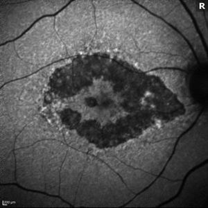
 AZOOR
AZOOR
Mar 19 2015 by Niloofar Piri, MD
#1: Fundus autofluorescence OD in a patient with AZOOR demonstrates characteristic peripapillary hypoAF as well as concentric rings of hypo and hyper AF in posterior pole .
Imaging device: Heidelberg Spectralis
Condition/keywords: acute zonal occult outer retinopathy (AZOOR)
-
 Branch Retinal Artery Occlusion With Calcium Embolus at the Disc - Fundus Autofluorescence Imaging (FAF)
Apr 7 2018 by Rameez N Hussain, MD
Acute branch retinal artery occlusion with a calcium embolus at the disc which is hyper autofluorescent in fundus autofluorescence Imaging (FAF) -resembles an LED light source ('LED sign').
Photographer: DR RAMEEZ N HUSSAIN
Imaging device: Heidelberg Spectralis
Condition/keywords: branch retinal artery occlusion (BRAO), embolus, fundus autofluorescence (FAF), retinal edema
-
 Epiretinal Membrane/Macular Pucker With Combined Hamartoma of Retina and RPE
Jul 8 2015 by Emmanuel Chang, MD PhD FACS FASRS
10-year-old with history of progressive severe distortion in the left eye over the past year.
Photographer: Retina and Vitreous of Texas
Imaging device: Heidelberg Autofluorescence
Condition/keywords: combined hamartoma, epiretinal membrane (ERM), retinal pigment epithelium (RPE) hamartoma
-
 Green Goblin Detachment
Jan 13 2022 by Netan Choudhry, MD, FRCS(C) FASRS
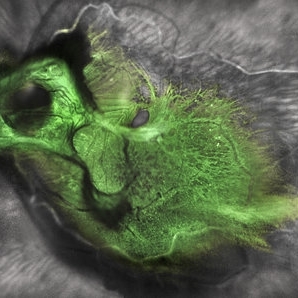
Tractional retinal detachment with macular hole in a 76-year-old female.
Photographer: John Golding BA, Vitreous Retina Macula Specialists of Toronto, OCTane Imaging Lab
Imaging device: Multicolor fundus photo taken on the Spectralis OCT2 (Heidelberg Engineering GmbH).
Condition/keywords: macular hole, Multispectral imaging, tractional retinal detachment
-
 Hourglass in an Eye
Apr 22 2025 by KRISHNENDU NANDI, MS
A twenty-five-year-young male presented with a decrease in vision in the right eye following a blunt trauma with a football. On examination the BCVA in the right eye was CFCF and the left eye was 6/6, N6. The anterior segment was within normal limits. AT was 12 and 10 mm of Hg in the right and left eyes, respectively. Fundus examination reveals subhyaloid haemorrhage in the right eye with an attached retina. The fundus of the left eye was within normal limits. YAG laser hyaloidotomy was done with an energy of 2 mJ in the right eye. After 3 weeks the BCVA in the right eye improved to 6/9, N6.
Photographer: Dr. Krishnendu Nandi
Imaging device: Topcon
Condition/keywords: Trauma, YAG HYALOIDOTOMY, Young Male
-
 Iris
Iris
Apr 29 2019 by Stephanie Moolman
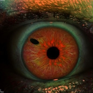
Multi-color images after Yag PI of iris.
Photographer: Stephanie Moolman, Dr Marissa Willemse, Pretoria, South Africa
Imaging device: Heidelberg Spectralis
Condition/keywords: glaucoma, iris, multicolor, NdYAG laser, peripheral iridotomy
-
 Lady in a dress
Feb 9 2023 by Shelby Helton
Fluorescein Angiography on a 67-year-old male with significant RPE changes secondary to a severe subretinal hemorrhage that required a vitrectomy with subretinal TPA in 2013.
Photographer: Shelby Helton
Imaging device: Heidelberg Spectralis
Condition/keywords: wet age-related macular degeneration (wet AMD)
-
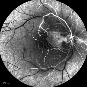 Central Retinal Artery Occlusion & Cilioretinal Artery Sparing
Dec 22 2012 by Hamid Ahmadieh, MD
FA image of the right eye of a 34-year-old man with sudden drop of vision due to CRAO. The macula is involved despite cilioretinal artery sparing .
Photographer: Zohre Salimi; Labbafinejad Medical Center, Shahid Beheshti University of Medical Sciences , Tehran
Imaging device: Heidelberg HRA
Condition/keywords: central retinal artery occlusion (CRAO), cilioretinal sparing
-
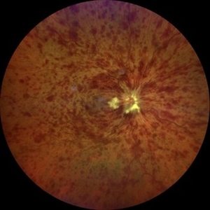 Central Retinal Vein Occlusion associated with disc edema
Oct 19 2023 by Gabriel Costa Andrade, PhD
53-year-old woman with an acute CRVO. The patient has a history of breast cancer undergoing treatment with systemic chemotherapy. Notice the peripapillary cotton wool spots, superficial flame shaped hemorrhages and deeper dot and blot hemorrhages in all 4 quadrants.
Photographer: Gabriel Andrade
Condition/keywords: central retinal vein occlusion (CRVO), macular edema, Retina
-
 Cuticular Drusen
Jan 17 2024 by John Lee
Heidelberg SD-OCT of a 65-year-old woman with age-related macular degeneration demonstrating classic sawtooth appearance of cuticular drusen.
Photographer: Natasha Vinson
Imaging device: Heidelberg Spectralis
Condition/keywords: age-related macular degeneration (AMD), cuticular drusen
-
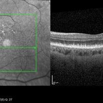
 Dry AMD, Advanced Atrophic without Subfoveal Involvement
Oct 12 2021 by Kelli Nyenhuis
OCT Heidelberg photograph of a 79-year-old woman with AMD advancing that has been observed over the last 10 years.
Photographer: Kelli Nyenhuis, OMA
Imaging device: Heidelberg
Condition/keywords: dry age-related macular degeneration (dry AMD)
-
 Erosion of Segmental Buckle
Feb 25 2022 by Roger A. Goldberg, MD, MBA
Erosion of sharp edge of segmental scleral buckle seen 15 years after being placed for repair of a retinal detachment
Photographer: Melissa Bartlett, Bay Area Retina Associates
Imaging device: Optos
Condition/keywords: retinal defect, scleral buckle
-
 Foveoschisis secondary to high myopia
Mar 13 2015 by Niloofar Piri, MD
Infrared and HD-OCT of the right eye in a 55-year-old African American female with high myopia (more than -6.00 D), BCVA: 20/25 OU Cartwheel appearance of the fovea in the infrared imaging is visible. HD- OCT demonstartes schisis in different layers of the retina (both NFL and OPL; notice stretching of the Muller cells); VMT is also present . Outer retinal layers are preserved which explains the good vision . She had the same findings in OS.
Photographer: Niloofar Piri, MD
Imaging device: Heidelberg Spectralis
Condition/keywords: high myopia, retinoschisis
 Loading…