Search results (78 results)
-
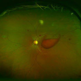
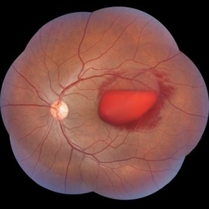
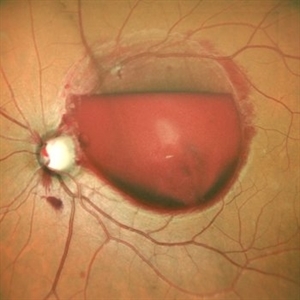 Valsalva Retinopathy
Jul 30 2025 by Akansha Sharma
Color fundus photograph of a 33 year old male with subhyaloid hemorrhage suggestive of valsalva retinopathy.
Photographer: DR. AKANSHA SHARMA
Condition/keywords: subhyaloid hemorrhage, valsalva retinopathy
-
 Valsalva Retinopathy
Jul 30 2025 by Akansha Sharma
Color fundus photograph of a 33 year old male with subhyaloid hemorrhage suggestive of valsalva retinopathy.
Photographer: DR. AKANSHA SHARMA
Condition/keywords: subhyaloid hemorrhage, valsalva retinopathy
-
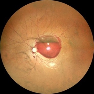
 Fishing Fundus
Jul 16 2025 by Moazzam Parvez
Fundus photograph of a 31 year old woman with sub ILM hemorrhage following her yoga sessions which involves breath holding exercises .
Photographer: Moazzam Parvez , Netralayam , Kolkata
Imaging device: Topcon Maestro 2
Condition/keywords: SUB ILM hemorrhage, valsalva retinopathy
-
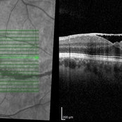 Optical Coherence Tomography of Valsalva Retinopathy at 2 Months
Oct 28 2024 by Andrew Jin, MD
OCT of a 30 year old man with resolving valsalva retinopathy 2 months after presentation.
Condition/keywords: OCT, valsalva retinopathy
-
 Optical Coherence Tomography of Valsalva Retinopathy at Presentation
Oct 28 2024 by Andrew Jin, MD
OCT of a 30 year old man who presented with valsalva retinopathy
Condition/keywords: OCT, valsalva retinopathy
-
 Valsalva Retinopathy
Oct 28 2024 by Andrew Jin, MD
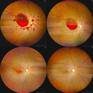
These fundus photos show the progression of subhyaloid hemorrhage due to valsalva retinopathy in a 38-year-old woman with hyperemesis gravidarum. She was managed conservatively with observation alone. The photos depict the initial presentation after 1 day of vision loss, at 1 month, and at 6 months of follow-up. Presenting visual acuity was counting fingers and returned to 20/20.
Condition/keywords: fundus photograph, valsalva retinopathy
-
 Valsalva Retinopathy
Oct 28 2024 by Andrew Jin, MD
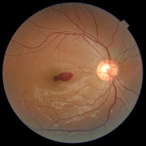
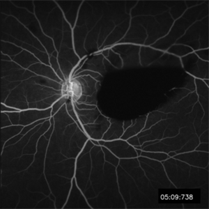
These photos depict the fundus photo and corresponding fluorescein angiogram for a 43 year old man with emesis after food poisoning. Note the blockage from the central preretinal hemorrhage and scattered peripheral intraretinal hemorrhages.
Condition/keywords: fluorescein angiogram (FA), fundus photograph, valsalva retinopathy
-
 Valsalva Retinopathy
Oct 28 2024 by Andrew Jin, MD
These photos depict the fundus photo and corresponding fluorescein angiogram for a 43 year old man with emesis after food poisoning. Note the blockage from the central preretinal hemorrhage and scattered peripheral intraretinal hemorrhages.
Condition/keywords: fluorescein angiogram (FA), fundus photograph, valsalva retinopathy
-
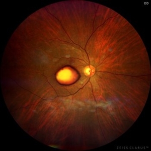
 Valsalva Retinopathy
Sep 10 2024 by KANWALJEET HARJOT MADAN, M.S. (Ophthalmology); FAICO (Vitreous - Retina)
These are fundus pictures of a young 32 years male who presented with sudden decrease in vision in right eye after lifting heavy weights. His vision in right eye was hand movements close to face. He had no systemic history or any history of trauma. He was diagnosed to have Valsalva Retinopathy. His hematological investigations were normal. He refused any active intervention. He was kept under observation and his vision improved to 20/20 within 4 weeks.
Photographer: Dr. Kanwaljeet Harjot Madan, M.S. (Ophthalmologist) Fellow in Vitrous & Retina. Thind Eye Hospital, Jalandhar City. Punjab. India
Imaging device: Zeiss Clarus
Condition/keywords: valsalva retinopathy
-
 Subhyaloid Hemorrhage
Jul 31 2024 by Arthi Mohankumar , MS,MRCS ED, FICO,FAICO
A 35 year old male presented with complaints of seeing a black spot in left eye for past one day after working out in the gym the previous day. He has history of uncontrolled diabetes and hypertension. Fundus exam of the left eye revealed a sub hyaloid hemorrhage nasal to the disc with minimal background Diabetic and hypertensive changes. His baseline CBG was 200 mg/dl and BP was 170/100 He was suggested observation initially considering the nasal location. But patient found the scotoma very disturbing and eventually underwent yag hyaloidotomy
Photographer: Arthi Mohankumar
Condition/keywords: Sub hyaloid haemorrhage, valsalva retinopathy
-
 Valsalva Retinopathy
Apr 19 2024 by Akansha Sharma
Color fundus photograph of a 24 year old male with valsalva retinopathy.
Photographer: Dr. Akansha Sharma, Bharati Eye Hospital
Condition/keywords: valsalva retinopathy
-
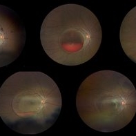 Valsalva retinopathy progression
Oct 4 2023 by Niloofar Piri, MD
Progression fundus photograph images of a 22 yo female with Valsalva retinopathy secondary to violent emesis. Note the sub ILM layered hemorrhage and gradual decrease then disappearance over time. Last image is at 6 month follow up with 20/20 vision. Of note the silhouette of ILM separation is still visible.
Condition/keywords: valsalva retinopathy
-
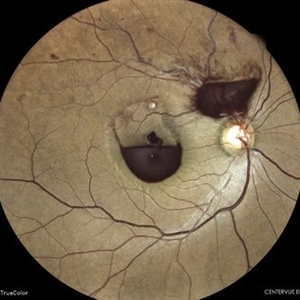
 Subhyaloid Haemorrhage - Valsalva Retinopathy
Jul 30 2023 by Maneesh M Bapaye, MD, MBA
A 25 years old paitient presented with sudden loss of vision following sudden rise intra abdominal pressure, Montage photo
Photographer: Dr.Maneesh Bapaye
Imaging device: Zeiss Fundus Camera
Condition/keywords: subhyaloid hemorrhage, valsalva retinopathy
-
 SUBHYALOID HEMORRHAGE IN A CASE OF VALSALVA RETINOPATHY
Jun 6 2023 by Akansha Sharma
colour fundus photograph of a 40 year old female with subhyaloid hemorrhage in a case of valsalva retinopathy
Photographer: Dr. Denish Patel, Dr. Akansha Sharma, Dr. Urmil Shah, Bharati Eye Hospital, Ahmedabad, Gujarat
Condition/keywords: SHH, subhyaloid hemorrhage
-
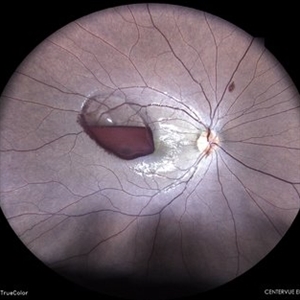
 VALSALVA RETINOPATHY
Jun 6 2023 by Akansha Sharma
COLOUR FUNDUS PHOTOGRAPH OF A 21 YEAR OLD MALE WITH VALSALVA RETINOPATHY
Photographer: Dr. Urmil Shah, Dr. Denish Patel, Dr. Akansha Sharma, Bharati Eye Clinic, Ahmedabad, Gujarat
Condition/keywords: Sub hyaloid haemorrhage, SUB RETINAL HEMORRHAGE, valsalva retinopathy
-
 SUBHYALOID HEMORRHAGE IN A CASE OF VALSALVA RETINOPATHY
May 31 2023 by Akansha Sharma
COLOUR FUNDUS PHOTOGRAPH OF A 20 YEAR OLD MALE WITH SUBHYALOID HEMORRHAGE IN A CASE OF VALSALVA RETINOPATHY
Photographer: Dr. Urmil Shah, Dr. Akansha Sharma, Dr. Denish Patel
Condition/keywords: Sub hyaloid Haemorrhage, valsalva retinopathy
-
 Valsalva Retinopathy
Feb 24 2023 by Harold Rodriguez
Valsalva Retinopathy of a 29 year old female.
Photographer: Harold Rodriguez OCT-C, OSC.
Condition/keywords: valsalva retinopathy
-
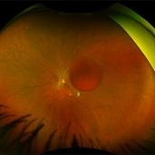
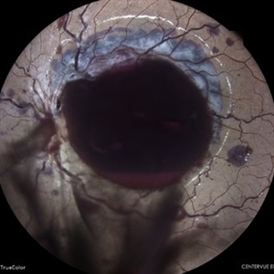
 Valsalva Retinopathy
Nov 18 2022 by Niloofar Piri, MD
Sudden vision loss immediately after severe vomiting. Color fundus photo demonstrates large sub ILM hemorrhage consistent with valsalva retinopathy.
Photographer: Sean Kelso, Saint Louis University
Condition/keywords: SUB ILM hemorrhage, sub internal limiting membrane haemorrhage, valsalva retinopathy
-
 Valsalva Retinopathy
Nov 18 2022 by Niloofar Piri, MD
21 yo female presented with decaresed central vision and scotoma immediately after severe vomiting. Color fundus phtograph demonstrates large sub ILM layered hemorrhage in the macula consistent with valsalva retinopathy. Notice the sacttered blot retinal hemorrhages in mid-periphery.
Photographer: Rocio Bentivegna, MD, Saint Louis University
Condition/keywords: sub ILM hemorrhage, valsalva retinopathy
-
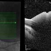
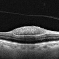 OCT of a sub-internal limiting membrane hemorrhage in Valsalva retinopathy
Jul 29 2022 by JORGE SOBERANES
Optical coherence tomography of a 70-year-old man with a sub-internal limiting membrane due to Valsalva retinopathy
Photographer: Jorge I. Soberanes, Asociación para Evitar la Ceguera en México.
Imaging device: PLEX Elite 9000, Zeiss
Condition/keywords: OCT, sub-inner limiting membrane hemorrhage, swept source, valsalva retinopathy
-
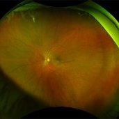
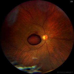 Sub-internal limiting membrane hemorrhage in Valsalva retinopathy
Jul 29 2022 by JORGE SOBERANES
A fundus photography of a 70-year-old man with premacular hemorrhage (Sub-internal limiting membrane) due to Valsalva retinopathy
Photographer: Jorge I. Soberanes, Asociación para Evitar la Ceguera en México.
Imaging device: Zeiss Clarus 700
Condition/keywords: premacular hemorrhage, sub-inner limiting membrane hemorrhage, valsalva retinopathy
-
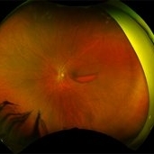
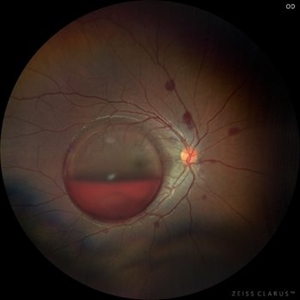
 Dehemoglobinized sub-internal limiting membrane hemorrhage
Jul 29 2022 by JORGE SOBERANES
Fundus photograph of a 70-year-old man with Valsalva retinopathy manifested as premacular hemorrhage (sub-ILM) in dehemoglobinized process.
Photographer: Jorge I. Soberanes, Asociación para Evitar la Ceguera en México.
Imaging device: Zeiss Clarus 700
Condition/keywords: dehemoglobinized hemorrhage, sub-inner limiting membrane hemorrhage, valsalva retinopathy
-
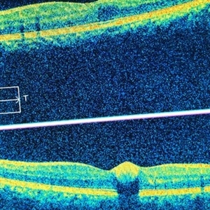 Valsalva Retinopathy
Feb 20 2022 by Ajay Indur Dudani, MBBS MS
OCT of. 24 yr girl with valsalva retinopathy and sub ILM macular haemorrhage
Photographer: Ajay DUDANI,Mumbai Retina Center,Mumbai,India
Imaging device: Zeiss Cirrus
Condition/keywords: optical coherence tomography (OCT)
 Loading…