Search results (18 results)
-
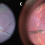 Cannula Tip Pressure
Mar 25 2025 by Robert Andrew Sisk, MD, FACS, FASRS
Color stills from surgical videos of subretinal delivery of gene augmentation therapy with A) voretigene neparvovec-ryzl and B) laru-zova. In the left panel, the cannula is slightly bent, and the retina and RPE are blanched white around the cannula tip engagement. The bleb was challenging to form in this patient with advanced retinal degeneration, and the bleb is shallow and mostly clear. In the right panel, the cannula tip is gently engaged, the cannula is straight, and it follows the retinotomy as the retina is elevated by the injection fluid.
Imaging device: Leica Proveo 8
Condition/keywords: gene therapy, genetic disorder, Leber's congenital amaurosis, retinitis pigmentosa, subretinal injection
-
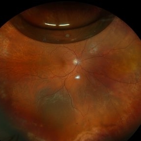 Recurrent Retinal Detachment with Single Break
Nov 2 2024 by Virginia Gebhart
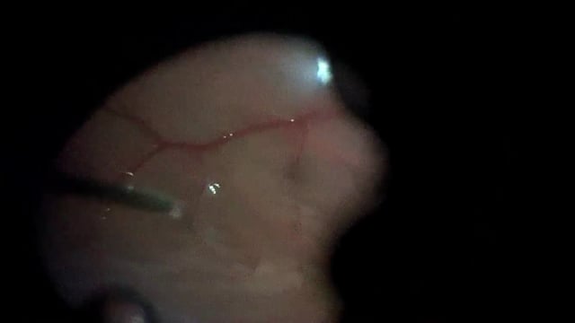
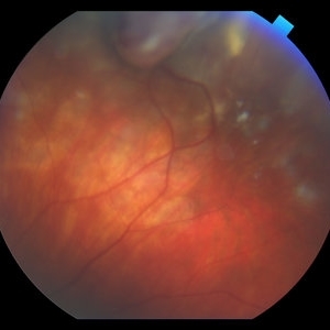
84 year old male with recurrent detachment s/p PPV/RD repair 2 weeks ago. Retinotomy is opened and appears to be the source of the fluid. Pt scheduled for emergency repair with scleral buckle.
Photographer: Virginia Gebhart
Imaging device: Optos California
-
 Idiopathic Uveal Effusion Syndrome
Aug 22 2024 by Jordyn Beckman
61 year old male with Idiopathic Uveal Effusion Syndrome with starry night appearance on fluorescein. 3 weeks s/p single external drainage retinotomy and 9 weeks of oral pred with recurrent choroidal effusions. Has since returned to surgery for secondary drainage retinotomy; subretinal fluid remain persistent.
Photographer: Jordyn Beckman
Imaging device: Optos California
Condition/keywords: chorioretinitis, Choroidal, exudative detachment, window defect
-
 Funnel Retinal Detachment
Jun 11 2023 by Ethan K Sobol, MD
Intraoperative view of a funnel retinal detachment with proliferative vitreoretinoapthy in an eye with previous open globe injury. PVR membranes were peeled, and the retina was flattened and re-attached with an inferior relaxing retinotomy and silicone oil tamponade
Condition/keywords: intraoperative, open funnel RD, open globe injury, proliferative vitreoretinopathy (PVR)
-
Iatrogenic Macular Hole and Subretinal Migration of PFCL
Feb 7 2023 by Aditya S Kelkar, MS, FRCS, FASRS,FRCOphth
The video demonstrates a surgical scenario where the fovea gives away by the force imparted by the jet of an injecting PFCL (Perfluorocarbon heavy Liquid) and the PFCL migrates subfoveally. Intraoperative OCT confirms the presence of a macular hole. The situation is managed by ILM peeling and mobilizing subfoveal PFCL peripherally by injecting another bubble of PFCL over the posterior pole. A peripheral drainage retinotomy is then created to aspirate the subretinal PFCL followed by fluid-air exchange, PFCL-air exchange, and endolaser around the retinotomy. Post-operative OCT at 3 weeks’ follow-up shows a sealed macular hole.
Condition/keywords: Iatrogenic macular hole, Intraoperative complications, Subretinal PFCL
-
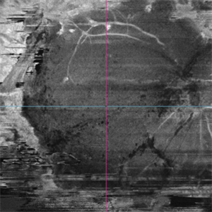
 360 retinotomy for combined closed funnel tractional and rhematogenous retinal detachment
Jan 1 2023 by Malek Yassine, MD
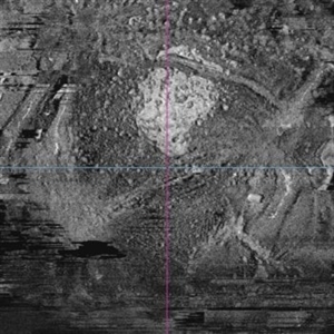
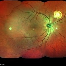
This is Fundus Autofluorecence, showing the residual hypoautofluorescent spots on the exposed choroid, relating to the previous panretinal photocoagulation, as well as the limits of the retinotomy with continuous laser which appeasr hypoautofluorecent with hyperautofluorecent margins.
Photographer: Malek Yassine, HMOED, Agadir, Morocco.
Imaging device: Zeiss Clarus
Condition/keywords: combined retinal detachment, rhegmatogenous retinal detachment, tractional retinal detachment
-
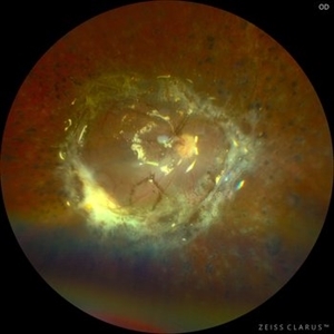 360 Retinotomy in a closed Funnel combined Tractional and rhegmatogenous retinal detachment
Jan 1 2023 by Malek Yassine, MD
This is the results at 6 months of a Bimanual 23 G-PPV with a very extensive and posterior 360 retinotomy for the management of a combined longstanding closed funnel RD, with submacular membranes, intraretinal PVR. Preop VA was a doubtful light perception. Borders of the retinotomy are stable at 6 months under 1300 Cs Silicon oil with some pigmented PVR developping the edges. Macula appears spared. Silicon oil emulsification droplets are well visualized beneath the superior temporal arcade.
Imaging device: Zeiss Clarus 700
Condition/keywords: combined retinal detachment, retinotomy, silicone oil
-
 OCT Angiography of a 360 retinotomy for closed funnel combined retinal detachment
Jan 1 2023 by Malek Yassine, MD
this is an OCTA image of 12X12 MM, showing all the 3 vascular plexi of the residual posterior retinal, with a good perfusion in the superior and central area, a ratification in the intermediate plexus in the inferior area, a non perfused temporal area, and some macular cysts. There's almost none macular translocation
Imaging device: Topcon Triton DRI-OCT
Condition/keywords: combined retinal detachment, OCT Angiography, retinotomy
-
 OCT en face of a 360 retinotomy for closed funnel combined retinal detachment
Jan 1 2023 by Malek Yassine, MD
Swept Source OCT en face at deep capillary plexus, shows foveal and parafoveal intraretinal cysts corresponding to macular edema under silicon oil
Imaging device: Topcon Triton DRI-OCT
Condition/keywords: combined retinal detachment, OCT EN FACE
-
 OCT en face of a 360 retinotomy for closed funnel combined retinal detachment
Jan 1 2023 by Malek Yassine, MD
Swept source OCT en face at the silicon oil - Retina Interface shows droplets of SO emulsification around the fovea and at the superior arcade, with some inferior striae corresponding to ERM formation
Imaging device: Topcon Triton DRI-OCT
Condition/keywords: oct en face
-
 Uveitis
Nov 25 2022 by Filip Kecer
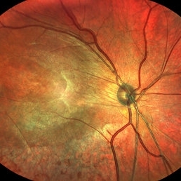
Multicolor composite of an 15 year old boy with cyclitis posterior, first diagnosed with uveitis intermedia retinoschisis o.dx., since then he underwent several procedures, such as PPV, ILM peeling, capsulectomy, retinotomy, transscleral cryo
Photographer: Filip Kecer, National Institute of Childrens Diseases
Imaging device: Spectralis, Heidelberg Engineering
Condition/keywords: cyclitis, epiretinal membrane (ERM), uveitis
-
 Circular & Radial Retinotomy for Retinal Detachment with PVR
Jan 26 2022 by Nikoloz Labauri, MD, FVRS
Intra-operative view of attached retina under PFCL. ILM & star folds were peeled off, circular and radial retinotomies are made and laser retinopexy applied.
Photographer: NIKOLOZ LABAURI MD
Condition/keywords: internal limiting membrane (ILM) peeling, laser retinopexy, PFCL liquid, proliferative vitreoretinopathy (PVR), star folds
-
 Spaghetti-Technique-for-Subretinal-PVR-Removal
Aug 27 2021 by Yodpong Chantarasorn, MD
An intraoperative image demonstrates extensive subretinal proliferative vitreoretinopathy that was removed by a spaghetti technique. The technique was aimed to minimize the traction on the retina surrounding access retinotomy, and thereby stabilized the retinotomy size.
Photographer: Yodpong Chantarasorn, Vajira Hospital, Navamindradhiraj University, Bangkok
Imaging device: Leica Proveo 8 (operating microscope)
Condition/keywords: proliferative vitreoretinopathy (PVR), re-attached retinal detachment (RRD), subretinal bands
-
 Surgical Management of a Symptomatic Full Thickness Macular Fold
Jul 19 2021 by Anton Orlin, MD
A balanced salt solution is injected into the subretinal space to focally detach the macula, ensuring that the fold is incorporated, in order to stretch and relax it. This can be done with a 38 or 41 gauge subretinal cannula, and is typically performed just within the macular arcade. Perfluorocarbon heavy liquid (PFCL) is then injected to flatten the macula and push the excess fluid to the periphery. A peripheral retinotomy is made, and the subretinal fluid is subsequently drained with air fluid exchange. The PFCL is removed and endolaser is applied to surround the retinotomy site. The eye was left with a gas tamponade.
Condition/keywords: macular fold, surgical management, video
-
 Bullous Retinal Detachment
Jul 9 2021 by Anton Orlin, MD
This is a color photograph of a right eye with a superior, bullous, macula-splitting retinal detachment. These features place the patient at a higher risk for macular fold formation postoperatively. To prevent fold formation, a surgeon should attempt for more complete subretinal fluid drainage during repair. This can be done with the use of perfluorocarbon liquid or by making a drainage retinotomy.
Condition/keywords: macular fold
-
 Post Vitrectomy
Apr 10 2018 by MOHAMED AHMED ALI, MD
Multi-color image of fundus show Draining retinotomy Post VIT.
Photographer: Mohamed A.Tawfik
-
 Vasoproliferative Tumor (VPT)
Apr 25 2017 by Christopher G Fuller, MD
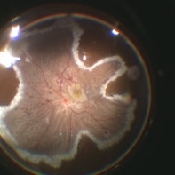
Fundus photograph of a presumptive vasoproliferative tumor (with resultant total exudative retinal detachment) in a 54-year-old white truck driver. Image is taken on post-operative day 4, after 25/27 gauge vitrectomy with drainage retinotomy, air-fluid exchange, endoscopic laser blanching of VPT, oil, and Ozurdex.
Photographer: Ray Garner, Texas Retina Associates [Lubbock, TX]
Condition/keywords: vasoproliferative retinopathy
-
 Vasoproliferative Tumor With Resultant Total Exudative RD Status Post Vitrectomy/Laser/Oil/Dexamethasone Intravitreal Implant
Apr 25 2017 by Christopher G Fuller, MD
Fundus photograph of a presumptive vasoproliferative tumor (with resultant total exudative retinal detachment) in a 54-year-old white truck driver. Image is taken on post-operative day 4, after 25/27 gauge vitrectomy with drainage retinotomy, air-fluid exchange, endoscopic laser blanching of VPT, oil, and dexamethasone intravitreal implant.
Photographer: Ray Gardner, Texas Retina Associates (Lubbock, TX)
Condition/keywords: exudative retinal detachment
 Loading…