 Initializing download.
-
 By Naveenam Srinivasa Muralidhar, MD
Co-author(s): Dr.Neha Bajpayee - Uploaded on Sep 11, 2023.
- Last modified by Joshua Friedman on Sep 12, 2023.
- Rating
- Appears in
- Miscellaneous
- Condition/keywords
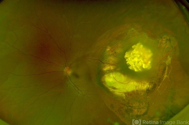
- Retinoma, spontaneously regressed retinoblastoma, optos
- Photographer
- Mr. Vedavyasa N K
- Imaging device
-
Fundus camera
Optos - Description
- Optos widefield image of left eye of a 31 year old male c/o defective vision in left eye since 6 years with esotropia of 30 degree. Fundus shows translucent greyish mass temporal to macula surrounded by zone of atrophy with pigmentation. Right eye fundus within normal limits.

")


