Search results (2718 results)
-
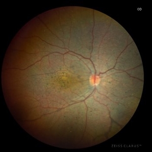
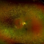
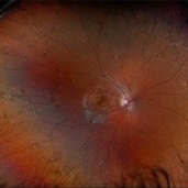
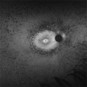
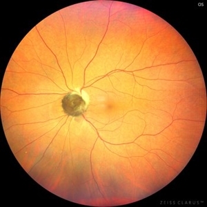 Optic Nerve Melanocytoma
May 4 2025 by KANWALJEET HARJOT MADAN, M.S. (Ophthalmology), FAICO (Vitreous - Retina)
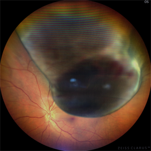
This is a fundus picture of a young 42-year male who visited for a routine eye exam. His BCVA was 20/20 in both eyes. Anterior segment examination was normal. His left eye showed grey-black pigmentation at the infero-nasal margin of the optic disc. Fundus of the right eye was normal. The patient was diagnosed to have optic disc melanocytoma on multimodal imaging and was advised regular follow-up. Optic nerve melanocytoma is typically a benign tumor made up of melanocytes and melanin. It can grow, but rarely transforms into a malignancy. Patients with Optic Nerve Melanocytoma should be periodically examined for evidence of growth, loss of visual field and optic nerve compression.
Photographer: Dr. Kanwaljeet Harjot Madan, Thind Eye Hospital, Jalandhar City (Punjab) INDIA.
Imaging device: Zeiss Fundus Camera
Condition/keywords: melanocytoma, melanoma, optic nerve
-
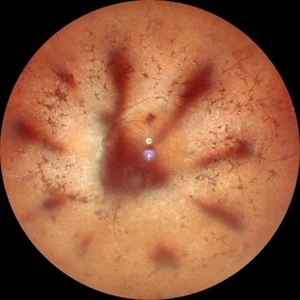 Retinitis Pigmentosa with Macular Hole with Posterior Subcapsular Cataract
Apr 28 2025 by Malvika Singh
Fundus photograph of the left eye of a 31 year old with retinitis pigmentosa, showing the shadow of posterior subcapsular cataract over the fundus.
Photographer: Dr Malvika Singh, Retina Foundation, Ahmedabad, India
Imaging device: Mirante SLO/OCT
Condition/keywords: posterior subcapsular cataract, retinitis pigmentosa
-
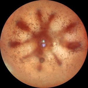 Retinitis Pigmentosa with Macular Hole with Posterior Subcapsular Cataract
Apr 28 2025 by Malvika Singh
Fundus photograph of the right eye of a 31 year old with retinitis pigmentosa with a macular hole, showing the shadow of posterior subcapsular cataract over the fundus.
Photographer: Dr Malvika Singh, Retina Foundation, Ahmedabad, India
Imaging device: Mirante SLO/OCT
Condition/keywords: macular hole, posterior subcapsular cataract, retinitis pigmentosa
-
 Retinocoroiditis Inactiva Por Toxoplasmosis
Apr 28 2025 by Paulina Araujo
Fundus photography demonstrates a 2-disc-diameter chorioretinal scar in the superior temporal arcade, consistent with inactive toxoplasmic retinochoroiditis. The lesion exhibits pigmented borders and central atrophy, with adjacent splinter hemorrhages and vascular sheathing. No vitreous inflammation or active satellite lesions are present.
Photographer: Paulina D.Araujo Martínez, Asociación para Evitar la Ceguera en México I.A.P., Hospital Dr Luis Sánchez Bulnes.
Condition/keywords: toxoplasmosis chorioretinitis
-
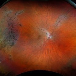
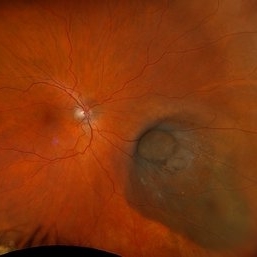
 Uveal Melanoma
Apr 26 2025 by Vishal Agrawal, MD, FRCS,FACS,FASRS
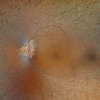
A 32 year-old male presented with complaints of perceiving a shadow in OS for 15-20 days. His BCVA was 20/20 OU. On Fundus examination, a large, elevated, well-defined, pigmented choroidal mass with few hemorrhages over the lesion was seen and a provisional diagnosis of uveal melanoma was made. urgent oncological consultation was recommended for further treatment.
Photographer: Dr Ayushi Gupta
Imaging device: Clarus 700
Condition/keywords: melanoma
-
 Optic Disc Drusen, RP
Apr 21 2025 by Virginia Gebhart
28 year old male with stable retinitis pigmentosa and optic disc drusen OU. Bardet-Biedl variant identified in previous genetic testing. BCVA 20/50 OD, 20/30 OS
Photographer: Virginia Gebhart, Retina Consultants of Carolina
Imaging device: Optos California
Condition/keywords: Drusen, optic disc drusen, retinitis pigmentosa
-
 Retinitis Pigmentosa
Apr 17 2025 by Virginia Gebhart
Fundus autofluorescence of 75 year old female with Retinitis Pigmentosa. Pt diagnosed at age 53. Diffuse RPE atrophy with minimal central sparing present in both eyes. Stable and unchanged compared to previous exams. BCVA 20/200 OD, NLP OS
Photographer: Virginia Gebhart, Retina Consultants of Carolina
Imaging device: Optos California
Condition/keywords: autofluorescence imaging, bone spicule, retinitis pigmentosa, RP
-
 LCA type 12
Apr 10 2025 by Joshua Friedman
LCA type 12 due to pathogenic mutations in RDH12. 13-year-old male with a visual acuity of 20/80 and 20/300 in the right and left eye, respectively. There is extensive pigment migration in the peripheral retina and macula. Like RPE65, there is widespread hypoautofluorescent signal, however, the peripapillary retina is uniquely spared in this form of LCA. On OCT, there is almost complete loss of the retina centrally.
Photographer: Stephen Tsang, MD, PhD
Condition/keywords: Leber Congenital Amaurosis
-
 LCA Type 8
Apr 10 2025 by Joshua Friedman
LCA Type 8 due to a pathogenic mutations in CRB1. 5-year-old male with a visual acuity of count fingers at 3 feet. Note the pseudopapilledema, para-arteriolar sparing, and nummular intraretinal pigment migration.
Photographer: Stephen Tsang, MD, PhD
Condition/keywords: Leber Congenital Amaurosis
-
 LCA type 10
Apr 10 2025 by Joshua Friedman
LCA type 10 due to mutations in CEP290. 36-year-old male with best corrected visual acuity of light perception in both eyes since childhood. On color fundus imaging, there is a mix of polymorphous white flecks and pigmentary changes. On autofluorescence imaging, there is almost complete loss of macular RPE. On OCT, there is complete loss of inner and outer retinal layers, the greatest losses occurring centrally.
Photographer: Stephen Tsang, MD, PhD
Condition/keywords: Leber Congenital Amaurosis
-
 Toxic Maculopathy (Elmiron)
Apr 9 2025 by Virginia Gebhart
79 year old male with toxic maculopathy from long term use of Elmiron (15+ yrs.) On exam there is stippled RPE changes, pigment clumping, and subretinal deposits. BCVA 20/100 | 20/40.
Photographer: Virginia Gebhart, Retina Consultants of Carolina
Imaging device: Optos California
Condition/keywords: autofluorescence imaging, cystoid macular degeneration, Elmiron Toxicity, Toxic Maculopathy
-
 Retinitis Pigmentosa
Apr 9 2025 by Virginia Gebhart
35 year old female with stable sectoral RP and high myopia OU. RP has not progressed in either eye since initial visit in 2021. Will continue to observe. VA 20/20 OU
Photographer: Virginia Gebhart, Retina Consultants of Carolina
Imaging device: Optos California
Condition/keywords: high myopia, retinitis pigmentosa
-
 New Choroidal Melanoma with Exudative Detachment
Apr 7 2025 by Virginia Gebhart
36 year old female referred for pigmented mass. Pt complains of flashes of light since last fall. Clinical exam and ultrasound findings consistent with choroidal melanoma with exudative detachment inferior. Pt will be scheduled for brachytherapy and possible tumor biopsy pending CT scan results.
Photographer: Virginia Gebhart, Retina Consultants of Carolina
Imaging device: Optos California
Condition/keywords: Choroidal melanoma, exudative detachment, melanoma, retinal detachment
-
 Retinitis Pigmentosa
Apr 1 2025 by Jordyn Beckman
63 year old woman with Retinitis Pigmentosa observed over time with peripheral loss. Over the span of 5 years BCVA changed from 20/25 to 20/50.
Photographer: Jordyn Beckman, Retina Consultants of Carolina, P.A.
Imaging device: Optos California
Condition/keywords: atrophy, bone spicules, retinitis pigmentosa
-
 Retinitis Pigmentosa
Mar 27 2025 by T. P . VIGNESH, MBBS,MS
Fundus photograph of a 52-year-old woman with retinitis pigmentosa with cystoid macular edema.
Photographer: Bharathi
Imaging device: EIDON
Condition/keywords: retinitis pigmentosa
-
 Collar Button Melanoma
Mar 27 2025 by Virginia Gebhart
62 year old male with large pigmented lesion with collar button. Pt states he was never aware of any lesion/nevus in the past. Fluid and orange pigment present, appears to be chronic. Pt will be scheduled for brachytherapy pending CT scan results.
Photographer: Virginia Gebhart, Retina Consultants of Carolina
Imaging device: Optos California
Condition/keywords: choroidal melanoma, collar button
-
 CHRPE
CHRPE
Mar 25 2025 by Toolie Winters
Ultra-wide field fundus photograph of a 78-year-old woman with extensive CHRPE lesions OS. Continued observation has been recommended at this time.
Photographer: Toolie Winters
Imaging device: Optos California
Condition/keywords: CHRPE, congenital hypertrophy of the retinal pigment epithelium (CHRPE), fundus photography, Optos, Optos California, pseudocolor, ultra-wide field imaging
-
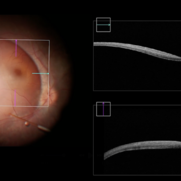 Completed Bleb with OCT Through Fovea
Mar 25 2025 by Robert Andrew Sisk, MD, FACS, FASRS
Color still from surgical video of subretinal delivery of laru-zova for X-linked retinitis pigmentosa. Live optical coherence tomography (OCT) with foveal tracking via the embedded software in the operating microscope allows monitoring foveal integrity for signs of stress. The contour of the fovea does not exceed the curvature of the bleb (e.g. no inversion). The tangential cannula angle facilitated steering of the bleb posteriorly. The bleb covers essentially the entire macula, which is the target area.
Imaging device: Zeiss Artevo 800
Condition/keywords: gene therapy, genetic disorder, optical coherence tomography (OCT), retinitis pigmentosa, subretinal injection
-
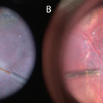 Cannula Tip Pressure
Mar 25 2025 by Robert Andrew Sisk, MD, FACS, FASRS
Color stills from surgical videos of subretinal delivery of gene augmentation therapy with A) voretigene neparvovec-ryzl and B) laru-zova. In the left panel, the cannula is slightly bent, and the retina and RPE are blanched white around the cannula tip engagement. The bleb was challenging to form in this patient with advanced retinal degeneration, and the bleb is shallow and mostly clear. In the right panel, the cannula tip is gently engaged, the cannula is straight, and it follows the retinotomy as the retina is elevated by the injection fluid.
Imaging device: Leica Proveo 8
Condition/keywords: gene therapy, genetic disorder, Leber's congenital amaurosis, retinitis pigmentosa, subretinal injection
-
 Choroidal Hemangioma
Mar 13 2025 by Virginia Gebhart
64 year old male referred for lesion in the STA with worsening SRF. Pt had been receiving injections for wetAMD q4weeks for 7 months. Reddish, elevated choroidal lesion, chronic SRF and pigment clumping consistent with hemangioma. FA/ICG/Bscan ultrasound also performed to confirm. Pt scheduled for PDT
Photographer: Virginia Gebhart, Retina Consultants of Carolina
Imaging device: Optos California
Condition/keywords: choroidal hemangioma, hemangioma, subretinal fluid
-
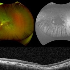
 Multimodal Imaging in CHRPE
Mar 6 2025 by Gerardo - Montante Montelongo, MD
Fundus photograph of an 83-year-old male with a history of Diabetes, smoking, cataract surgery on the right eye in 2022, and open-angle glaucoma. Asymptomatic. Indirect ophthalmoscopy revealed 80% excavation, peripapillary atrophy, and a hyperpigmented perifoveal lesion with 35% atrophy, 10% drusen, and 5.1 mm diameter, corresponding to a CHRPE. At multimodal imaging, FFA shows hypoautofluorescence of the lesion, OCT shows preservation of internal retinal layers, atrophy of external retinal layer, with an RPE disruption, and posterior shadowing. USG shows a flat hyperechoic lesion 5.1 mm in diameter and 1.32 mm in thickness, solid and with high internal reflectance.
Photographer: Gerardo Montante-Montelongo, MD, Mexican Institute of Ophthalmology
Imaging device: Clarus 700
Condition/keywords: congenital hypertrophy of the retinal pigment epithelium (CHRPE), multimodal imaging
-
 Hereditary Retinal Dystrophy
Feb 27 2025 by Kimberly Wakester
Optomap RGB image of a 7-year-old girl with Hereditary retinal dystrophy. Biological mother is a CHM gene carrier and biological father is diagnosed with RP. Patient had genetic testing and was also confirmed to be a CHM gene carrier and also has the TTC21B gene. There is linear pigmentary changes on clinical exam and fundus photos. Atypical appearance of Retinitis Pigmentosa. Patient will continue follow up care with repeat imaging.
Photographer: Kimberly Wakester, COA
Imaging device: Optos California
Condition/keywords: CHM gene, hereditary retinal dystrophy, linear pigmentary changes
-
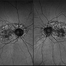
 Retinitis Pigmentosa
Feb 18 2025 by Drew Mitchell
FAF, Color, IR, OCT of Mild CME secondary to Retinitis Pigmentosa.
Photographer: Drew Mitchell OCT-C
Imaging device: Optos California
Condition/keywords: cystoid macular edema (CME), Optos, OPTOS CALIFORNIA, retinitis pigmentosa, RP
-
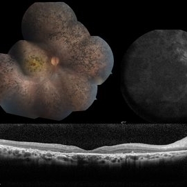
 Choroidal Melanoma
Feb 6 2025 by Virginia Gebhart
81 year old female with large pigmented collar button ciliochoroidal mass extending into the mid-vitreous cavity. Clinical exam and ultrasound finding consistent with melanoma. Due to size of tumor, pt scheduled for enucleation. CT scan of abdomen showed no evidence of metastatic disease.
Photographer: Virginia Gebhart, Retina Consultants of Carolina
Imaging device: Optos California
Condition/keywords: ciliochoroidal melanoma, collar button, melanoma
-
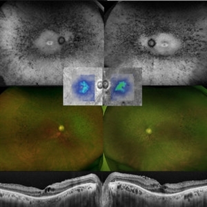
 Retinitis Pigmentosa Bullseye Appearing Autofluorescence
Feb 4 2025 by Isaac Agranoff
Fundus Autofluorescence of a 14-year-old boy with suspected RP. ERG performed afterwards was almost flat. VA measured at 20/30 but with extensive constriction of confrontational visual fields. Currently awaiting genetic testing.
Photographer: Isaac Agranoff
Imaging device: Optos California
Condition/keywords: fundus autofluorescence (FAF), retinitis pigmentosa, RP
 Loading…