Search results (2718 results)
-
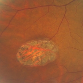
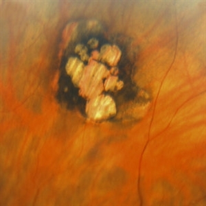
 Siegrist Streaks
Mar 29 2013 by Henry J. Kaplan, MD
Typical Siegrist streaks in hypertensive choridopathy; hyperpigmentations in a linear fashion along choroidal vessels , a rare finding.
Condition/keywords: hypertensive choroidopathy, Siegrist Streaks
-
 Congenital Hypertrophy of the Retinal Pigment Epithelium (CHRPE)
Mar 1 2014 by Homayoun Tabandeh, MD, FASRS
Congenital hypertrophy of the retinal pigment epithelium (CHRPE).
Condition/keywords: congenital hypertrophy of the retinal pigment epithelium (CHRPE)
-
 Vortex Vein In A Patient With A Blond Fundus
Oct 2 2013 by Jerald A. Bovino, MD
The vortex vein and vortex vein ampulla are visible in this patient with a blonde (lightly pigmented) fundus.
Condition/keywords: choroidal circulation, fundus photograph, vortex vein
-
 PED due to CSCR
Sep 2 2012 by Hamid Ahmadieh, MD
OCT image of a 37-year-old man with a serous PED secondary to CSCR.
Photographer: Hamid Ahmadieh, Ophthalmic Research Center, Labbafinejad Medical Center
Imaging device: Heidelberg Spectralis
Condition/keywords: central serous chorioretinopathy (CSCR), optical coherence tomography (OCT), pigment epithelial detachment (PED)
-
---thumb.jpg/image-square;max$300,300.ImageHandler) Inferior Sector Iris Atrophy With Depigmentation
Inferior Sector Iris Atrophy With Depigmentation
Aug 1 2013 by From the Collections of Thomas M. Aaberg, MD and Thomas M. Aaberg Jr., MD
Inferior sector iris atrophy with depigmentation.
Condition/keywords: depigmentation, inferior sector iris atrophy
-
 Kearns-Sayre Syndrome
Sep 18 2012 by Michael P. Kelly, FOPS
Retinal fundus photograph of a Kearns-Sayre Syndrome patient.
Photographer: Michael P. Kelly, FOPS Director, Duke Eye Labs, Duke University Hospital, Duke Eye Center
Imaging device: Canon 60UV
Condition/keywords: bilateral pigmentary retinopathy, cardiac conduction abnormalities, chronic progressive ophthalmoplegia, heart-block, Kearns-Sayre Syndrome, ptosis
-
 Scleral Indentation In A Normal Eye
Nov 9 2012 by Norman Byer
This shows the appearance of scleral indentation in a normal eye. Note the convex shadow which marks the posterior border of the indented area. It is caused in part by a small angle which separates the viewing axis from the illuminating axis thus allowing the observer to see slightly into the shadow beyond the illuminated crest of the indentation. It is also caused in part by viewing the pigment epithelial layer in a tangential manner. This shadow is of great diagnostic usefulness since it becomes a dark background against which many tiny retinal abnormalities can be seen beautifully by contrast. Two other particular advantages of scleral indentation will be demonstrated in the following photographs: First, the ability to see the extreme anterior part of the retina to the ora serrata and beyond, and second, the ability to examine any abnormality in multiple profiles depending on slight movements of the scleral depressor in various directions.
Condition/keywords: extreme anterior retina, posterior border, scleral indentation, shadow, tangential view of pigment epithelial layer
-
 Bear Tracks
Dec 31 2012 by Raj K. Maturi, MD
Photographer: Tom Steele, CRA Midwest Eye Institute Indianapolis, Indiana
Imaging device: Topcon 50ex 50 degree field
Condition/keywords: bear tracks, benign pigmented lesions, congenital hypertrophy of the retinal pigment epithelium (CHRPE), OD
-
 Cystic Retinal Tuft
Nov 9 2012 by Norman Byer
This is a rather poor photograph taken in 1969 but is important for comparison with the next slide pair. It shows a cystic retinal tuft in a 49-year-old woman and was taken without scleral indentation. The two pigment spots just inferior to the tuft represent a secondary degenerative change in the pigment epithelium.
Condition/keywords: cystic retinal tuft, degenerative changes of retinal pigment epithelium, pigmented spots
-
 Gonioscopy: Pigment Dispersion Glaucoma
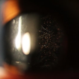
Jul 8 2013 by Jason S. Calhoun
Patient with no family history of glaucoma, came in with elevated IOP. During gonioscopy exam. brown pigment overlying the trabecular meshwork. Also, trans-illumination defects on the iris.
Photographer: Jason S. Calhoun, Department of Ophthalmology, Mayo Clinic Jacksonville, Florida
Condition/keywords: gonioscopy, pigment dispersion syndrome of iris
-
 Gonioscopy: Pigment Dispersion Glaucoma
Jul 8 2013 by Jason S. Calhoun
Patient with no family history of glaucoma, came in with elevated IOP. During gonioscopy exam. brown pigment overlying the trabecular meshwork. Also, trans-illumination defects on the iris.
Photographer: Jason S. Calhoun, Department of Ophthalmology, Mayo Clinic Jacksonville, Florida
Condition/keywords: gonioscopy, pigment dispersion syndrome of iris
-
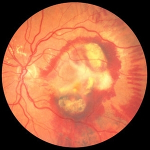 ARMD with Disciform Scar
Oct 16 2012 by Jeffrey G. Gross, MD, FASRS
ARMD with disciform scar, RPE contracture, and subretinal hemorrhage, CF.
Condition/keywords: disciform scar, retinal pigment epithelium (RPE) contracture, subretinal hemorrhage
-
 Ocular Melanocytosis Scleral Pigmentation
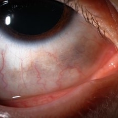
Jul 9 2014 by Susanna S. Park, MD, PhD
Slit lamp photograph of a 12-year-old boy with ocular melanocytosis showing scleral and episcleral pigmentation. The other eye is blue.
Photographer: Ellen Redenbo
Condition/keywords: melanocytoma
-
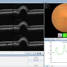
---thumb.JPG/image-square;max$300,300.ImageHandler) Retinal Pigment Epithelium Detachment
Retinal Pigment Epithelium Detachment
Jul 12 2013 by Jason S. Calhoun
Composite of HD-OCT and fundus photograph showing central RPE detachment. Patient proceeded with Eylea injection.
Photographer: Jason S. Calhoun, Department of Ophthalmology, Mayo Clinic Jacksonville, Florida
Condition/keywords: retinal pigment epithelium
-
 Enclosed Ora Bay On The Temporal Side
Nov 9 2012 by Norman Byer
This is a developmental abnormality in a 59-year-old man. It is an enclosed ora bay on the temporal side, an isolated island of normal pars plana epithelium. It is important not to confuse this entity with a retinal break. It has smooth, sloping borders not a sharp, thin, visible retinal edge as a retinal break would have. The border looks exactly like that of the ora serrata, and the grayish pigmented base has the same appearance as the normal pars plana.
Condition/keywords: developmental abnormality, enclosed ora bay, grayish pigmented base, horizontal nasal meridian, pars plana epithelium, smooth sloping borders, temporal retina
-
 Operculated Hole and CHRPE
Jan 16 2018 by Carolyn Daley
58-year-old woman with an operculated hole and CHRPE in the right eye. Patient is asymptomatic so no treatment was recommended at this time.
Photographer: Carolyn Daley
Imaging device: Optos ultra wide field image
Condition/keywords: congenital hypertrophy of the retinal pigment epithelium (CHRPE), operculated retinal hole, Optos, ultra-wide field imaging
-
 Spontaneous Flattening of Drusenoid PED
Jul 1 2014 by John S. King, MD
Consult to r/o ExAMD; observed; scans about a year apart.
Photographer: Wayne A Ladlee Jr
Imaging device: Cirrus
Condition/keywords: drusenoid PED, macular drusenoid lesion, pigment epithelial detachment (PED)
-
 Schaffer's Sign
Dec 23 2019 by Hashim Ali Khan, OD, FAAO
Brown iris pigment in vitreous of a pseudophakic eye without retinal detachment or breaks/ holes in retina.
Condition/keywords: detached vitreous, Schaffer's sign, vitreous pigment
-
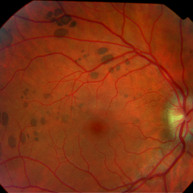
 Pigmented Peripheral Retinal Degeneration
Jun 27 2013 by Jason S. Calhoun
42-year-old male came in for routine eye exam and to follow up on peripheral retinal degeneration in both eyes. VA is 20/20, right eye and 20/25, left eye. Patient is asymptomatic with no visual complaints.
Photographer: Jason S. Calhoun, Mayo Clinic Jacksonville, Florida
Imaging device: TOPCON TRC 50-EX
Condition/keywords: peripheral retinal degeneration
-
 Congenital Hypertrophy of the Retinal Pigment Epithelium (CHRPE)
Mar 1 2014 by Homayoun Tabandeh, MD, FASRS
Congenital hypertrophy of the retinal pigment epithelium (CHRPE).
Condition/keywords: congenital hypertrophy of the retinal pigment epithelium (CHRPE)
-
 Traumatic Macular Hole with Retinal Detachment and PVR
Sep 27 2012 by Pauline T Merrill, MD, FASRS
Fundus photo of a 13-year-old boy s/p soccer ball injury 1 month previously. In addition to full-thickness macular hole and total retinal detachment with grade C PVR, note pigment granules visible in vitreous over optic nerve.
Photographer: Karen Parque, Illinois Retina Associates, Chicago, IL
Condition/keywords: proliferative vitreoretinopathy (PVR), traumatic macular hole
-
 Fibrovascular PED
Feb 21 2014 by Roy Schwartz, MD
72-year-old female with fibrovascular PED. Upper picture - PED with sub RPE hyper-reflective substance, in a multi-layered pattern, corresponding to fibrovascular PED. CME. Lower picture - PED flattened, a denser sub RPE hyperreflective substance is seen. CME resolved.
Condition/keywords: fibrovascular pigment epithelial detachment (PED), neovascular age-related macular degeneration (AMD), optical coherence tomography (OCT), ranibizumab
-
 Melanocytoma with Choroidal Melanoma
Oct 8 2012 by Susanna S. Park, MD, PhD
Fundus photograph of a 75-year-old woman with a slowly growing pigmented lesion.
Photographer: Ellen Redenbo, University of California Davis Eye Center
Condition/keywords: melanocytoma
-
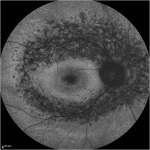 Retinitis Pigmentosa - Fundus Autofluorescence
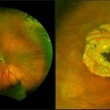
Sep 20 2014 by Rameez N Hussain, MD
Fundus autofluorescence of retinitis pigmentosa showing hyperautofluorescent rings or foveal hyperautofluorescence.
Photographer: Dr.Rameez N Hussain, MD, Central Imaging Center, Vitreo Retinal Services, Giridhar Eye Institute, Cochin, India
Imaging device: Heidelberg Blue Peak Autofluorescence imaging.
Condition/keywords: bone spicule, cystoid macular edema (CME), fundus autofluorescence (FAF), retinitis pigmentosa
-
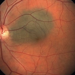 Choroidal Nevus
May 8 2014 by S. Natarajan, MD, FASRS, FRCS (GLASGOW) , FICO, D.Sc, FELA
Fundus photograph of a 48-year-old female in for a routine eye checkup with vision 6/6 OU. Showed solitary pigmented juxtapapilary nevus of 3*2 disc diameters size.
Photographer: ADITYA JYOT EYE HOSPITAL,MUMBAI INDIA
Condition/keywords: choroidal nevus
 Loading…