Search results (308 results)
-
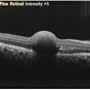
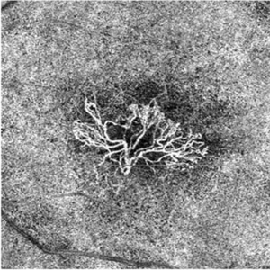
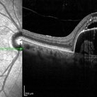
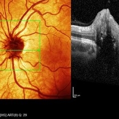
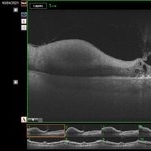
 Myelinated Nerve Fibers
Apr 18 2025 by DR Rohit Gupta
The **myelinated nerve fibers of the optic disc** (also known as **medullated nerve fibers**) are retinal nerve fibers that retain their myelin sheath as they pass through the optic nerve head. Normally, retinal nerve fibers are unmyelinated to allow for light transparency, but in some cases, myelination extends anteriorly into the retina, appearing as a striking white, feathery patch on the optic disc or peripapillary retina. ### **Key Features:** 1. **Appearance:** - Dense, white, striated patches with feathery edges. - Typically located at the superior or inferior pole of the optic disc. - May obscure retinal vessels underneath. 2. **Clinical Significance:** - Usually **benign** and asymptomatic. - **Congenital** (present at birth or early childhood). - Rarely associated with **visual field defects** (e.g., scotomas corresponding to the area of myelination). - Occasionally linked with **high myopia** or **amblyopia** if extensive. 3. **Pathophysiology:** - Failure of oligodendrocytes or Schwann cells to stop myelination at the lamina cribrosa. - Normally, myelination stops at the optic nerve head, but in this condition, it extends into the retina. 4. **Diagnosis:** - **Fundoscopy:** Classic white, feathery appearance. - **Optical Coherence Tomography (OCT):** Shows thickened retinal nerve fiber layer (RNFL). - **Visual Field Testing:** May detect defects if large. 5. **Differential Diagnosis:** - Optic disc edema - Cotton wool spots - Retinoblastoma (rarely, but must be ruled out in children) 6. **Management:** - No treatment required if asymptomatic. - Monitor for amblyopia in children. - Rare cases with significant visual impairment may need further evaluation. ### **Fun Fact:** Myelinated nerve fibers are seen in **~0.5-1%** of the population and are usually an incidental finding.
Photographer: Dr Rohit gupta
Imaging device: Samsung S21
Condition/keywords: Medulated Nerve fibre, Medullated Nerve fibres, myelinated nerve fibers, Myelinated Nerve Fibres, optic disc drusen
-
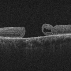
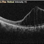
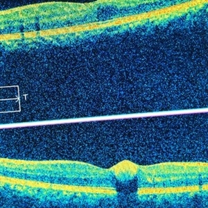
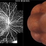
 Sub-Internal Limiting Membrane Hemorrhage
Apr 17 2025 by Malvika Singh
OCT of a 41 year-old, male, with a central retinal vein occlusion and a foveal sub-internal limiting membrane hemorrhage.
Photographer: Dr Malvika Singh, Retina Foundation, Ahmedabad, India
Imaging device: Mirante SLO/OCT
Condition/keywords: optical coherence tomography (OCT), SUB ILM hemorrhage
-
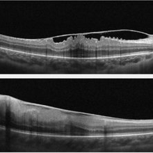
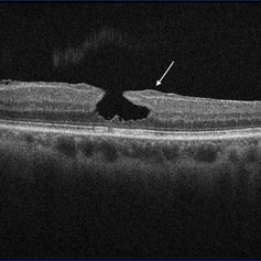
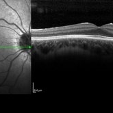
 Completed Bleb with OCT Through Fovea
Mar 25 2025 by Robert Andrew Sisk, MD, FACS, FASRS
Color still from surgical video of subretinal delivery of laru-zova for X-linked retinitis pigmentosa. Live optical coherence tomography (OCT) with foveal tracking via the embedded software in the operating microscope allows monitoring foveal integrity for signs of stress. The contour of the fovea does not exceed the curvature of the bleb (e.g. no inversion). The tangential cannula angle facilitated steering of the bleb posteriorly. The bleb covers essentially the entire macula, which is the target area.
Imaging device: Zeiss Artevo 800
Condition/keywords: gene therapy, genetic disorder, optical coherence tomography (OCT), retinitis pigmentosa, subretinal injection
-
 OCT in Adult Vitelliform Dystrophy
Jun 25 2024 by Tejaswita Verma
OCT image of a 62 year old female with 6/12 vision in both eyes showing sub retinal fluid with RPE granularity s/o Adult vitelliform macular dystrophy.
Photographer: DR. TEJASWITA VERMA
Imaging device: MIRANTE
Condition/keywords: adult vitelliform dystrophy, optical coherence tomography (OCT)
-
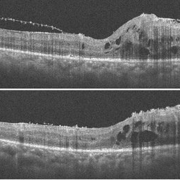
 Bullseye Maculopathy
Jan 22 2024 by Kali Jend
Optical coherence tomography of a 73-year-old female with Bullseye Macular Changes affecting her left eye. Patient reports having a family history of this condition and denies prior Plaquenil or Elmiron use. Compared to previous imaging, the patient's condition progressed in the left eye from 2020 to 2023. Patient has a history of fluctuating Diabetic Macular Edema and a current Epiretinal Membrane as well. Patient's vision was Ncc20/60 at the time the image was taken.
Photographer: Kali Jend
Imaging device: Heidelberg Spectralis
Condition/keywords: bullseye maculopathy, epiretinal membrane (ERM), heidelberg spectralis, left eye, macular pucker, OCT, optical coherence tomography (OCT)
-
 Neovascular-network-OCTA
Jan 2 2024 by Tahsin Khundkar, MD
En Face optical coherence tomography (OCT).- angiography shows a large choroidal neovascular membrane in the outer retina to choriocapillaris slab.
Photographer: Jeffrey Zeigler, Concord Eye Center
Imaging device: Zeiss
Condition/keywords: neovascular membrane, wet age-related macular degeneration (wet AMD)
-
 Chronic Full Thickness Macular Hole
Oct 25 2023 by Jessica Hampton, BS
Optical-coherence tomography image of a 65-year old woman with a chronic full-thickness macular hole in the left eye, recurred following three attempts at repair with pars plana vitrectomy, membrane peel, and gas tamponade.
Photographer: Dr. Diana Do, Stanford Medicine, Byers Eye Institute
Condition/keywords: full thickness macular hole, optical coherence tomography (OCT)
-
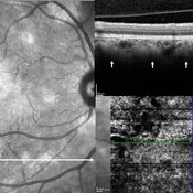 Choroidal Nodules in Neurofibromatosis
Sep 6 2023 by Maria Filipa Madeira
Macular near-infrared reflectance (NIR) imaging, optical coherence tomography (OCT) B-scan and OCT angiography (OCTA) of a 54-year-old woman with neurofibromatosis type 1. Choroidal abnormalities were asymptomatic and not visible on funduscopic exam, but had a striking appearance on retinal imaging. B-scan (horizontal arrow) showed hyperreflective nodules in the deeper choroid (vertical arrows) underlying the multiple hyperreflective patches on NIR, in correlation with hyperflow areas of the deep choroidal plexus in OCTA.
Photographer: Maria Filipa Madeira, Centro Hospitalar de Lisboa Ocidental, Hospital de Egas Moniz
Imaging device: Heidelberg Spectralis
Condition/keywords: choroid, neurofibromatosis
-
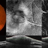 Retinal Arterial Macroaneurysm
Apr 8 2023 by Yousef A Fouad, MD, FRCS (Glasg.)
Multimodal imaging of a retinal arterial macroaneurysm in the right eye of a 73-year-old male patient with uncontrolled hypertension. Fundus photography shows hemorrhage surrounding an arterial branch of the upper temporal arcade. Optical coherence tomography (OCT) through the lesion shows inner retinal hyperreflectivity with back shadowing, and adjacent cystoid macular edema in the outer retina. En face OCT centered on the lesion delineates the fusiform dilatation of the affected vessel, and OCT angiography confirms the presence of blood flow within the aneurysmal dilatation.
Photographer: Yousef Fouad, Ain Shams University, Egypt
Condition/keywords: arteriolar macroaneurysm, enface imaging, macroaneurysm, macroarterial aneurysm, OCT Angiography, OCTA
-
 Pigment Epithelium Detachment, Secondary to AMD
Mar 17 2023 by Ceara Donovan
Optical coherence tomography of a 76 year old woman with a Pigment Epithelium Detachment, Secondary to AMD affecting her right eye. Patient had no significant response to Avastin, Eylea, Lucentis 0.5, or Vabysmo and was switched to Beovu. Following Beovu intravitreal injection her edema improved on OCT. Patient's vision was sc20/200+1 at the time the image was taken.
Photographer: Ceara Donovan
Imaging device: Heidelberg Spectralis
Condition/keywords: exudative age-related macular degeneration, heidelberg spectralis, macular degeneration, optical coherence tomography (OCT), pigment epithelial detachment (PED), Sub-retinal fluid
-
 Idiopathic Choroidal Neovascularization
Mar 2 2023 by Corey Grant
Optical coherence tomography and ultra-wide field fundus photograph of a 51 year old male with idiopathic choroidal neovascularization affecting his right eye. The patient had no symptoms at the time of the appointment and his vision was Dcc20/20-2 OU. The physcian stated that there wasn't active exudation on the exam or ocular imaging and based on the clinical findings, he has recommended we defer any treatments.
Photographer: Corey Grant
Imaging device: Heidelberg Spectralis, OPTOS California
Condition/keywords: choroidal neovascularization (CNV), CNVM, fundus photograph, OCT, optical coherence tomography (OCT), Optos, Right Eye, ultra-wide field imaging
-
 Juvenile X-linked Retinoschisis
Nov 8 2022 by Vaidehi Sathaye
OCT image of RE of a 9 year male patient with Juvenile X-linked Retinoschisis
Photographer: Dr. Vaidehi Sathaye
Imaging device: Mirante
Condition/keywords: foveal schisis, optical coherence tomography (OCT)
-
 COATS DISEASE OCT
Sep 2 2022 by FLOR ANGELICA JACOME GUTIERREZ
Macular OCT in a coats disease with epiretinal membrane.
Photographer: Dr. Guillermo Salcedo Villanueva
Condition/keywords: Coats' disease, optical coherence tomography (OCT)
-
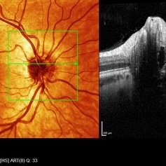 Right eye SD- OCT-RNFL of optic nerve head drusen showing hypo reflective centre with hyper reflective margins.
Aug 5 2022 by Kavitha Duraipandi, MD DNB FICO FRCS
A 20 year old patient referred to the clinic with blurred disc margins to rule out papilledema.
Photographer: Natalie Fox- Bussell
Condition/keywords: optic nerve drusen, optical coherence tomography (OCT)
-
 Left eye SD- OCT-RNFL of optic nerve head drusen showing hypo reflective centre with hyper reflective margins.
Aug 5 2022 by Kavitha Duraipandi, MD DNB FICO FRCS
A 20 year old patient referred to the clinic with blurred disc margins to rule out papilledema.
Photographer: Natalie Fox- Bussell
Condition/keywords: optic nerve drusen, optical coherence tomography (OCT)
-
 Vitreomacular Traction
Jun 15 2022 by Zach Seim
Optical Coherence Tomography (OCT) of a 69 year old male with Vitreomacular Traction affecting his right eye. Patient was referred to this office for Choroidal Melanoma in his right eye in May 2021. The patient was treated with Brachytherapy in July 2021 and this OCT was taken at a follow-up appointment in May 2022. Patient's vision was 20/30-2 at the time this OCT was taken. Patient states that his vision was better since his last visit, and that he sees floaters occasionally.
Photographer: Zach Seim
Imaging device: Heidelberg Spectralis
Condition/keywords: heidelberg spectralis, OD, optical coherence tomography (OCT), right eye, subretinal fluid, vitreomacular adhesion, vitreomacular interface disorders, vitreomacular traction (VMT)
-
 Valsalva Retinopathy
Feb 20 2022 by Ajay Indur Dudani, MBBS MS
OCT of. 24 yr girl with valsalva retinopathy and sub ILM macular haemorrhage
Photographer: Ajay DUDANI,Mumbai Retina Center,Mumbai,India
Imaging device: Zeiss Cirrus
Condition/keywords: optical coherence tomography (OCT)
-
 Lamellar hole
Feb 19 2022 by Hong-Uyen Hua, MD
Optical coherence tomography (OCT) image of a lamellar hole. The OCT shows the three mandatory features of a lamellar hole: 1) presence of irregular foveal contour 2) presence of foveal cavity with undermined edges, and 3) the apparent loss of foveal tissue. It also demonstrates lamellar-hole-associated epiretinal proliferation (LHEP) (arrow).
Condition/keywords: lamellar macular hole
-
 Post Retinal Reattachment Surgery Epiretinal Membrane
Sep 14 2021 by Ogugua Ndubuisi Okonkwo, MD, FRCS (Edin), FASRS
Postoperative optical coherence tomography (OCT) of the right eye in a 65-year-old male who had retinal reattachment surgery for a macular hole retinal detachment. This OCT scan shows epiretinal membrane and intraretinal cystic fluid spaces.
Photographer: Oreoluwa Olabode , Eye Foundation Hospital, Lagos.
Imaging device: Optovue Avanti RTVue.
Condition/keywords: epiretinal membrane (ERM), macular hole retinal detachment, Retinal Reattachment surgery
-
 Persistence of Sub Retinal Fluid Post Retinal Reattachment Surgery
Sep 14 2021 by Ogugua Ndubuisi Okonkwo, MD, FRCS (Edin), FASRS
Postoperative Optical Coherence Tomography (OCT) of the right eye of a 35-year-old male showing persistence of subretinal fluid in the original area of the retinal detachment.
Photographer: Oreoluwa , Eye Foundation Hospital, Lagos
Imaging device: Optovue Avanti RTVue
Condition/keywords: re-attached retinal detachment (RRD), subretinal fluid
-
 Rhegmatogenous Retinal Detachment- Macula-On
Sep 14 2021 by Ogugua Ndubuisi Okonkwo, MD, FRCS (Edin), FASRS
Preoperative Optical Coherence Tomography (OCT) of the right eye of a 35-year-old male showing detached retina ( subretinal fluid elevates the retina), sparing the macula.
Photographer: Oreoluwa Olabode , Eye Foundation Hospital, Lagos
Imaging device: Optovue Avanti RTVue
Condition/keywords: re-attached retinal detachment (RRD)
-
 Retinal Hemorrhage
Sep 2 2021 by Avris Romario Diparaja Siahaan
Swept source OCT angiography of a 58-year-old man with hemorrhage in his left eye.
Photographer: Nanda Lessi Hafni Eka Putri, MD (Ophthalmologist) & Ryan Mishbahuddin (Nurse), Ciawi General Hospital (Rumah Sakit Umum Daerah Ciawi)
Imaging device: DRI OCT Triton Plus (Topcon)
Condition/keywords: fundus photograph, optical coherence tomography (OCT)
-
 Diabetic Retinopathy
Sep 2 2021 by Avris Romario Diparaja Siahaan
Swept source OCT angiography (montage photography) and fundus photography of a 61-year-old woman with proliferative diabetic retinopathy in her right eye.
Photographer: Nanda Lessi Hafni Eka Putri, MD (Ophthalmologist) & Ryan Mishbahuddin (Nurse), Ciawi General Hospital (Rumah Sakit Umum Daerah Ciawi)
Imaging device: DRI OCT Triton Plus (Topcon)
Condition/keywords: diabetic retinopathy, fundus photograph, optical coherence tomography (OCT)
-
 CSNB-OCT-OD
Aug 23 2021 by Jennifer Carstens
OCT/infrared image showing myopic fundus with normal retinal structure in patient with CACNA1F associated X-linked CSNB (OD).
Photographer: Jing Zhang, Ophthalmic Photographer
Condition/keywords: congenital stationary night blindness (CSNB), infrared image, optical coherence tomography (OCT)
-
 CSNB-OCT-OS
Aug 23 2021 by Jennifer Carstens
OCT/infrared image showing myopic fundus with normal retinal structure in patient with CACNA1F associated X-linked CSNB (OS).
Photographer: Jing Zhang, Ophthalmic Photographer
Condition/keywords: congenital stationary night blindness (CSNB), infrared image, optical coherence tomography (OCT)
 Loading…