 Initializing download.
-
 By Hong-Uyen Hua, MD
Bascom Palmer Eye Institute
Co-author(s): Abdul-Hadki Kaakour, Cleveland Clinic Cole Eye Institute; Aleksandra Rachitskaya, Cleveland Clinic Cole Eye Institute - Uploaded on Feb 19, 2022.
- Last modified by Joshua Friedman on Feb 22, 2022.
- Rating
- Appears in
- Miscellaneous
- Condition/keywords
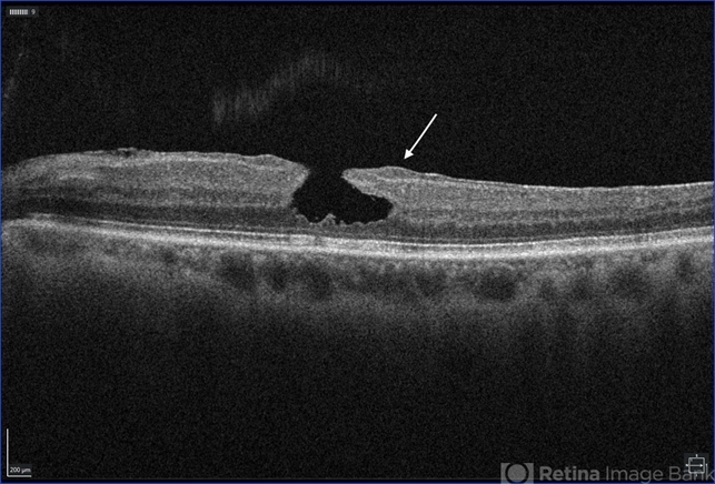
- lamellar macular hole
- Description
- Optical coherence tomography (OCT) image of a lamellar hole. The OCT shows the three mandatory features of a lamellar hole: 1) presence of irregular foveal contour 2) presence of foveal cavity with undermined edges, and 3) the apparent loss of foveal tissue. It also demonstrates lamellar-hole-associated epiretinal proliferation (LHEP) (arrow).


