Search results (103 results)
-
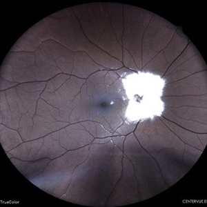
 Myelinated Nerve Fibers
Jun 4 2025 by Paulina Araujo
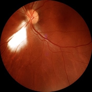
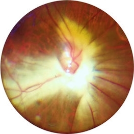
The 55-degree central fundus photograph of the left eye reveals myelination of the nerve fiber layer along the inferior nasal arcade.
Photographer: Paulina D.Araujo Martínez, Asociación para Evitar la Ceguera en México I.A.P., Hospital Dr Luis Sánchez Bulnes.
Condition/keywords: myelinated nerve fibers
-
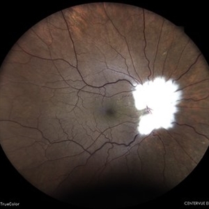
 Myelinated Retinal Nerve Fiber Layer
May 20 2025 by Ignacio Leonardo Pueyo Bestue, MD
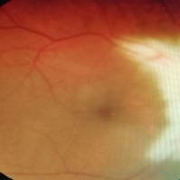
Fundus photo of an 80-year-old woman with myelinated RNFL, 20/20 vision, and mild hyperopia
Photographer: Pueyo-Bestue, I.L., MD, Universite Libre de Bruxelles, Ophthalmology Department
Condition/keywords: myelinated nerve fiber layer
-
 Myelinated Nerve Fibers
Apr 18 2025 by DR Rohit Gupta
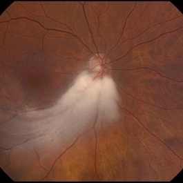
The **myelinated nerve fibers of the optic disc** (also known as **medullated nerve fibers**) are retinal nerve fibers that retain their myelin sheath as they pass through the optic nerve head. Normally, retinal nerve fibers are unmyelinated to allow for light transparency, but in some cases, myelination extends anteriorly into the retina, appearing as a striking white, feathery patch on the optic disc or peripapillary retina. ### **Key Features:** 1. **Appearance:** - Dense, white, striated patches with feathery edges. - Typically located at the superior or inferior pole of the optic disc. - May obscure retinal vessels underneath. 2. **Clinical Significance:** - Usually **benign** and asymptomatic. - **Congenital** (present at birth or early childhood). - Rarely associated with **visual field defects** (e.g., scotomas corresponding to the area of myelination). - Occasionally linked with **high myopia** or **amblyopia** if extensive. 3. **Pathophysiology:** - Failure of oligodendrocytes or Schwann cells to stop myelination at the lamina cribrosa. - Normally, myelination stops at the optic nerve head, but in this condition, it extends into the retina. 4. **Diagnosis:** - **Fundoscopy:** Classic white, feathery appearance. - **Optical Coherence Tomography (OCT):** Shows thickened retinal nerve fiber layer (RNFL). - **Visual Field Testing:** May detect defects if large. 5. **Differential Diagnosis:** - Optic disc edema - Cotton wool spots - Retinoblastoma (rarely, but must be ruled out in children) 6. **Management:** - No treatment required if asymptomatic. - Monitor for amblyopia in children. - Rare cases with significant visual impairment may need further evaluation. ### **Fun Fact:** Myelinated nerve fibers are seen in **~0.5-1%** of the population and are usually an incidental finding.
Photographer: Dr Rohit gupta
Imaging device: Samsung S21
Condition/keywords: Medulated Nerve fibre, Medullated Nerve fibres, myelinated nerve fibers, Myelinated Nerve Fibres, optic disc drusen
-
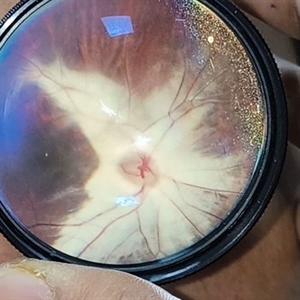
 Myelinated Nerve Fibres With Combined Hamartoma of Retina and RPE
Jul 31 2024 by Tejaswita Verma
Fundus image of a 20 year old female who presented with metamorphopsia ,slightly blurred vision. BCVA was 6/9, epiretinal membrane present on central fundus examination with myelinated nerve fibres.
Photographer: DR. TEJASWITA VERMA
Imaging device: MIRANTE
Condition/keywords: combined hamartoma of retina and RPE, myelinated nerve fibers
-
 Right Myelinated Nerve Fibre, LE Normal
May 6 2024 by Anupama Janardhanan
Fundus photograph of a 24 year old male patient with Anisometropia and Right Eye High Myopia associated with Diffuse circumpapillary myelinated Nerve Fibre and Left Eye normal fundus.
Photographer: Dr. Anupama Janardhanan, Aravind Eye hospital, Tirunelveli, India
Imaging device: Heidelberg Spectralis
Condition/keywords: anisometropia, myelinated nerve fibers, myopic degeneration
-
 Myelinated Nerve Fibres
Jan 30 2024 by Akansha Sharma
Color fundus photograph of a 15 year old male with myelinated nerve fibres all around the disc in the right eye.
Photographer: Dr. Akansha Sharma, Bharati Eye Hospital
Condition/keywords: MNF, myelinated nerve fibers
-
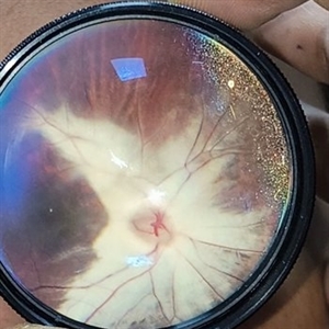
 Myelinated Nerve Fibre (MNF)
Sep 12 2023 by Ben Serar
Fundus photograph of RE showing Myelinated Nerve Fibre along superior disc margin
Condition/keywords: MNF, myelinated nerve fiber
-
 Myelinated Nerve Fibre (MNF)
Jun 17 2023 by Harsh Vardhan Singh, MS
Fundus photograph of 32-year-old male having good best corrected visual acuity in both eyes with right eye having high myopia & MNF as incidental finding
Photographer: Dr Harsh Vardhan Singh, Assistant Professor, AIIMS, Guwahati
Condition/keywords: medullated nerve fibers, MNF, myelinated nerve fiber layer, myelinated nerve fibers, Nerve fiber layer arrangements, NFL
-
 MYELINATED NERVE FIBRES
May 31 2023 by Akansha Sharma
COLOUR FUNDUS PHOTOGRAPH OF A 32 YEAR OLD FEMALE WITH MYELINATED NERVE FIBRES
Photographer: Dr. Urmil Shah, Dr. Akansha Sharma, Dr. Denish Patel
Condition/keywords: myelinated nerve fibers
-
 Myelinated Nerve Fibers
Apr 26 2023 by Kalyan Singh
Young male, presented to our OPD side for diminution of vision in fellow eye.
Photographer: Dr Kalyan Singh, Junior resident , Department of ophthalmology, GSVM MEDICAL COLLEGE KANPUR
Imaging device: One plus 10 R
Condition/keywords: myelinated nerve fibers
-
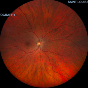
 Isolated myelinated nerve fiber layers
Mar 5 2023 by Niloofar Piri, MD
Fundus photograph of the right eye demonstrating patches of isolated myelinated nerve fiber layers along inferior arcade as well as nasal retina
Photographer: Sean Kelso, Saint Louis University
Condition/keywords: myelinated nerve fiber layer, myelinated nerve fibers
-
 Medullated nerve fibres
Mar 5 2023 by Kalyan Singh
History of trauma 4-5 years back and presented to our side with unilateral diminution of vision.
Photographer: Kalyan Singh, GSVM medical college, Kanpur
Imaging device: Smartphone (1 plus 10 R)
Condition/keywords: myelinated nerve fibers, trauma
-
 Myelinated Nerve Fiber Layer
Nov 24 2022 by Eder Díaz Dorado
Fundus photograph of an 35-year-old woman with myelinated nerve fiber layer
Photographer: Eder Díaz Dorado, Hospital Central Militar CDMX
Imaging device: Smartphone
Condition/keywords: myelinated nerve fibers
-
 Myelinated Nerve Fiber
Nov 10 2022 by Tandava Krishnan
Color fundus photo of the Left eye of a patient showing myelinated nerve fiber
Condition/keywords: myelinated nerve fibers
-
 Myelinated nerve fibre
Nov 10 2022 by Tandava Krishnan
Red free fundus photograph of the left eye of a patient with myelinated nerve fibre. Myelinated nerve fibres are autofluorescent and can be clearly made out on red free imaging
Condition/keywords: myelinated nerve fibers
-
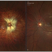
 Normal Fundus Photo, OD in Pt. with Myelinated NFL, OS
Oct 27 2021 by Charles Hurth
Fundus photograph of a 35-year-old woman with myelinated nerve fiber layer in her left eye who presented to the adult strabismus clinic with exotropia of her left eye.
Photographer: Charles Hurth, IV, DO, Saint Louis University
Condition/keywords: normal eye
-
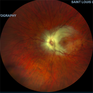 Myelinated Nerve Fiber Layer, OS
Oct 27 2021 by Charles Hurth
Fundus photograph of a 35-year-old woman with myelinated nerve fiber layer in her left eye who presented to the adult strabismus clinic with exotropia of her left eye.
Photographer: Charles Hurth, IV, DO, Saint Louis University
Condition/keywords: myelinated nerve fiber layer
-
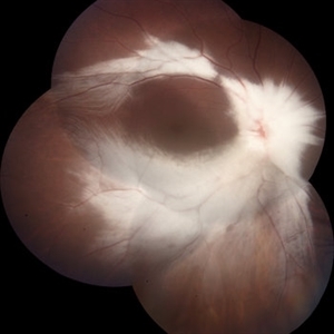
 Extensive Myelinated Nerve Fibres
May 20 2021 by Anmol Naik
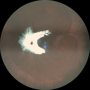
A 21-year-old Indian male presented with incidentally discovered subnormal vision in the left eye. On examination, he had esotropia with high myopia of -14 dioptres. Fundus examination revealed extensively myelinated nerve fibres around the optic disc extending along the arcade but sparing the fovea. The association of myelinated nerve fibres with high myopia and amblyopia is well documented but the causal association between these is unproven. Early detection of refractive error and aggressive therapy to prevent amblyopia has been reported with some success.
Photographer: Anmol Naik, Nakshatra Superspeciality Eye Hospital, Pune, India.
Imaging device: Zeine slit-lamp mounted Fundus imaging system
Condition/keywords: amblyopia, high myopia, myelinated nerve fibers
-
 Myelinated Nerve Fiber
May 5 2021 by Priya Rasipuram Chandrasekaran, MBBS, DO, DNB, FRCS
A 31-year-old male presented with a decreased vision of 20/125 N24 with -6.50 DS/-3.50 cyl 90 in the left eye. Fundus examination revealed peripapillary MNF progressing superiorly, obscuring disc and vessels and sparing the macula. OCT of ONH showed hyper reflective NFL and an abrupt ending of RPE and inner retinal layers (IRL) with underlying shadowing at the beginning of hyper reflectivity. The absence of photoreceptor integrity line (PIL) in the macula is believed to cause refractory amblyopia in such patients.
Condition/keywords: myelinated nerve fibers
-
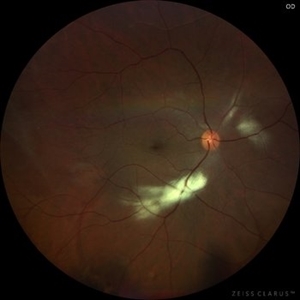
 Straatsma Syndrome
Nov 17 2020 by Linda A Cernichiaro- Espinosa, MD
5-year-old male with leukocoria, high myopia and myelinated nerve fibers (Straatsma Syndrome). OCT showed a foveal depression and intact photoreceptors. Amblyopia management was started.
Photographer: Guillermo Salcedo-Villanueva, MD
Imaging device: Zeiss Clarus
Condition/keywords: high myopia, leukocoria, myelinated nerve fibers, pediatric retina
-
 Myelinated Nerve Fiber Layer
Aug 2 2020 by Kelly Hannan
Myelinated nerve fiber layer.
Imaging device: heidelberg spectralis
Condition/keywords: myelinated nerve fiber layer
-
 Myelinated Nerve Fiber (mNFL)
Jun 21 2020 by Dhaivat Shah
Myelinated nerve fiber layer (mNFL) is a benign clinical entity that results from an embryologic developmental anomaly. Myelination along the visual pathway is noted around the eighth month of gestation, and typically reaches the posterior globe around the time of birth with virtually all fibers reaching complete myelination by age 7 months till the lamina cribrosa. Sometimes, due to altered neuro hormonal signals, this process of myelination extends past the lamina cribrosa and is visible on fundus examination as distinct white patches on the inner retinal surface. On infrared and red-free imaging, mNFL appears white, which is likely due to the high lipid content of myelin. Myelin blocks detection of underlying fluorescent material, thus appearing dark on fundus autofluorescence. On optical coherence tomography , it appears as a thickened and hyperreflective retinal nerve fiber layer. mNFL is typically benign but can be mistaken for other potentially serious conditions like neoplastic infiltration or infection. Hence, it is crucial to recognize the benign nature of mNFL to avoid superfluous medical testing.
Photographer: Ms Srishti Sharma
Imaging device: Choithram Netralaya
Condition/keywords: myelinated nerve fibers
-
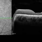 Plateau Fovea with Inner Retinal Thinning
May 27 2020 by Olivia Rainey
Optical coherence tomography of the left eye of a 20-year-old male with Alport Syndrome. The patient did not present with any ocular or visual symptoms, yet the distinct "plateau contour" of his fovea was noted on OCT during his visit. The patient presented with 20/25 vision at the time of his visit. There was myelinated nerve fiber layer noted in both eyes, but these features had remained stable from his appointment three years prior. The physician noted that myelinated nerve fiber was a congenital change, and had not affected his vision or health of the eye, nor is a feature of Alport Syndrome.
Photographer: Olivia Rainey, OCT-C, COA
Imaging device: Heidelberg Spectralis
Condition/keywords: Alports disease, Heidelburg Spectralis, inner retinal thinning, left eye, optical coherence tomography (OCT), plateau fovea
-
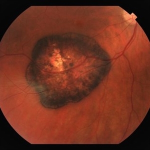 CHRPE & Myelinated RNFL
May 21 2020 by John S. King, MD
47-year-old white female, asymptomatic, sent to evaluate a scar OD. 20/40 cc, normotensive, examination significant for a flat, solitary lesion with pigmented borders and depigmented center with early lacunae forming, along with myeliated RNFL at the temporal edge of the lesion.
Photographer: Kay Dalby
Imaging device: Topcon
Condition/keywords: congenital hypertrophy of the retinal pigment epithelium (CHRPE), myelinated nerve fiber layer
-
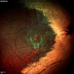 Myelinated Nerve Fiber With Epiretinal Membrane With Lamellar Macular Hole
May 4 2020 by SWATI INDURKHYA
Heidelberg HRA + OCT Spectralis Multicolor (30 degree) retinal image of the left eye of a 28-year-old male showing myelinated nerve fibre (MNF) with epiretinal membrane (ERM) and lamellar macular hole (LMH).
Photographer: Rakesh PR, Giridhar Eye Institute, Kerala, India
Imaging device: Heidelberg Spectralis HRA + OCT
Condition/keywords: epiretinal membrane (ERM), myelinated nerve fibers
 Loading…