 Initializing download.
-
 By John S. King, MD
Retina Associates, PA - Uploaded on May 21, 2020.
- Last modified by Caroline Bozell on May 21, 2020.
- Rating
- Appears in
- Miscellaneous
- Condition/keywords
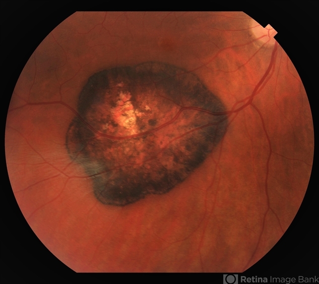
- congenital hypertrophy of the retinal pigment epithelium (CHRPE), myelinated nerve fiber layer
- Photographer
- Kay Dalby
- Imaging device
-
Fundus camera
Topcon - Description
- 47-year-old white female, asymptomatic, sent to evaluate a scar OD. 20/40 cc, normotensive, examination significant for a flat, solitary lesion with pigmented borders and depigmented center with early lacunae forming, along with myeliated RNFL at the temporal edge of the lesion.


")
---thumb.jpg/image-square;max$79,0.ImageHandler "Macular CHRPE")
")
---thumb.jpg/image-square;max$79,0.ImageHandler "ARMD RPE Defect / Myelinated NFL")
---thumb.jpg/image-square;max$79,0.ImageHandler "ARMD RPE Defect / Myelinated NFL")
---thumb.jpg/image-square;max$79,0.ImageHandler "ARMD RPE Defect / Myelinated NFL")
---thumb.jpg/image-square;max$79,0.ImageHandler "ARMD RPE Defect / Myelinated NFL")
---thumb.jpg/image-square;max$79,0.ImageHandler "ARMD RPE Defect / Myelinated NFL")
---thumb.jpg/image-square;max$79,0.ImageHandler "ARMD RPE Defect / Myelinated NFL")
")