File number: 53844
Comments
-
 Suber S. Huang, MD, MBA, FASRS (May 26 2020)
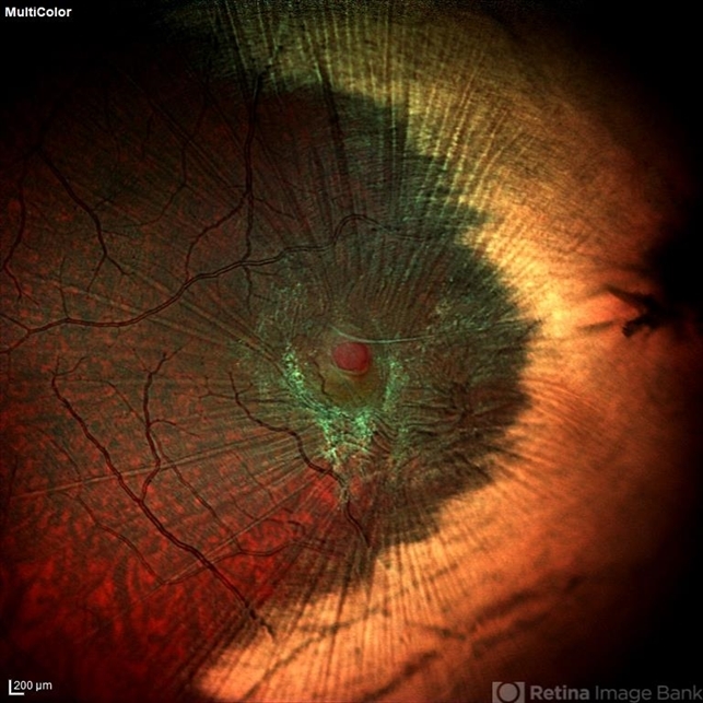
spectacular capture of a rare presentation.
Question: is there any possibility that this could have arisen from distant trauma with peripapillary choroidal rupture, traumatic macular hole, and secondary macular pucker? The yellow-white areas do not strictly follow the nerve fiber layer and the presence of striae from epiretinal traction confuse the picture. Please add a detailed history to the description.
Sign in to comment.
 Initializing download.
-
 By SWATI INDURKHYA
GIRIDHAR EYE INSTITUTE
Co-author(s): Dr Giridhar Anantharaman, Giridhar Eye Institute, Kerala, India, Dr Mahesh Gopalakrishnan , Giridhar Eye Institute, Kerala, India , Dr Shivam Madan, Giridhar Eye Institute, Kerala, India, Dr Dinesh Rungta, Giridhar Eye Institute, Kerala, India - Uploaded on May 4, 2020.
- Last modified by Caroline Bozell on May 5, 2020.
- Rating
- Appears in
- Miscellaneous
- Condition/keywords
- myelinated nerve fibers, epiretinal membrane (ERM)
- Photographer
- Rakesh PR, Giridhar Eye Institute, Kerala, India
- Imaging device
- Heidelberg Spectralis HRA + OCT
- Description
- Heidelberg HRA + OCT Spectralis Multicolor (30 degree) retinal image of the left eye of a 28-year-old male showing myelinated nerve fibre (MNF) with epiretinal membrane (ERM) and lamellar macular hole (LMH).


---thumb.JPG/image-square;max$79,0.ImageHandler "ERM")
---thumb.jpg/image-square;max$79,0.ImageHandler "ERM")


