Search results (127 results)
-
 Systemic Lupus Erythematosus (SLE) Vasculitis
Jan 29 2025 by Kimberly Wakester
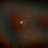
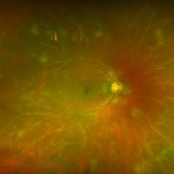
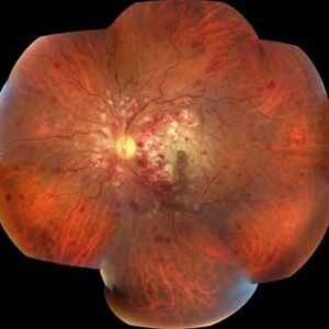
Fundus photographs of an 13-year-old boy with Systemic Lupus Erythematosus (SLE) Vasculitis in both eyes s/p PRP laser. Patient is doing well s/p PRP Laser OU and with continued use of oral medications. Patient will be monitored with follow up exams to check for recurring vasculitis or recurring/worsening NVE/NVD. Patient is to continue ongoing management with Rheumatologist.
Photographer: Kimberly Wakester, COA
Imaging device: Optos California
Condition/keywords: NVD, NVE, occlusive vasculitis, pan-retinal photocoagulation (PRP), Systemic Lupus Erythematosus (SLE) Vasculitis
-
 Systemic Lupus Erythematosus (SLE) Vasculitis
Jan 29 2025 by Kimberly Wakester
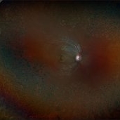
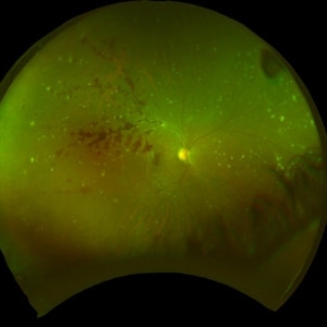
Fundus photographs of an 13-year-old boy with Systemic Lupus Erythematosus (SLE) Vasculitis in both eyes s/p PRP laser. Patient is doing well s/p PRP Laser OU and with continued use of oral medications. Patient will be monitored with follow up exams to check for recurring vasculitis or recurring/worsening NVE/NVD. Patient is to continue ongoing management with Rheumatologist.
Photographer: Kimberly Wakester, COA
Imaging device: Optos California
Condition/keywords: NVD, NVE, occlusive vasculitis, pan-retinal photocoagulation (PRP), Systemic Lupus Erythematosus (SLE) Vasculitis
-
 APLA syndrome with Systemic lupus erythematosus retinopathy
Jan 14 2024 by Hemanth Murthy, MBBS, MD, FASRS
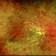
22 year female presented with loss of vision in right eye since 2 days. Vision was CF. Fundus showed cotton wool patches in posterior pole with large blotchy haemorrhage with fuzzy appearance of arteries and sheathing of veins and dull fovea. OCT showed inner layer hyper reflectivity with SSRD and cystoid edema. APLA was positive and ANA profile positive for SLE
Photographer: Mr Veda Vyas
Imaging device: Optos Daytona
Condition/keywords: APLA Syndrome, systemic lupus erythematosus (SLE) retinopathy
-
 Central Retinal Vein Occlusion with Macular Edema in Antiphospholipid Syndrome
Dec 24 2023 by Nikhil K Bommakanti, MD
A man in his thirties presented with a central retinal vein occlusion with macular edema in the right eye. Vision improved from 20/70 to 20/25 after 1 treatment with intravitreal bevacizumab. Laboratory testing revealed the presence of lupus anticoagulant.
Condition/keywords: antiphospholipid antibody syndrome, central retinal vein occlusion (CRVO), cystoid macular degeneration, macular edema
-
 Cilioretinal Artery and Lupus Retinopathy
Mar 31 2022 by Franco Benvenuto, MD
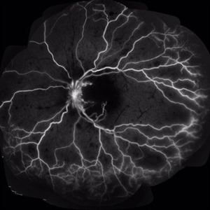
Right eye fundus fluorescein angiography of a 30-year-old female patient presented with diminution of vision in both eyes since 3 months. Fundus examination revealed cotton-wool spots, vasculitis and the presence of a cilioretinal artery in the right eye. Laboratory investigations were positive for antinuclear antibodies and antidouble stranded/native DNA antibodies.
Photographer: Franco Benvenuto, Universidad de Buenos Aires, Argentina; Universidad de Guadalajara, México.
Condition/keywords: cilioretinal artery, systemic lupus erythematosus (SLE) retinopathy, systemic lupus erythematosus (SLE) vasculitis
-
 Retinal Lupus Vasculitis
Sep 25 2021 by Denis Jusufbegovic, MD
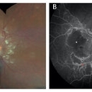
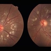
27-year-old woman with a history of systemic lupus erythematosus (SLE) presented with decreased vision to counting fingers at 2’ in the right eye. Funduscopic examination of the right eye (A) demonstrated retinal thickening and whitening of the macula, numerous cotton-wool spots, intra-retinal hemorrhages, sclerotic vessels, and vascular sheathing. Fluorescein angiography (B) demonstrated extensive vessel wall leakage (red asterisk) and large areas of capillary non-perfusion (white asterisk). These findings were consistent with severe vaso-occlusive retinopathy, a serious ophthalmologic manifestation of SLE. She was also diagnosed with concomitant cerebral lupus vasculitis. She was treated with intravenous methylprednisolone followed by oral prednisone taper and aspirin therapy. Mycophenolate mofetil was titrated to 1500 mg twice daily. Upon follow-up vision improved to 20/200.
Imaging device: Zeiss Clarus 500
Condition/keywords: cerebral lupus vasculitis, cotton wool spots, systemic lupus erythematosus (SLE) vasculitis, vaso-occlusive disease
-
 Lupus Retinopathy
Mar 14 2021 by Marco Antonio Sauza
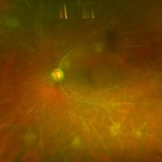
Fundus photo of a young female patient with LES after pregnancy.
Photographer: Marco Sauza
Condition/keywords: systemic lupus erythematosus (SLE) retinopathy
-
 Lupus Retinopathy
Mar 14 2021 by Marco Antonio Sauza
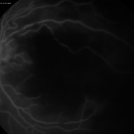
FA in a 13-year-old female with ischemic LES retinopathy.
Photographer: Marco sauza
Condition/keywords: systemic lupus erythematosus (SLE) retinopathy
-
 Lupus Retinopathy
Mar 14 2021 by Marco Antonio Sauza
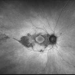
Red free retina photo in a teenage female with complete vision loss (NLP) with ischemic lupus retinopathy.
Photographer: Marco Sauza
Imaging device: Zeiss
Condition/keywords: systemic lupus erythematosus (SLE) retinopathy
-
 Lupus Retinopathy
Mar 14 2021 by Marco Antonio Sauza
FA with ischemic lupus retinopathy in a 21-year-old female with LES after pregnancy.
Photographer: Marco Sauza
Imaging device: Zeiss
Condition/keywords: systemic lupus erythematosus (SLE) retinopathy
-
 Macular Ischemia in Lupus Retinopathy
Mar 14 2021 by Marco Antonio Sauza
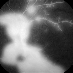
FA in a female with 21-year-old with LES after pregnancy.
Photographer: Marco Sauza
Imaging device: Zeiss
Condition/keywords: systemic lupus erythematosus (SLE) retinopathy
-
 Lupus Retinopathy
Mar 14 2021 by Marco Antonio Sauza
FA of a 13-year-old female with ischemic retinopathy and LES.
Photographer: Marco Sauza
Imaging device: Zeiss
Condition/keywords: systemic lupus erythematosus (SLE) retinopathy
-
 Lupus Retinopathy
Mar 14 2021 by Marco Antonio Sauza
FA photo of a 13-year-old female with ischemic retinopathy and LES.
Photographer: Marco sauza
Imaging device: Zeiss
Condition/keywords: systemic lupus erythematosus (SLE) retinopathy
-
 Lupus Retinopathy
Mar 14 2021 by Marco Antonio Sauza
Fluorescein angiography photo of and 13-year-old female with ischemic retinopathy with LES.
Photographer: Marco Sauza
Imaging device: Zeiss fundus camera
Condition/keywords: systemic lupus erythematosus (SLE) retinopathy
-
 Plaquenil Toxicity
Jun 20 2019 by Olivia Rainey
Ultra-wide field fundus autofluorescence photograph of a 60-year-old female with plaquenil toxicity affecting both eyes. Patient was taking plaquenil for management of Lupus, but discontinued use in 2011, but continues to be affected with severe progression of toxicity. Patient developed macular edema affecting her right eye and has received 5 Avastin injections.
Photographer: Olivia Rainey
Imaging device: Optos
Condition/keywords: autofluorescence imaging, focal pigmentary changes, fundus autofluorescence (FAF), macular edema, Optos, plaquenil toxicity, ultra-wide field imaging
-
 Retinal Vasculitis
Apr 9 2019 by Nikisha Kothari, MD
Fundus photograph of a 56-year-old female with SLE occlusive vasculitis.
Photographer: Orly Catz
Imaging device: Optos
Condition/keywords: systemic lupus erythematosus (SLE) vasculitis
-
 Vasculitis-OD
Apr 9 2019 by Nikisha Kothari, MD
Fundus photograph of a 56-year-old female with SLE vasculitis.
Photographer: Orly Catz
Imaging device: Optos
Condition/keywords: systemic lupus erythematosus (SLE) vasculitis
-
 Central Retinal Artery Occlusion
Aug 28 2018 by Gabriela Lopezcarasa Hernandez, MD
35-year-old women with CRAO and vasculitis due to systemic lupus.
Photographer: MARCO ANTONIO SAUZA CASTILLEJOS M.D., MEXICO.
Condition/keywords: central retinal artery occlusion (CRAO), vasculitis
-
 Central Retinal Artery Occlusion
Aug 28 2018 by Gabriela Lopezcarasa Hernandez, MD
35-year-old women with CRAO and vasculitis due to systemic lupus.
Photographer: MARCO ANTONIO SAUZA CASTILLEJOS M.D., MEXICO.
Condition/keywords: central retinal artery occlusion (CRAO), vasculitis
-
 Central Retinal Artery Occlusion
Aug 28 2018 by Gabriela Lopezcarasa Hernandez, MD
CRAO related to systemic LUE.
Photographer: MARCO ANTONIO SAUZA CASTILLEJOS M.D., MEXICO.
Condition/keywords: central retinal artery occlusion (CRAO), lupus
-
 SLE Retinopathy
Jul 10 2018 by Deepak Bhojwani, MS
Colour fundus montage image of a 33-year-old young lady with history of Systemic Lupus Erythematosus of 6 years showing classic SLE retinopathy with multiple cotton wool spots , few haemorrhages and multiple small vessel sheathing s/o SLE vasculitis.
Photographer: Deepak Bhojwani
Condition/keywords: systemic lupus erythematosus (SLE) retinopathy, systemic lupus erythematosus (SLE) vasculitis
-
 Lupus Hemorrhagic Occlusive Vasculitis
Apr 23 2018 by Frank Chin
Fundus photograph of the right eye of a 24-year-old woman with history of systemic lupus erythematosus who presented with decreased visual acuity for 2-3 days found to have lupus hemorrhagic occlusive vasculitis with mild disc elevation, diffuse punctate cotton wool spots and dot blot hemorrhages, and a hemorrhage occlusive vasculitis along the superior branch of the superotemporal arcade involving the artery and vein.
Photographer: Frank Chin, MD, George Washington University
Imaging device: Optos 200Tx
Condition/keywords: blot hemorrhages, cotton wool spots, occlusive vasculitis, systemic lupus erythematosus (SLE) vasculitis
-
 SLE Retinopathy
Nov 14 2016 by Mitzy E Torres Soriano, MD
25-year-old female patient with systemic lupus erythematosus. Photographs show cotton wool spots, intraretinal hemorrhages and vascular tortuosity. FA demonstrated retinal vasculitis and OCT revealed cystoid macular edema. In this case diagnosis of SLE was made after ocular manifestation.
Photographer: Grupo Laser Vision, Rosario, Argentina
Condition/keywords: cotton wool spots, occlusive retinal vasculitis, occlusive vasculitis, systemic lupus erythematosus, vasculopathy
-
 Acute Necrotizing Retinal Vasculitis as Onset of Systemic Lupus Erythematosus.
Sep 3 2016 by ADRIANO FERREIRA
28-year-old white man was referred to the rheumatology clinic with gradually and rapid deterioration of the vision (both eyes). In this picture, we can observe vasculitis (leakage from vessels) and diffuse ischemia in the left eye.
Photographer: Claudio Zett Lobo
Imaging device: HRA-Spectralis
Condition/keywords: systemic lupus erythematosus (SLE) vasculitis, vasculitis
-
 Acute Necrotizing Retinal Vasculitis as Onset of Systemic Lupus Erythematosus.
Sep 3 2016 by ADRIANO FERREIRA
A 28-year-old white man was referred to the rheumatology clinic with gradually and rapid deterioration of the vision (both eyes). In this picture we can observe cotton wool spots in the papillomacular area and extensive hemorrhages in the left eye.
Photographer: Claudio Zett Lobo
Imaging device: TRC50DXi TOPCON
Condition/keywords: systemic lupus erythematosus (SLE) vasculitis, vasculitis
 Loading…