Search results (127 results)
-
 Central Retinal Artery Occlusion
Aug 28 2018 by Gabriela Lopezcarasa Hernandez, MD
CRAO related to systemic LUE.
Photographer: MARCO ANTONIO SAUZA CASTILLEJOS M.D., MEXICO.
Condition/keywords: central retinal artery occlusion (CRAO), lupus
-
 Lupus Retinopathy
Mar 27 2014 by Jason S. Calhoun
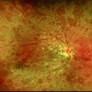
Female patient in for evaluation on lupus retinopathy. Has poor vision in the right eye. VA is hand motion in the right eye. Fundus photos show fibrosis along the temporal arcades and narrowing of the arteries. No macular edema found.
Photographer: Jason S. Calhoun, Mayo Clinic Jacksonville, Department of Ophthalmology
Imaging device: TOPCON TRC 50-EX
Condition/keywords: lupus, retinopathy
-
 Lupus Retinopathy
Mar 27 2014 by Jason S. Calhoun
Female patient in for evaluation on lupus retinopathy. Has poor vision in the right eye. VA is hand motion in the right eye. Fundus photos show fibrosis along the temporal arcades and narrowing of the arteries. No macular edema found.
Photographer: Jason S. Calhoun, Mayo Clinic Jacksonville, Department of Ophthalmology
Imaging device: TOPCON TRC 50-EX
Condition/keywords: lupus, retinopathy
-
 Lupus Vasculitis
Feb 13 2013 by From the Collections of Thomas M. Aaberg, MD and Thomas M. Aaberg Jr., MD
Obliterative peripheral vasculitis.
Condition/keywords: ischemia, lupus
-
---thumb.jpg/image-square;max$300,300.ImageHandler) Lupus Vasculitis Angiogram
Lupus Vasculitis Angiogram
Feb 13 2013 by From the Collections of Thomas M. Aaberg, MD and Thomas M. Aaberg Jr., MD
FA, lupus vasculitis angiogram.
Condition/keywords: lupus, obliterative peripheral vasculitis, retinal ischemia
-
 Macular Infarction in Systemic Lupus Erythematosus
Sep 21 2012 by Allen Chiang, MD, FASRS
Fluorescein angiogram of a 46-year old male with macular infarction, which was the presenting sign of his systemic lupus erythematosus. The fellow eye looked identical.
Imaging device: Topcon
Condition/keywords: lupus, macular ischemia
-
 Macular Infarction in Systemic Lupus Erythematosus
Sep 21 2012 by Allen Chiang, MD, FASRS
Fundus photograph of a 46-year old male with macular infarction, which was the acute presenting sign of his systemic lupus erythematosus. The fellow eye looked identical.
Imaging device: Topcon
Condition/keywords: lupus, macular ischemia
-
 Retinal Vasculopathy With Retinal Vasculitis and Ischemia
Jul 23 2014 by John S. King, MD
Purtscher's-like retinopathy.
Photographer: UPMC
Condition/keywords: lupus, systemic lupus erythematosus (SLE) retinopathy, systemic lupus erythematosus (SLE) vasculitis
-
 SLE Retinal Vasculopathy With Retinal Vasculitis and Ischemia
Jul 23 2014 by John S. King, MD
OD: 45 sec and 3 min OS: 1:30 min and 3 min
Photographer: UPMC
Condition/keywords: lupus, systemic lupus erythematosus (SLE) retinopathy, systemic lupus erythematosus (SLE) vasculitis
-
 SLE Retinal Vasculopathy With Retinal Vasculitis and Ischemia
Jul 23 2014 by John S. King, MD
Discoid lesions.
Photographer: UPMC
Condition/keywords: lupus, systemic lupus erythematosus (SLE) retinopathy, systemic lupus erythematosus (SLE) vasculitis
-
---thumb.jpg/image-square;max$300,300.ImageHandler) SLE Retinopathy
SLE Retinopathy
Feb 26 2013 by Henry J. Kaplan, MD
Cotton wool spots, right eye. #1
Condition/keywords: lupus, systemic lupus erythematosus (SLE) retinopathy, systemic lupus erythematosus (SLE) vasculitis
-
---thumb.jpg/image-square;max$300,300.ImageHandler) Lupus Anticoagulant Disorder
Lupus Anticoagulant Disorder
Feb 26 2013 by Henry J. Kaplan, MD
Vasoocclusive retinopathy associated with Lupus anticoagulate factor.
Condition/keywords: lupus anticoagulate factor, vasoocclusive retinopathy
-
---thumb.jpg/image-square;max$300,300.ImageHandler) Lupus Anticoagulant Disorder
Lupus Anticoagulant Disorder
Feb 26 2013 by Henry J. Kaplan, MD
Occluded retinal vessels and vitreous hemorrhage apparent.
Condition/keywords: lupus anticoagulate factor
-
 Lupus Hemorrhagic Occlusive Vasculitis
Apr 23 2018 by Frank Chin
Fundus photograph of the right eye of a 24-year-old woman with history of systemic lupus erythematosus who presented with decreased visual acuity for 2-3 days found to have lupus hemorrhagic occlusive vasculitis with mild disc elevation, diffuse punctate cotton wool spots and dot blot hemorrhages, and a hemorrhage occlusive vasculitis along the superior branch of the superotemporal arcade involving the artery and vein.
Photographer: Frank Chin, MD, George Washington University
Imaging device: Optos 200Tx
Condition/keywords: blot hemorrhages, cotton wool spots, occlusive vasculitis, systemic lupus erythematosus (SLE) vasculitis
-
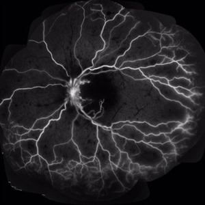
 Lupus Retinopathy
Mar 14 2021 by Marco Antonio Sauza
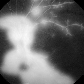
FA with ischemic lupus retinopathy in a 21-year-old female with LES after pregnancy.
Photographer: Marco Sauza
Imaging device: Zeiss
Condition/keywords: systemic lupus erythematosus (SLE) retinopathy
-
 Lupus Retinopathy
Mar 14 2021 by Marco Antonio Sauza
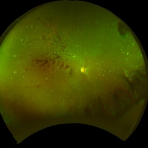
Red free retina photo in a teenage female with complete vision loss (NLP) with ischemic lupus retinopathy.
Photographer: Marco Sauza
Imaging device: Zeiss
Condition/keywords: systemic lupus erythematosus (SLE) retinopathy
-
 Lupus Retinopathy
Mar 14 2021 by Marco Antonio Sauza
FA in a 13-year-old female with ischemic LES retinopathy.
Photographer: Marco sauza
Condition/keywords: systemic lupus erythematosus (SLE) retinopathy
-
 Lupus Retinopathy
Mar 14 2021 by Marco Antonio Sauza
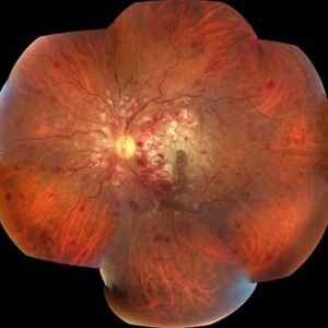
Fundus photo of a young female patient with LES after pregnancy.
Photographer: Marco Sauza
Condition/keywords: systemic lupus erythematosus (SLE) retinopathy
-
 Lupus Retinopathy
Mar 14 2021 by Marco Antonio Sauza
Fluorescein angiography photo of and 13-year-old female with ischemic retinopathy with LES.
Photographer: Marco Sauza
Imaging device: Zeiss fundus camera
Condition/keywords: systemic lupus erythematosus (SLE) retinopathy
-
 Lupus Retinopathy
Mar 14 2021 by Marco Antonio Sauza
FA photo of a 13-year-old female with ischemic retinopathy and LES.
Photographer: Marco sauza
Imaging device: Zeiss
Condition/keywords: systemic lupus erythematosus (SLE) retinopathy
-
 Lupus Retinopathy
Mar 14 2021 by Marco Antonio Sauza
FA of a 13-year-old female with ischemic retinopathy and LES.
Photographer: Marco Sauza
Imaging device: Zeiss
Condition/keywords: systemic lupus erythematosus (SLE) retinopathy
-
 Acute Necrotizing Retinal Vasculitis as Onset of Systemic Lupus Erythematosus.
Sep 3 2016 by ADRIANO FERREIRA
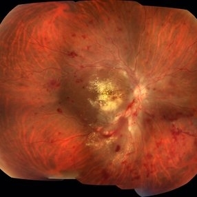
A 28-year-old white man was referred to the rheumatology clinic with gradually and rapid deterioration of the vision (both eyes). In this picture, we can observe cotton wool spots in the papillomacular area and extensive hemorrhages in posterior polo and in the middle periphery. Hard exudates are present in macular area (macular edema)
Photographer: Claudio Zett Lobo
Imaging device: TRC50DXi TOPCON
Condition/keywords: systemic lupus erythematosus (SLE) vasculitis, vasculitis
-
 Acute Necrotizing Retinal Vasculitis as Onset of Systemic Lupus Erythematosus.
Sep 3 2016 by ADRIANO FERREIRA
A 28-year-old white man was referred to the rheumatology clinic with gradually and rapid deterioration of the vision (both eyes). In this picture we can observe cotton wool spots in the papillomacular area and extensive hemorrhages in the left eye.
Photographer: Claudio Zett Lobo
Imaging device: TRC50DXi TOPCON
Condition/keywords: systemic lupus erythematosus (SLE) vasculitis, vasculitis
-
 Acute Necrotizing Retinal Vasculitis as Onset of Systemic Lupus Erythematosus.
Sep 3 2016 by ADRIANO FERREIRA
28-year-old white man was referred to the rheumatology clinic with gradually and rapid deterioration of the vision (both eyes). In this picture, we can observe vasculitis (leakage from vessels) and diffuse ischemia in the left eye.
Photographer: Claudio Zett Lobo
Imaging device: HRA-Spectralis
Condition/keywords: systemic lupus erythematosus (SLE) vasculitis, vasculitis
-
 APLA syndrome with Systemic lupus erythematosus retinopathy
Jan 14 2024 by Hemanth Murthy, MBBS, MD, FASRS
22 year female presented with loss of vision in right eye since 2 days. Vision was CF. Fundus showed cotton wool patches in posterior pole with large blotchy haemorrhage with fuzzy appearance of arteries and sheathing of veins and dull fovea. OCT showed inner layer hyper reflectivity with SSRD and cystoid edema. APLA was positive and ANA profile positive for SLE
Photographer: Mr Veda Vyas
Imaging device: Optos Daytona
Condition/keywords: APLA Syndrome, systemic lupus erythematosus (SLE) retinopathy
 Loading…