 Initializing download.
-
 By Denis Jusufbegovic, MD
Indiana University, Glick Eye Institute
Co-author(s): Shivam Patel - Uploaded on Sep 25, 2021.
- Last modified by Jennifer Carstens on Sep 29, 2021.
- Rating
- Appears in
- Miscellaneous
- Condition/keywords
- systemic lupus erythematosus (SLE) vasculitis, cotton wool spots, vaso-occlusive disease
- Imaging device
- Zeiss Clarus 500
- Description
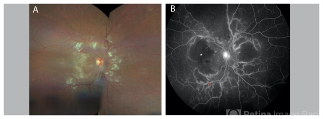
- 27-year-old woman with a history of systemic lupus erythematosus (SLE) presented with decreased vision to counting fingers at 2’ in the right eye. Funduscopic examination of the right eye (A) demonstrated retinal thickening and whitening of the macula, numerous cotton-wool spots, intra-retinal hemorrhages, sclerotic vessels, and vascular sheathing. Fluorescein angiography (B) demonstrated extensive vessel wall leakage (red asterisk) and large areas of capillary non-perfusion (white asterisk). These findings were consistent with severe vaso-occlusive retinopathy, a serious ophthalmologic manifestation of SLE. She was also diagnosed with concomitant cerebral lupus vasculitis. She was treated with intravenous methylprednisolone followed by oral prednisone taper and aspirin therapy. Mycophenolate mofetil was titrated to 1500 mg twice daily. Upon follow-up vision improved to 20/200.


