Search results (37 results)
-
 Commotio Retinae
Jun 10 2025 by CUI YUELING
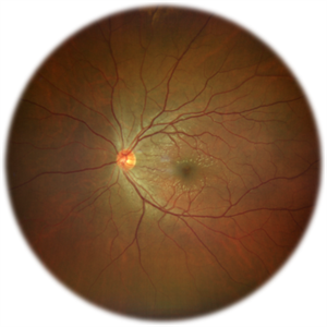
The patient presented 2 hours after sustaining a left eye injury caused by a stick. Visual acuity in the left eye was 0.2 without improvement upon correction, and intraocular pressure measured 15 mmHg. Examination of the anterior segment revealed ciliary conjunctival injection accompanied by patchy subconjunctival hemorrhage. The corneal surface remained smooth, and the anterior chamber was deep with hyphema characterized by blood-tinged aqueous humor predominantly settled inferiorly. The pupil was slightly irregular, approximately 3 mm in diameter, with a superotemporal notch; pupillary light reflex was intact. The lens appeared clear. Fundus examination showed well-defined optic disc margins with normal coloration and a cup-to-disc ratio of 0.2. Retinal arteries and veins were normally distributed with an artery-to-vein ratio of 2:3. At the posterior pole, the foveal reflex exhibited concentric ripple-like changes centered on the fovea, accompanied by localized pigment attenuation and reduced reflex intensity. Irregular reflectivity was noted in the superotemporal and inferotemporal nerve fiber layers.
Photographer: Yueling Cui
Imaging device: Zeiss Clarus 500
Condition/keywords: commotio retinae
-
Posterior Hyphema
Apr 29 2025 by Gustavo Uriel Fonseca Aguirre
This kinetic B-mode ultrasound scan (inferior transverse view) reveals combined vitreous and subhyaloid hemorrhage, accompanied by a mobile posterior hyphema level. The dynamic evaluation shows dependent blood shifting with positional changes, confirming fresh hemorrhage without organization.
Condition/keywords: diabetic retinopathy
-
 Advanced Proliferative Diabetic Retinopathy
Apr 9 2025 by Gustavo Uriel Fonseca Aguirre
B-mode ultrasound of a patient with long-standing poorly controlled diabetes demonstrates characteristic findings of advanced proliferative diabetic retinopathy. The examination reveals moderate vitreous hemorrhage appearing as diffuse hyperechoic opacities throughout the vitreous cavity, along with a posterior hyaloid membrane densely infiltrated by hemorrhagic material, showing irregular thickening and increased reflectivity. A mild subhyaloid hemorrhage is visible as a subtle hyphema-like space anterior to the retinal surface. The study documents a total tractional retinal detachment, evidenced by rigid retinal folds with clear insertion points of vitreous strands, accompanied by a significant subretinal hemorrhage seen as a prominent hyperechoic collection beneath the elevated retina. These findings collectively illustrate the severe vitreoretinal interface pathology characteristic of end-stage diabetic eye disease, with predominant tractional components and distinct echographic stratification of hemorrhagic layers - from anterior vitreous involvement to deeper subretinal blood accumulation.
Photographer: Gustavo U. Fonseca Aguirre, Hospital Conde de Valenciana, Ciudad de México
Condition/keywords: diabetic retinopathy, tractional retinal detachment, Vitreous hemorrhage
-
Eye of the Hurricane
Apr 9 2025 by Gustavo Uriel Fonseca Aguirre
Ultrasound biomicroscopy of a post-operative eye (status post trabeculectomy and phacoemulsification) reveals a patent ostium on the right side, along with an intraocular lens in position. A hyphema is observed displaying small convection currents, creating a circular motion pattern due to the temperature gradient between the iris and cornea. Notably, the blood flow can be seen circulating toward the trabeculectomy site.
Condition/keywords: hyphema, trabeculectomy
-
 Eye of the Hurricane
Apr 8 2025 by Gustavo Uriel Fonseca Aguirre
Ultrasound biomicroscopy of a post-operative eye (status post trabeculectomy and phacoemulsification) reveals a patent ostium on the right side, along with an intraocular lens in position. A hyphema is observed displaying small convection currents, creating a circular motion pattern due to the temperature gradient between the iris and cornea. Notably, the blood flow can be seen circulating toward the trabeculectomy site.
Photographer: Gustavo U. Fonseca Aguirre, Hospital Conde de Valenciana, Ciudad de México
Condition/keywords: Hyphema, trabeculectomy
-
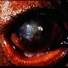
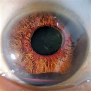 Rubeosis and Hyphema
Nov 19 2022 by Gareth Lema, MD, PhD
Florid rubeosis and hyphema.
Photographer: Gareth Lema, MD, PhD, New York Eye and Ear of Mount Sinai
Imaging device: Cell phone with a macro lens and muscle light for illumination.
Condition/keywords: Hyphema, Rubeosis
-
 The Effects of Blunt Trauma
Feb 27 2022 by Jesus Lozano, MD
Axial Head Ct. 60 year old man with a history of blunt trauma and lost of vision after the event. VA HM. Iop 25mmhg. Cornea clear. Complete hyphema. BMode US: diffuse Vitreous Hemorrhage with a Dislocated Lens. Retina attached.
Photographer: Dr. Jesus Lozano. Retina Specialist. Hillel Yaffe Medical Center,Israel.
Imaging device: Axial Head CT
Condition/keywords: blunt trauma, hyphema, lens dislocation
-
 Slide 7-73
Feb 25 2019 by Lancaster Course in Ophthalmology
A dense, fibrous plaque occupies the anterior chamber as the result of organization of a hyphema.
Condition/keywords: anterior chamber, hyphema, plaque
-
 Subconjuntival IOL After Blunt Trauma
Jun 27 2018 by Gabriel Costa Andrade, PhD
A 73-year-old male patient was referred to our ophthalmic emergency department with complaints of redness, pain, and diminution of vision in his left eye, after fall from height. The patient underwent small incision cataract surgery with polymethylmethacrylate (PMMA) IOL implantation in both the eyes 15 years back through superior sclerocorneal incision under local anesthesia. His best-corrected visual acuity was perception of light in the left eye. Ophthalmic examination using slit lamp biomicroscopy of the left eye revealed diffuse subconjunctival hemorrhage with no conjunctival laceration and inferior bulbar conjunctiva showed traumatic pseudophacocele with a sign “golden half ring,” suggesting the presence of PCIOL in subconjunctival space.There was total hyphema obscuring the view of rest of the ocular structures in his left eye.
Photographer: Gabriel Andrade, RETINA CLINIC, São Paulo, BRAZIL
Condition/keywords: dislocated intraocular lens (IOL), trauma
-
 Hyphema
May 13 2016 by Nichole Lewis
Hyphema and anterior chamber air bubble.
Photographer: Nichole Lewis
Condition/keywords: hyphema
-
 24 Hours Post Scleral Wound Closure+ Scleral Buckle+25 g Vitrectomy+Silicon Oil
Jan 23 2015 by Carlos Quezada-Ruiz, MD, FASRS
24 hours post op fundus photograph of a 43-year-old man who had perforating injury to the right eye with a small piece of plastic while he was hammering. OD LP, subconjunctival hemorrhage, clear cornea, hyphema, irido and ciclodyalisis as well as a luxated lens with traumatic cataract and a dense vitreous hemorrhage. B-US showed rhegmatogenous retinal detachment with a tear and a big inferior hemorrhagic choroidal detachment. 360 peritomy revealed 2-entry scleral wounds were found in zone II (M V and M VI) and closure was performed. 25 G PPV was performed with the infusion canal placed in the AC through the limbus. Lensectomy and removal of a dense recent vitreous hemorrhage revealed a white detached retina with an exit wound through the temporal inferior segment of the optic nerve with a nasal GRT and sub retinal hemorrhage as well as temporal inferior choroidal, PVD was induced and PFOs helped stabilizing the retina while vitrectomy and sub-retinal hemorrhage was removed through the GRT. Fluid air exchange was made and 360 endolaser over the buckle indentation was done and silicon oil was used as endotamponade. This picture was taken 24 hrs after the surgery.
Photographer: Lilibeth Rodriguez, Instituto de la Visión. Torreon, Mexico.
Condition/keywords: central retinal artery occlusion (CRAO), giant retinal tear, trauma
-
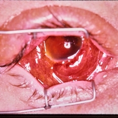 Trauma
Jan 8 2015 by H. Michael Lambert, MD
Surgical view of repaired lid laceration with hyphema.
Condition/keywords: trauma
-
 Trauma
-
 Trauma
-
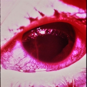
 Blood in the Anterior Chamber
Jul 13 2013 by Jason S. Calhoun
Trauma to a patient. Slit lamp shows anterior chamber full of blood mixed with the aqueous humor.
Photographer: Jason S. Calhoun, Department of Ophthalmology, Mayo Clinic Jacksonville, Florida
Imaging device: TOPCON D-90 SL NIKON CAMERA
Condition/keywords: hyphema
-
 Silicone Oil In Anterior Chamber With Hyphema
Jul 11 2013 by Jason S. Calhoun
Patient who had previous retinal detachment surgery. A hyphema is present with a drop of silicone oil in the anterior chamber.
Photographer: Jason S. Calhoun, Department of Ophthalmology, Mayo Clinic Jacksonville, Florida
Condition/keywords: hyphema, silicone oil
-
 Gonioscopy, Blood in the Anterior Chamber from Hyphema
Jul 8 2013 by Jason S. Calhoun
Patient with blunt trauma to the right eye due to a BB gun incident. Patient was present with a hyphema at 8-o'clock about 1mm thick. Gonioscopy photos were then taken to show blood from the hyphema entered into the anterior chamber. Patient had no angle recession in the right eye.
Photographer: Jason S. Calhoun, Department of Ophthalmology, Mayo Clinic Jacksonville, Florida
Condition/keywords: angle recession, gonioscopy
 Loading…