Search results (37 results)
-
 Black Ball Hyphema
-
 Hyphema
-
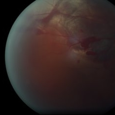
 Retinal Lesion
Nov 9 2012 by Norman Byer
This 30-year-old man sustained a severe blow to his brow region which resulted in a variety of injuries including hyphema and vitreous and retinal hemorrhages. This photograph shows a retinal lesion, which is either a tiny area of elevated full thickness retina or a traumatic retinal cyst.
Condition/keywords: elevated retinal lesion, retinal hemorrhage, traumatic retinal cyst
-
 Silicone Oil In Anterior Chamber With Hyphema
Jul 11 2013 by Jason S. Calhoun
Patient who had previous retinal detachment surgery. A hyphema is present with a drop of silicone oil in the anterior chamber.
Photographer: Jason S. Calhoun, Department of Ophthalmology, Mayo Clinic Jacksonville, Florida
Condition/keywords: hyphema, silicone oil
-
 Gonioscopy, Blood in the Anterior Chamber from Hyphema
Jul 8 2013 by Jason S. Calhoun
Patient with blunt trauma to the right eye due to a BB gun incident. Patient was present with a hyphema at 8-o'clock about 1mm thick. Gonioscopy photos were then taken to show blood from the hyphema entered into the anterior chamber. Patient had no angle recession in the right eye.
Photographer: Jason S. Calhoun, Department of Ophthalmology, Mayo Clinic Jacksonville, Florida
Condition/keywords: angle recession, gonioscopy
-
 Silicone Oil In Anterior Chamber With Hyphema
Jul 11 2013 by Jason S. Calhoun
Patient who had previous retinal detachment surgery. A hyphema is present with a drop of silicone oil in the anterior chamber.
Photographer: Jason S. Calhoun, Department of Ophthalmology, Mayo Clinic Jacksonville, Florida
Condition/keywords: hyphema, silicone oil
-
 Gonioscopy, Blood in the Anterior Chamber from Hyphema
Jul 8 2013 by Jason S. Calhoun
Patient with blunt trauma to the right eye due to a BB gun incident. Patient was present with a hyphema at 8-o'clock about 1mm thick. Gonioscopy photos were then taken to show blood from the hyphema entered into the anterior chamber. Patient had no angle recession in the right eye.
Photographer: Jason S. Calhoun, Department of Ophthalmology, Mayo Clinic Jacksonville, Florida
Condition/keywords: angle recession, gonioscopy
-
 Gonioscopy, Blood in the Anterior Chamber from Hyphema
Jul 8 2013 by Jason S. Calhoun
Patient with blunt trauma to the right eye due to a BB gun incident. Patient was present with a hyphema at 8-o'clock about 1mm thick. Gonioscopy photos were then taken to show blood from the hyphema entered into the anterior chamber. Patient had no angle recession in the right eye.
Photographer: Jason S. Calhoun, Department of Ophthalmology, Mayo Clinic Jacksonville, Florida
Condition/keywords: angle recession, gonioscopy
-
 24 Hours Post Scleral Wound Closure+ Scleral Buckle+25 g Vitrectomy+Silicon Oil
Jan 23 2015 by Carlos Quezada-Ruiz, MD, FASRS
24 hours post op fundus photograph of a 43-year-old man who had perforating injury to the right eye with a small piece of plastic while he was hammering. OD LP, subconjunctival hemorrhage, clear cornea, hyphema, irido and ciclodyalisis as well as a luxated lens with traumatic cataract and a dense vitreous hemorrhage. B-US showed rhegmatogenous retinal detachment with a tear and a big inferior hemorrhagic choroidal detachment. 360 peritomy revealed 2-entry scleral wounds were found in zone II (M V and M VI) and closure was performed. 25 G PPV was performed with the infusion canal placed in the AC through the limbus. Lensectomy and removal of a dense recent vitreous hemorrhage revealed a white detached retina with an exit wound through the temporal inferior segment of the optic nerve with a nasal GRT and sub retinal hemorrhage as well as temporal inferior choroidal, PVD was induced and PFOs helped stabilizing the retina while vitrectomy and sub-retinal hemorrhage was removed through the GRT. Fluid air exchange was made and 360 endolaser over the buckle indentation was done and silicon oil was used as endotamponade. This picture was taken 24 hrs after the surgery.
Photographer: Lilibeth Rodriguez, Instituto de la Visión. Torreon, Mexico.
Condition/keywords: central retinal artery occlusion (CRAO), giant retinal tear, trauma
-
 Gonioscopy, Blood in the Anterior Chamber from Hyphema
Jul 8 2013 by Jason S. Calhoun
Patient with blunt trauma to the right eye due to a BB gun incident. Patient was present with a hyphema at 8-o'clock about 1mm thick. Gonioscopy photos were then taken to show blood from the hyphema entered into the anterior chamber. Patient had no angle recession in the right eye.
Photographer: Jason S. Calhoun, Department of Ophthalmology, Mayo Clinic Jacksonville, Florida
Condition/keywords: angle recession, gonioscopy
-
 Silicone Oil In Anterior Chamber With Hyphema
Jul 11 2013 by Jason S. Calhoun
Patient who had previous retinal detachment surgery. A hyphema is present with a drop of silicone oil in the anterior chamber.
Photographer: Jason S. Calhoun, Department of Ophthalmology, Mayo Clinic Jacksonville, Florida
Condition/keywords: hyphema, silicone oil
-
 Gonioscopy, Blood in the Anterior Chamber from Hyphema
Jul 8 2013 by Jason S. Calhoun
Patient with blunt trauma to the right eye due to a BB gun incident. Patient was present with a hyphema at 8-o'clock about 1mm thick. Gonioscopy photos were then taken to show blood from the hyphema entered into the anterior chamber. Patient had no angle recession in the right eye.
Photographer: Jason S. Calhoun, Department of Ophthalmology, Mayo Clinic Jacksonville, Florida
Condition/keywords: angle recession, gonioscopy
-
 Silicone Oil In Anterior Chamber With Hyphema
Jul 11 2013 by Jason S. Calhoun
Patient who had previous retinal detachment surgery. A hyphema is present with a drop of silicone oil in the anterior chamber.
Photographer: Jason S. Calhoun, Department of Ophthalmology, Mayo Clinic Jacksonville, Florida
Condition/keywords: hyphema, silicone oil
-
 Hyphema, Total
-
 Blood in the Anterior Chamber
Jul 13 2013 by Jason S. Calhoun
Trauma to a patient. Slit lamp shows anterior chamber full of blood mixed with the aqueous humor.
Photographer: Jason S. Calhoun, Department of Ophthalmology, Mayo Clinic Jacksonville, Florida
Imaging device: TOPCON D-90 SL NIKON CAMERA
Condition/keywords: hyphema
-
 Gonioscopy, Blood in the Anterior Chamber from Hyphema
Jul 8 2013 by Jason S. Calhoun
Patient with blunt trauma to the right eye due to a BB gun incident. Patient was present with a hyphema at 8-o'clock about 1mm thick. Gonioscopy photos were then taken to show blood from the hyphema entered into the anterior chamber. Patient had no angle recession in the right eye.
Photographer: Jason S. Calhoun, Department of Ophthalmology, Mayo Clinic Jacksonville, Florida
Condition/keywords: angle recession, gonioscopy
-
 Subconjuntival IOL After Blunt Trauma
Jun 27 2018 by Gabriel Costa Andrade, PhD
A 73-year-old male patient was referred to our ophthalmic emergency department with complaints of redness, pain, and diminution of vision in his left eye, after fall from height. The patient underwent small incision cataract surgery with polymethylmethacrylate (PMMA) IOL implantation in both the eyes 15 years back through superior sclerocorneal incision under local anesthesia. His best-corrected visual acuity was perception of light in the left eye. Ophthalmic examination using slit lamp biomicroscopy of the left eye revealed diffuse subconjunctival hemorrhage with no conjunctival laceration and inferior bulbar conjunctiva showed traumatic pseudophacocele with a sign “golden half ring,” suggesting the presence of PCIOL in subconjunctival space.There was total hyphema obscuring the view of rest of the ocular structures in his left eye.
Photographer: Gabriel Andrade, RETINA CLINIC, São Paulo, BRAZIL
Condition/keywords: dislocated intraocular lens (IOL), trauma
-
 Hyphema
-
 Hyphema
May 13 2016 by Nichole Lewis
Hyphema and anterior chamber air bubble.
Photographer: Nichole Lewis
Condition/keywords: hyphema
-
 Choroidal rupture Subretinal and Vitreous Hemorrhage Secondary to Blunt Trauma
Dec 30 2012 by Humberto Ruiz-Garcia, MD
Fundus photograph of a 23-year-old male, who suffered blunt trauma while working out with resistance rubber bands. The patient presented with "3-ball" hyphema which solved 48 hours with head up positioning and topical steroid and cyclopegic.
Photographer: Pedro Ruiz-Orozco, MD, Clinica Santa Lucia, Guadalajara, Mexico
Condition/keywords: choroidal rupture, vitreous hemorrhage
-
 GHPC
GHPC
May 10 2013 by Howard Schatz, MD
III GHPC, 8-year-old white male, blunt trauma, had hyphema.
Condition/keywords: blunt trauma, geographic helicoid peripapillary choroidopathy (GHPC), hyphema
 Loading…