Search results (23 results)
-
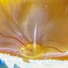
 Remnant of Hyaloid Artery
Jul 2 2021 by Nivesh Gupta
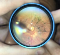
Fundus photograph of a 1 month old male with remnant of hyaloid artery.
Photographer: Dr. Nivesh Gupta, Retina foundation
Imaging device: NIDEK SLO MIRANTE
Condition/keywords: hyaloid artery, persistence of the hyaloid artery, tunica vasculosa lentis
-
 Tunica Vasculosa Lentis
Jul 2 2021 by Nivesh Gupta
Photograph of a 1 month old male with tunica vasculosa lentis.
Photographer: Dr. Nivesh Gupta, Retina foundation
Imaging device: NIDEK SLO MIRANTE
Condition/keywords: hyaloid artery, persistence of the hyaloid artery, tunica vasculosa lentis
-
 Bergmeister's Papilla
Sep 29 2020 by Dhaivat Shah
Bergmeister's papilla is a small tuft of glial tissue which arises from the center of the optic disc, and represents a remnant of the fetal hyaloid artery. The hyaloid artery provides nutrition to the lens during development, and runs forward to the lens from the optic disc. At birth the hyaloid artery regresses, and is normally completely regressed by the time of birth. Bergmeister's papilla is frequently observed as an incidental clinical finding if this artery has an incomplete regression posteriorly. However, in the severe forms it can be associated with cataracts, persistence of the primitive vitreous, microphthalmia, vitreous hemorrhages and sometimes tractional retinal detachment, due to contraction of the residual fibro vascular tissue. Therefore, careful monitoring of vitreous thickening in the peripapillary areas, both by examining the ocular fundus, and especially by SD-OCT, is of considerable importance. Here we have one such of a 30 year old young male who came in for a routine checkup, in whom we noted a Bergmeister’s papilla. Due to its benign nature, patient was reassured and was asked to follow up yearly.
Condition/keywords: Bergmeister's Papillae
-
 Vascular Primary Vitreous
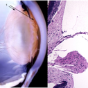
Sep 8 2020 by J. Sebag, MD, FACS, FRCOphth, FARVO
The hyaloid artery (“3”, left) feeds the vasa hyaloidea propria (“2”, left) which anastomosis with the tunica vasculosa lentis (“1”, left). The histologic section to the right is stained with H & E (bar = 100 uM) [Left: from Yee at al.: Vitreous cytokines and regression of the fetal hyaloid vasculature. In: Vitreous – in Health & Disease. Springer, New York, 2014; pg. 42 (image © Springer Nature, reprinted with permission) Right: from Sebag J: Vitreous and vitreo-retinal interface. In: Ryan’s Retina 6th edition (A. Schachat, ed.) Elsevier, 2018; pg. 546.
Condition/keywords: hyaloid artery, vitreous
-
 Vitreous Structure in Youth
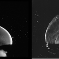
Sep 1 2020 by J. Sebag, MD, FACS, FRCOphth, FARVO
Dark-field slit microscopy was performed on fresh, unfixed, post-mortem human eyes that had undergone dissection to peel off the sclera, choroid, and retina. The vitreous body remains attached to the anterior segment which is seen below, while the posterior pole is above in these images. These horizontal optical sections demonstrate intense light scattering by the posterior vitreous cortex and the remnant of the hyaloid artery destined to be Cloquet’s Canal (left image), but no other light scattering within the vitreous body in either the 33 GW human fetus (left image) or this 6 year-old child (right image). [from Sebag J: The Vitreous - Structure, Function, and Pathobiology. Springer-Verlag, New York, 1989, left image pg. 77; right image pg. 79; images © Springer Nature, reprinted with permission]
Condition/keywords: vitreous
-
 PHPV from Smartphone Fundoscopy.
May 5 2020 by Gabriel Castilho S Barbosa, MD
Here we show a persistent hyperplastic primary vitreous (PHPV) documented with smartphone fundoscopy technique.
Condition/keywords: hyaloid artery, persistent hyperplastic primary vitreous (PHPV)
-
 Bergmeister Papilla
Feb 20 2020 by Nisarg Joshi, MD
Gross pathology photo of a Bergmeister Papilla. It is a remnant of incompletely resorbed hyaloid vasculature from ocular development. This glial tissue is seen emminating from the optic nerve, which also shows glaucomatous cupping. The eye was enucleated due to a choroidal melanoma.
Photographer: Nisarg Joshi, MD, Geisinger Medical Center
Imaging device: Digital camera
Condition/keywords: Bergmeister's Papillae, hyaloid artery, persistent fetal vasculature (PFV)
-
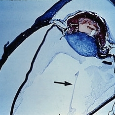 Slide 8-4
Mar 4 2019 by Lancaster Course in Ophthalmology
Persistence and hyperplasia of the primary vitreous (PHPV). The dense fibrovascular tissue molds the posterior surface of the lens and has drawn the ciliary processes and peripheral retina toward the center of the mass. A section of the hyaloid artery is present in the vitreous cavity (arrow). (A.F.l.P. No. 744398)
Condition/keywords: ciliary, fibrovascular tissue, hyaloid artery, persistent hyperplastic primary vitreous (PHPV)
-
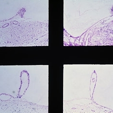 Slide 8-3
Mar 4 2019 by Lancaster Course in Ophthalmology
Examples of Bergmeister's papilla. Note the strands of vitreous, composed of glial cells, attached to the papilla. The presence of a small vessel within the Bergmeister's papilla (lower right) suggests that its origin is related to the hyaloid artery. (E. P. Nos. 31888,38589, 29360, and 32239)
Condition/keywords: Bergmeister's Papillae, glial cells, vitreous
-
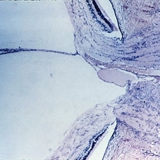 Slide 8-2
Mar 4 2019 by Lancaster Course in Ophthalmology
Hyaloid artery extending into the vitreous cavity from the optic nerve head. (E.P. No. 16127)
Condition/keywords: hyaloid artery, optic nerve head, vitreous cavity
-
 Hyaloid Artery
May 3 2018 by Alexandr Stepanov
Hyaloid artery
Photographer: Alexandr Stepanov MD, PhD, FEBO, Faculty Hospital Hradec Kralove, Czech Republic
Condition/keywords: hyaloid artery
-
 Hyaloid Artery, Anterior Segment
May 3 2018 by Alexandr Stepanov
Hyaloid artery, anterior segment.
Photographer: Alexandr Stepanov MD, PhD, FEBO, Faculty Hospital Hradec Kralove, Czech Republic
Condition/keywords: hyaloid artery
-
 Remnant of Hyaloidal Artery
Feb 5 2014 by Gerardo Garcia-Aguirre, MD
Video of the fundus of the left eye of a 14-year-old asymptomatic female, where a prepapillary vitreous opacity is observed. The opacity is attached to the origin of the retinal vessels in the optic nerve head, and is considered to be a remnant of the hyaloidal artery.
Photographer: Gerardo Garcia-Aguirre, MD
Condition/keywords: persistence of the hyaloid artery
-
 Remnant of Hyaloidal Artery
Feb 5 2014 by Gerardo Garcia-Aguirre, MD
Fundus photograph of the left eye of a 14-year-old asymptomatic female. The photograph is focused on the posterior vitreous where a prepapillary vitreous opacity is observed (white arrows). The opacity is attached to the origin of the retinal vessels in the optic nerve head.
Photographer: Gerardo Garcia-Aguirre, MD
Condition/keywords: persistence of the hyaloid artery
-
 Remnant of Hyaloidal Artery
Feb 5 2014 by Gerardo Garcia-Aguirre, MD
Fundus photograph of the left eye of a 14-year-old asymptomatic female. The photograph is focused on the retina, and a prepapillary vitreous opacity is observed (white arrows). The opacity is attached to the origin of the retinal vessels in the optic nerve head.
Photographer: Gerardo Garcia-Aguirre, MD
Condition/keywords: persistence of the hyaloid artery
-
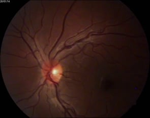
 Remnant of Hyaloidal Artery
Feb 5 2014 by Gerardo Garcia-Aguirre, MD
Fundus photograph of the left eye of a 14-year-old asymptomatic female. The photograph is focused on the posterior vitreous where a prepapillary vitreous opacity is observed (see next picture where opacity is marked by arrows). The opacity is attached to the origin of the retinal vessels in the optic nerve head.
Photographer: Gerardo Garcia-Aguirre, MD
Condition/keywords: persistence of the hyaloid artery
-
 Remnant of Hyaloidal Artery
Feb 5 2014 by Gerardo Garcia-Aguirre, MD
Fundus photograph of the left eye of a 14-year-old asymptomatic female. The photograph is focused on the retina, and a prepapillary vitreous opacity is observed (white arrows). The opacity is attached to the origin of the retinal vessels in the optic nerve head.
Photographer: Gerardo Garcia-Aguirre, MD
Condition/keywords: persistence of the hyaloid artery
-
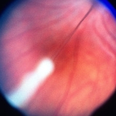
 Persistence Of The Hyaloid Artery
Sep 25 2013 by Alexandre Durao Alves Pereira, MD
Persistence of the hyaloid artery.
Photographer: Alexandre Pereira
Condition/keywords: persistence of the hyaloid artery
-
 Pesistent Hyperplastic Primary Vítreos
Jun 24 2013 by Ximena Mira Lorenzo, MD
Fundus photograph of a 1-year-old child .
Photographer: Ximena Mira Lorenzo MD.
Imaging device: Visucam Lite Zeiss
Condition/keywords: hyaloid artery, persistent fetal vasculature (PFV)
-
 PHPV
PHPV
May 2 2013 by Henry J. Kaplan, MD
The same patient ; notice the persistant hyaloid artery which is changed to fibrotic tissue anteriorly; #2.
Condition/keywords: persistent fetal vasculature (PFV), persistent hyperplastic primary vitreous (PHPV)
-
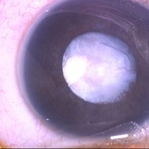
 PHPV
PHPV
May 2 2013 by Henry J. Kaplan, MD
The same patient; hyaloid artery has changed to fibrous tissue anteriorly; #3.
Condition/keywords: hyaloid artery, persistent fetal vasculature (PFV), persistent hyperplastic primary vitreous (PHPV)
-
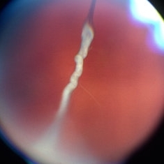
 Bergmeister's Papillae
Mar 29 2013 by Henry J. Kaplan, MD
Remnants of fetal hyaloid artery as fibrous tuft called Bergmeister`s papillae on the optic disc.
Condition/keywords: Bergmeister's Papillae, hyaloid artery
-
 Aggressive Posterior Retinopathy of Prematurity
Oct 9 2012 by Audina M. Berrocal, MD FASRS
Aggressive posterior Type 1 ROP with bleeding from regression of the posterior hyaloid artery
Photographer: Ditte Hess CRA, BPEI
Imaging device: RETCAM
Condition/keywords: retinopathy of prematurity (ROP)
 Loading…