 Initializing download.
-
 By J. Sebag, MD, FACS, FRCOphth, FARVO
VMR Institute for Vitreous Macula Retina - Uploaded on Sep 1, 2020.
- Last modified by Caroline Bozell on Sep 1, 2020.
- Rating
- Appears in
- Vitreous
- Condition/keywords
- vitreous
- Description
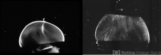
- Dark-field slit microscopy was performed on fresh, unfixed, post-mortem human eyes that had undergone dissection to peel off the sclera, choroid, and retina. The vitreous body remains attached to the anterior segment which is seen below, while the posterior pole is above in these images. These horizontal optical sections demonstrate intense light scattering by the posterior vitreous cortex and the remnant of the hyaloid artery destined to be Cloquet’s Canal (left image), but no other light scattering within the vitreous body in either the 33 GW human fetus (left image) or this 6 year-old child (right image). [from Sebag J: The Vitreous - Structure, Function, and Pathobiology. Springer-Verlag, New York, 1989, left image pg. 77; right image pg. 79; images © Springer Nature, reprinted with permission]


