 Initializing download.
-
 By J. Sebag, MD, FACS, FRCOphth, FARVO
VMR Institute for Vitreous Macula Retina - Uploaded on Sep 8, 2020.
- Last modified by Caroline Bozell on Sep 8, 2020.
- Rating
- Appears in
- Vitreous
- Condition/keywords
- hyaloid artery, vitreous
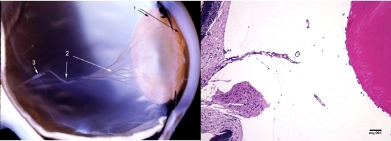
- Description
- The hyaloid artery (“3”, left) feeds the vasa hyaloidea propria (“2”, left) which anastomosis with the tunica vasculosa lentis (“1”, left). The histologic section to the right is stained with H & E (bar = 100 uM) [Left: from Yee at al.: Vitreous cytokines and regression of the fetal hyaloid vasculature. In: Vitreous – in Health & Disease. Springer, New York, 2014; pg. 42 (image © Springer Nature, reprinted with permission) Right: from Sebag J: Vitreous and vitreo-retinal interface. In: Ryan’s Retina 6th edition (A. Schachat, ed.) Elsevier, 2018; pg. 546.


