 Initializing download.
-
 By César Adrián Gomez Valdivia, MD
HOL
Co-author(s): @eyemissu2 - Uploaded on Sep 30, 2025.
- Last modified by Joshua Friedman on Oct 1, 2025.
- Rating
- Appears in
- Miscellaneous
- Condition/keywords
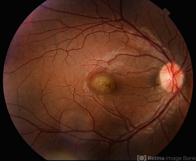
- Idiopathic Choroidal Neovascularization
- Photographer
- @eyemissu2
- Imaging device
- TOPCON TRX
- Description
- At the foveal area, there is a yellowish-greenish elevated lesion with indistinct borders, corresponding to a subfoveal choroidal neovascular membrane (CNV). There are subtle overlying changes including mild retinal pigment epithelium (RPE) disruption, and small hemorrhagic spots suggesting active leakage. Surrounding the lesion, there are faint retinal folds or striae, likely due to localized subretinal fibrosis or traction.


