Search results (94 results)
-
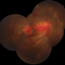
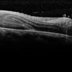
 Choroidal Melanoma with Exudative Detachment
Apr 7 2025 by Virginia Gebhart
Autofluorescence image of 36 year old female showing demarcation line of fluid/detachment from new choroidal melanoma. Pt will be scheduled for brachytherapy pending CT scan results.
Photographer: Virginia Gebhart, Retina Consultants of Carolina
Imaging device: Optos California
Condition/keywords: Autoflourescenceautofluorescence imagingchoroidal melanomamelanomaretinal detachment
-
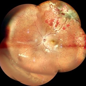
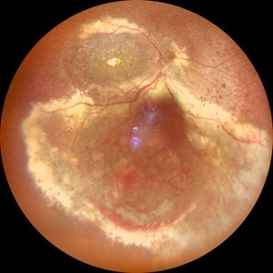
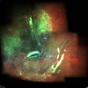
 New Choroidal Melanoma with Exudative Detachment
Apr 7 2025 by Virginia Gebhart
36 year old female referred for pigmented mass. Pt complains of flashes of light since last fall. Clinical exam and ultrasound findings consistent with choroidal melanoma with exudative detachment inferior. Pt will be scheduled for brachytherapy and possible tumor biopsy pending CT scan results.
Photographer: Virginia Gebhart, Retina Consultants of Carolina
Imaging device: Optos California
Condition/keywords: Choroidal melanomaexudative detachmentmelanomaretinal detachment
-
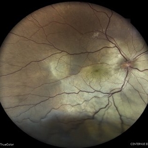
 Regressing Choroidal Melanoma
Mar 10 2025 by Virginia Gebhart
56 year old male 4 months s/p plaque brachytherapy for choroidal melanoma. Tumor is regressing, there is an exudative detachment with worsening SRF. Treated with Avastin to promote hopeful improvement of the SRF
Photographer: Virginia Gebhart, Retina Consultants of Carolina
Imaging device: Optos California
Condition/keywords: brachytherapyChoroidal melanomaexudative detachmentmelanoma
-
 Coats` Disease
Dec 18 2024 by Thirumalesh Mochi Basavaraj, MD
Fundus photo graph of a 6 year old child with exudative retinal detachment with sub retinal lipid exudation and peripheral telengectasia.
Photographer: Puttaswamy N K
Condition/keywords: exudative detachmentperipheral telangiectasia
-
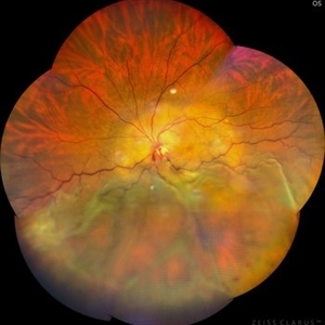
 New Choroidal Melanoma with Exudative Detachment
Oct 16 2024 by Virginia Gebhart
56 year old male with a large pigmented tumor with an exudative detachment inferior and shallow fluid through the macula. Pt states they have been having symptoms for over a year. Scheduled for brachytherapy.
Photographer: Virginia Gebhart, Retina Consultants of Carolina
Imaging device: Optos California
Condition/keywords: Choroidal melanomaexudative detachmentmelanoma
-
 Idiopathic Uveal Effusion Syndrome
Aug 22 2024 by Jordyn Beckman
61 year old male with Idiopathic Uveal Effusion Syndrome with starry night appearance on fluorescein. 3 weeks s/p single external drainage retinotomy and 9 weeks of oral pred with recurrent choroidal effusions. Has since returned to surgery for secondary drainage retinotomy; subretinal fluid remain persistent.
Photographer: Jordyn Beckman
Imaging device: Optos California
Condition/keywords: chorioretinitisChoroidalexudative detachmentwindow defect
-
 Von Hippel Lindau Syndrome
Jun 9 2024 by Anjana Mirajkar, MS Ophthalmology
A widefield montage of a 23 year old female of LE case of VHL syndrome showing some hemorrhages with traction superiorly in a silicon oil filled eye with central settled retina. Cryo and laser marks are noted in periphery.
Photographer: Dr. Anjana Mirajkar -Retina Foundation, Ahmedabad
Imaging device: Mirante-Nidek
Condition/keywords: cryotherapyexudative detachmentlaser photocoagulationvitreous hemorrhageVon Hippel-Lindau
-
 Exudative Retinal Detachment
May 27 2024 by Akansha Sharma
Color fundus photograph of a 38 year old male with breast carcinoma leading to intraocular metastasis as represented by an exudative retinal detachment.
Photographer: Dr. Akansha Sharma, Bharati Eye Hospital
Condition/keywords: Disc Edemaexudative detachmentmacular puckerMETASTATSIS
-
 Exudative Retinal Detachment With Choroidal Metastasis
May 1 2024 by Vishal Agrawal, MD, FRCS,FACS,FASRS
Left eye fundus picture of a 65-year-old female with choroidal metastases and exudative retinal detachment. The patient is under treatment for breast carcinoma.
Photographer: Dr Ayushi
Imaging device: Clarus 700
Condition/keywords: choroidal metastasisexudative detachment
-
 Large PEHCR causing an exudative inferior detachment in a patient with AMD
Apr 15 2024 by David A Reichstein, MD
(A) Ultra-widefield color fundus photograph demonstrates a temporal PEHCR causing minimal intra- and subretinal hemorrhage along with lipid exudation. There is an associated inferior detachment due to the dependent nature of the exudation. Note the lipid exudation at the posterior edge of the detachment indicating chronicity of the lesion. Drusen in the macula are also appreciated. (B) Ultra-widefield FA in early stage demonstrates hypofluorescence temporally and inferiorly. (C) Ultra-widefield color fundus photograph taken after 1 year of monthly anti-VEGF therapy demonstrates resolution of the exudative detachment and resultant chorioretinal scarring.
Condition/keywords: peripheral exudative hemorrhagic chorioretinopathy (PEHCR)
-
 Von Hippel-Lindau
Feb 6 2024 by Thirumalesh Mochi Basavaraj, MD
8 Year old child with Multiple Capillary Haemangiomas with Exudative retinal detachment.
Photographer: Puttaswamy
Condition/keywords: exudative detachmentVon Hippel-Lindau
-
 Exudative RD
Feb 6 2024 by Thirumalesh Mochi Basavaraj, MD
26 yr old male with a exudative retinal detachment with the retina just behind the crystalline lens in a case of optic disc capillary hemangioma.
Photographer: Puttaswamy N K
Condition/keywords: exudative detachment
-
 PEHCR (Peripheral Exudative Hemorrhagic Chorioretinopathy)
May 12 2023 by Niloofar Piri, MD
Ultrawide fundus photograph of the left eye demonstrating extensive peripheral hemorrhagic exudative detachment in a 79 yo Caucasian female with prior history of non-exudative AMD. Recent diagnosis of Acute myeloid leukemia with low platelet count which might have contributed to the above presentatuon. Please note the temporal subretinal hemorrhage as well as RPE atrophy and hyperplasia in the macula.
Photographer: Rocio Bentivegna, MD, Saint Louis University; Jessica Maddox, COA, Saint Louis University
Condition/keywords: peripheral exudative hemorrhagic chorioretinopathy (PEHCR)
-
 Exudative Retinal Detachment secondary to Leber's Miliary Aneurysm in a case of Retinitis Pigmentosa
Oct 13 2022 by Vaidehi Sathaye
Fundus Photograph of RE of a 23 year male patient , with an Exudative Retinal Detachment secondary to Leber's Miliary Aneurysm in a case of Retinitis Pigmentosa.
Photographer: Dr. Vaidehi Sathaye
Imaging device: Mirante
Condition/keywords: exudative detachmentLeber's miliary aneurysmretinitis pigmentosa
-
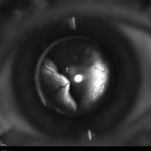
 Ciliochoroidal Melanoma with Total Serous Retinal Detachment
Jun 20 2021 by Jesus Lozano, MD
64-year-old woman with a ciliochoroidal melanoma after SRS with a exudative retinal detachment.
Photographer: Dr. Jesús Lozano Gutiérrez. Hadassah Medical Center, Israel.
Imaging device: Slit lamp BI 900 camera
Condition/keywords: cataractexudative detachment
-
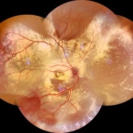 Coats' Disease Montage
Feb 5 2021 by Akansha Sharma
Fundus photograph of a 5-year-old male child who presented with unilateral diminution of vision since one month.
Photographer: Dr. Nivesh Gupta, M.S., Retina Foundation, Ahmedabad
Condition/keywords: angiomatosis retinaeCoats' diseaseexudative detachmentsubretinal exudates
-
 Ultra-Widefield Fundus Image of Coats' Disease with Exudative Retinal Detachment
Dec 22 2020 by Kushal S Delhiwala, MBBS, MS, FMRF,FICO, FAICO
Ultra-widefield fundus image of right eye showing Coats' disease with exudative retinal detachment (stage 3) in a 4 year old male complaining of squinting right eye. Telangiectatic vessels prominent superotemporal periphery.
Photographer: Kushal Delhiwala, Netralaya superspeciality eye hospital, Ahmedabad, Gujarat,India
Imaging device: Optos Daytona
Condition/keywords: bullous retinal detachmentCoats' diseaseexudative detachmentultra-wide field imaging
-
 Bartonella Posterior Granulomatous Mass with Exudative Detachment Post 2 Months
Sep 26 2020 by Swati Agarwal-Sinha, MD, FASRS
Color fundus photo of the left eye two months post-treatment showing resolved exudative detachment, fibrosed granuloma at the optic disc, tractional detachment at the posterior pole and extensive diffuse subretinal exudates extending all around up to the retinal periphery. No retinal vascular changes are noted.
Photographer: Harry Rosa, University of Florida
Condition/keywords: Bartonella bacteriaexudative detachment
-
 Bartonella Posterior Granulomatous Mass with an Exudative Retinal Detachment.
Sep 26 2020 by Swati Agarwal-Sinha, MD, FASRS
Color fundus photo of right eye showing normal optic disc, vessels, macula. Left eye two days after presentation with diffuse exudative retinal detachment with a large inflammatory retinal granuloma obscuring the optic disc.
Photographer: Harry Rosa, University of Florida
Condition/keywords: Bartonella bacteriachildhoodexudative detachment
-
 Total Rhegmatogenous and Tractional Retinal Detachment Following Choroidal Melanoma Laser Ablation Treatment
Sep 22 2020 by Sophia El Hamichi, MD
A 69-year-old female, with a history of choroidal melanoma in her left eye with exudative detachment, underwent tumor laser ablation. She then developed a complex combined tractional and rhegmatogenous retinal detachment with a giant retinal tear.
Photographer: Belinda Rodriguez, Murray Ocular Oncology and Retina, Miami
Condition/keywords: tractional retinal detachment
-
 Total Rhegmatogenous and Tractional Retinal Detachment Following Choroidal Melanoma Laser Ablation Treatment
Sep 22 2020 by Sophia El Hamichi, MD
A 69-year-old female, with a history of choroidal melanoma in her left eye with exudative detachment, underwent tumor laser ablation. She then developed a complex combined tractional and rhegmatogenous retinal detachment with a giant retinal tear.
Photographer: Belinda Rodriguez, Murray Ocular Oncology and Retina, Miami
Condition/keywords: anterior segmentmelanoma
-
 Large Retinal Fold Masking the Optic Nerve
Sep 22 2020 by Sophia El Hamichi, MD
A 69-year-old female, with a history of choroidal melanoma in her left eye with exudative detachment, underwent tumor laser ablation. She then developed a complex combined tractional and rhegmatogenous retinal detachment with a giant retinal tear. The patient underwent surgical repair of her retinal detachment with pars plana vitrectomy and silicone oil. In the post-op, the patient developed large retinal folds masking the optic nerve depicted in the OCT photograph.
Photographer: Belinda Rodriguez, Murray Ocular Oncology and Retina, Miami
Condition/keywords: giant retinal tearmelanomapars plana vitrectomy (PPV)retinal foldsilicone oil
-
 Large Retinal Fold
Sep 22 2020 by Sophia El Hamichi, MD
A 69-year-old female, with a history of choroidal melanoma in her left eye with exudative detachment, underwent tumor laser ablation. She then developed a complex combined tractional and rhegmatogenous retinal detachment with a giant retinal tear. The patient underwent surgical repair of her retinal detachment with pars plana vitrectomy and silicone oil. In the post-op, the patient developed large retinal folds masking the optic nerve depicted in the fundus photograph.
Photographer: Belinda Rodriguez, Murray Ocular Oncology and Retina, Miami
Condition/keywords: melanomapars plana vitrectomy (PPV)retinal foldsilicone oil
-
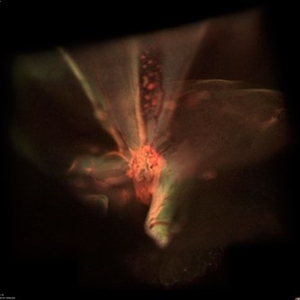
 Fluorescein Angiogram of Coats's Disease With Exudative Retinal Detachment
Dec 9 2019 by Sophia El Hamichi, MD
A 3-year-old male presenting a complex Coats' disease of the left eye with exudative retinal detachment, abnormal telangiectatic vasculature with peripheral nonperfusion and leakage.
Photographer: Abby Orcutt-Hayes, Murray Ocular Oncology and Retina
Condition/keywords: Coats' diseaseexudative detachmentfluorescein angiogram (FA)montageretinal macrocyst
-
 Coats' Disease With Exudative Retinal Detachment and Retinal Macrocyst
Dec 9 2019 by Sophia El Hamichi, MD
A 3-year-old male with a presentation of a complex Coats' disease in the left eye with exudative retinal detachment, abnormal telangiectatic vasculature, and inferotemporal retinal macrocyst/retinoschisis.
Photographer: Abby Orcutt-Hayes, Murray Ocular Oncology and Retina
Imaging device: RetCam
Condition/keywords: Coats' diseaseexudative detachmentmontageretinal macrocyst
 Loading…