Search results (257 results)
-
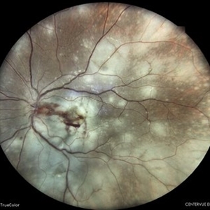
 Acute syphilitic posterior placoid chorioretinitis
Apr 24 2022 by Aniruddha K Agarwal, MD
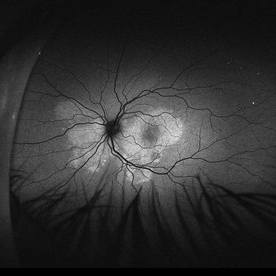
Green-light fundus autofluorescence (FAF) of the right eye from a 55-year-old man with risk factors for sexually trasnmitted diseases who presented to the retina clinic for a central scotoma. Funduscopy revealed a placoid lesion in the posterior pole. FAF highlights a hyperautofluorescent placoid lesion involving the macula with granular hyperfluorescence. The patient tested positive for syphilis and received intravenous penicillin treatment.
Photographer: Esther CIANCAS, MD, PhD, Gema CRESPO-RODRÍGUEZ, RN
Imaging device: Zeiss Clarus fundus camera
Condition/keywords: chorioretinitis, IUSG, syphilis, uveitis
-
 Acute Syphilitic Posterior Placoid Chorioretinitis
Oct 16 2024 by César Adrián Gómez Valdivia, MD
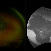
Fundus autofluorescence image of an acute syphilitic posterior placoid chorioretinitis found in a HIV positive 28 YO male patient with suspected neurosyphilis. A beautiful butterfly autofluorescence pattern can be appreciated.
Photographer: @eyemissu2
Imaging device: California ICG OPTOS
Condition/keywords: acute syphilitic posterior placoid chorioretinitis, chorioretinitis, syphilis
-
 Candida-chorioretinitis
Apr 30 2023 by Vishal Sanjay Jadhav, MS Ophthalmology
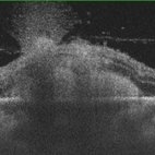
Optical coherence tomography with enhanced depth imaging of a patient with candida chorioretinitis showing 'rain-cloud' sign
Photographer: -
Condition/keywords: candida, chorioretinitis, rain cloud
-
 Chorioretinitis
Oct 30 2025 by Kimberly Wakester
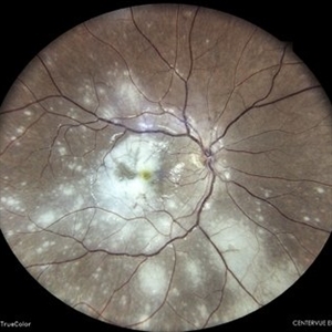
Optomap RGB of an 37-year-old-woman with stable Chorioretinitis in the right eye. Patient is to return in 6 months for dilated exam and repeat diagnostic testing.
Photographer: Kimberly Wakester, COA, OCT-C
Imaging device: Optos California
Condition/keywords: chorioretinitis
-
 Chorioretinitis
Dec 16 2025 by Kimberly Wakester
Optomap RGB of a 37 year-old woman with Chorioretinitis. The chorioretinitis remains largely stable in both eyes on exam and compared to prior photos. Clinical and diagnostic findings in both eyes continue to be most consistent with punctate inner choroidopathy (PIC) /Multifocal choroiditis and panuveitis (MCP). Will continue follow up care every 6 months with dilated exam and repeat testing.
Photographer: Kimberly Wakester, COA, OCT-C
Imaging device: Optos California
Condition/keywords: chorioretinitis, multifocal chorioretinitis (MCP), punctate inner choroidopathy (PIC)
-
---thumb.jpg/image-square;max$300,300.ImageHandler) Chorioretinitis
Chorioretinitis
Aug 15 2013 by From the Collections of Thomas M. Aaberg, MD and Thomas M. Aaberg Jr., MD
Pigmentary disturbance.
-
---thumb.jpg/image-square;max$300,300.ImageHandler) Chorioretinitis
Chorioretinitis
Aug 15 2013 by From the Collections of Thomas M. Aaberg, MD and Thomas M. Aaberg Jr., MD
Pigmentary disturbance.
-
---thumb.jpg/image-square;max$300,300.ImageHandler) Chorioretinitis
Chorioretinitis
Aug 15 2013 by From the Collections of Thomas M. Aaberg, MD and Thomas M. Aaberg Jr., MD
Pigmentary disturbance.
-
 Chorioretinitis
May 17 2014 by Avris Romario Diparaja Siahaan
Fundus photograph of a 34-year-old man with chorioretinitis (toxo) on his right eye.
Photographer: Prayid Listiyanto, Klinik Mata Nusantara
Imaging device: Topcon TRC 50 DX Type IA
Condition/keywords: fundus photograph, toxo chorioretinitis
-
 Chorioretinitis with Overlying Vitreous Stranding/Vitritis
Mar 23 2023 by Isaac Agranoff
Fundus photograph of a 37-year-old woman presenting with chorioretinitis with overlying vitreous stranding/vitritis that has remained unchanged for multiple years. Patient presented with irritation and blurred vision and her vision was 20/40 OD. The OCT revealed evidence of low-grade inflammation and the recommend treatment was anti-inflammatory eye drops at this time and to obtain second opinion with another physician in the office.
Photographer: Isaac Agranoff, Technician
Imaging device: Optos California
Condition/keywords: chorioretinal scar, chorioretinitis, inflammation, Optos, ultra-wide field imaging, vitritis
-
 Choroiditis
Sep 29 2023 by PUSHPANJALI BADOLE
36 yr old female with recent decline in right eye vision, with left eye having poor vision since childhood. Fundus examination revealed BE healed chororetinitis with RE OCT s/o maculoschisis with full thickness macular hole.
Photographer: NITIN DESLE, ISHA NETRALAYA, KALYAN
Condition/keywords: chorioretinitis
-
 Cytomegalovirus Chorioretinitis
Nov 23 2021 by Raymundo García Vásquez
Fundus photograph of a 76-year-old male patient with cytomegalovirus chorioretinitis.
Photographer: Universidad de Monterrey
Imaging device: iPhone 12 and VOLK 20D
Condition/keywords: chorioretinitis, cytomegalovirus (CMV)
-
 Idiopathic Uveal Effusion Syndrome
Aug 22 2024 by Jordyn Beckman
61 year old male with Idiopathic Uveal Effusion Syndrome with starry night appearance on fluorescein. 3 weeks s/p single external drainage retinotomy and 9 weeks of oral pred with recurrent choroidal effusions. Has since returned to surgery for secondary drainage retinotomy; subretinal fluid remain persistent.
Photographer: Jordyn Beckman
Imaging device: Optos California
Condition/keywords: chorioretinitis, Choroidal, exudative detachment, window defect
-
 Multifocal Chorioretinitis
Apr 9 2024 by Akansha Sharma
Color fundus photograph of a 34 year old male patient with multifocal chorioretinitis.
Photographer: Dr. Akansha Sharma, Bharati Eye Hospital
Condition/keywords: chorioretinal inflammations, chorioretinitis
-
 Multifocal Chorioretinitis
Apr 9 2024 by Akansha Sharma
Color fundus photograph of a 34 year old male patient with multifocal chorioretinitis with subretinal bleed.
Photographer: Dr. Akansha Sharma, Bharati Eye Hospital
Condition/keywords: chorioretinal inflammations, chorioretinitis, subretinal hemorrhage
-
 Recurrent Toxoplasmosis
Apr 8 2019 by Gary R. Cook, MD, FACS
56-year-old white male with recurrent activation of an old toxoplasmosis lesion; V.A. = 20/25-2; Toxo titer = 1:512.
Imaging device: Topcon VT-50
Condition/keywords: chorioretinitis, toxo chorioretinitis, toxoplasmosis
-
 Recurrent Toxoplasmosis
Apr 8 2019 by Gary R. Cook, MD, FACS
16-year-old white male with a small, new toxoplasmosis lesion in the inferonasal macula OD 2 years following his initial lesion (inferotemporal one); V.A. = 20/20+1.
Imaging device: Topcon VT-50
Condition/keywords: acute toxoplasmosis, chorioretinitis, ocular toxoplasmosis, toxoplasmosis
-
 Relentless Placoid Chorioretinitis
Jan 22 2021 by Renata Garcia Franco, Md
20-year-old male with reduction of vision in both eyes, scotoma and metamorphopsia. Widespread multiple chorioretinal lesions with RPE hyperplasia, which appear from posterior pole to peripheral retina.
Photographer: Fatima Hernandez, Instituto de la Retina del Bajio SC
Imaging device: Zeiss
Condition/keywords: chorioretinitis
-
 Relentless Placoid Chorioretinitis
Jan 22 2021 by Renata Garcia Franco, Md
20-year-old male with reduction of vision in both eyes, scotoma and metamorphopsia. Widespread multiple chorioretinal lesions with RPE hyperplasia, which appear from posterior pole to peripheral retina and inactive choroidal neovascular membrane.
Photographer: Fatima Hernandez, Instituto de la Retina del Bajio SC
Imaging device: Zeiss
Condition/keywords: chorioretinitis
-
 Syphilitic Chorioretinitis
Apr 8 2019 by Gary R. Cook, MD, FACS
68-year-old patient with diffuse 'salt and pepper' chorioretinitis OD secondary to acquired syphilis; V.A. = 20/25
Imaging device: Topcon VT-50
Condition/keywords: chorioretinitis, pseudo retinitis pigmentosa, syphilis
-
 Syphilitic Chorioretinitis
Apr 8 2019 by Gary R. Cook, MD, FACS
68-year-old patient with 'salt and pepper'-type diffuse chorioretinitis OD secondary to acquired syphilis; V.A. = 20/25
Imaging device: Topcon VT-50
Condition/keywords: chorioretinitis, pseudo retinitis pigmentosa, syphilis
-
 Syphilitic Chorioretinitis
Apr 8 2019 by Gary R. Cook, MD, FACS
Mid-phase (48 seconds) fluorescein angiogram image of the right eye of a 68-year-old female with diffuse salt-and-pepper-type chorioretinitis secondary to syphilis; V.A. = 20/25
Imaging device: Topcon VT-50
Condition/keywords: chorioretinitis, FA mid phase, fluorescein angiogram (FA), pseudo retinitis pigmentosa, syphilis
-
 Syphilitic Chorioretinitis
Apr 8 2019 by Gary R. Cook, MD, FACS
Late-phase (10 minutes) fluorescein angiogram image of the posterior pole of the right eye of a 68 -year-old female patient with a diffuse, bilateral salt-and-pepper chorioretinitis secondary to syphilis; V.A. = 20/25
Imaging device: Topcon VT-50
Condition/keywords: chorioretinitis, FA late phase, fluorescein angiogram (FA), pseudo retinitis pigmentosa, syphilis
-
 Tularemia Chorioretinitis
Apr 8 2019 by Gary R. Cook, MD, FACS
Old chorioretinal scars following Tularemia infection in the right eye of a 60ish-year-old white male. He was squirrel hunting as a young man, and shot a squirrel but did not kill it. When he picked it up, it bit him, from which he contracted tularemia sepsis.
Imaging device: Topcon VT-50
Condition/keywords: chorioretinitis, disseminated chorioretinitis, tularemia sepsis
-
 Tularemia Chorioretinitis
Apr 8 2019 by Gary R. Cook, MD, FACS
Old chorioretinal scars following Tularemia infection in the left eye of a 60ish-year-old white male. He was squirrel hunting as a young man, and shot a squirrel but did not kill it. When he picked it up, it bit him, from which he contracted tularemia sepsis.
Imaging device: Topcon VT-50
Condition/keywords: chorioretinitis, disseminated chorioretinitis, tularemia sepsis
 Loading…