Search results (219 results)
-
Endophthalmitis
Apr 9 2025 by Gustavo Uriel Fonseca Aguirre
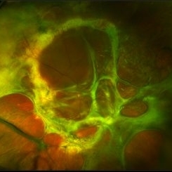
B-mode ultrasound video of a vitrectomized eye reveals characteristic vitreous cavity membranes secondary to endophthalmitis. The real-time imaging demonstrates these inflammatory membranes exhibit semi-rigid dynamics, displaying viscoelastic behavior with limited displacement during ocular movements while maintaining structural integrity. This restricted mobility pattern, showing both resistance to kinetic forces and slow recoil, represents a pathognomonic feature of advanced fibrotic organization in endophthalmitis.
Condition/keywords: endophthalmitis
-
 Endophtalmitis
Apr 3 2025 by Gustavo Uriel Fonseca Aguirre
B-mode ultrasound imaging of a vitrectomized eye showing vitreous cavity membranes secondary to endophthalmitis.
Photographer: Gustavo U. Fonseca Aguirre, Hospital Conde de Valenciana, Ciudad de México
Condition/keywords: endophthalmitis
-
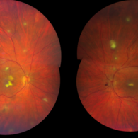
 Seedlings of Fungal Endophthalmitis
Mar 14 2025 by SHILPI H NARNAWARE, ICO ( Retina) , FAICO ( Vitreo-Retina)
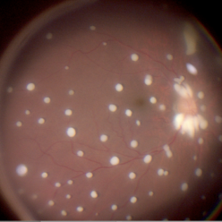
57 year diabetic female , was treated as a case of recurrent vitreous post cataract surgery. Patient was posted for vitrectomy 3 months post cataract surgery. Intra-operatively, multiple yellowish colonies were seen all over the posterior pole were seen, which were later found to be Aspergillus colonies.
Photographer: Shilpi Narnaware, Sarakshi Netralaya , Nagpur, Maharashtra , India
Imaging device: Ngenuity
Condition/keywords: endophthalmitis, fungal
-
 Fungal Endophthalmitis
Feb 25 2025 by Gaurav K. Shah, MD
This image shows the classic string of pearls with fluff infiltrates in the retina SP intraocular surgery.
Condition/keywords: fluff infiltrates, fungal endophthalmitis, string of pearls
-
 Endophthalmitis
Jul 5 2024 by Zach Seim
Optos Fundus Photograph of a 59 year old male with Fungal Endophthalmitis. Patient presented with VA Dsc 20/70.
Photographer: Zach Seim
Imaging device: Optos California
Condition/keywords: endophthalmitis, fundus photograph, fungal, optos, OPTOS CALIFORNIA
-
 Endophthalmitis
Jul 5 2024 by Zach Seim
OCT of a 59 year old male with Fungal Endophthalmitis. The fungus can be seen breaking through in the OCT.
Photographer: Zach Seim
Imaging device: Heidelberg OCT
Condition/keywords: endophthalmitis, fungal, OCT
-
 Fish Hook Eye Trauma
Jun 12 2024 by Miguel Brito, MD, FASRS
Fundus photograph of a 15-year-old boy post cataract aspiration, pars plana vitrectomy, suprachoroidal drainage, and retinal reattachment surgery secondary to traumatic endophthalmitis.
Photographer: Miguel Brito
Condition/keywords: endophthalmitis, PFCL, Retinal detachment under Silicon Oil, retinal fold
-
 Acute Endophthalmitis
Nov 14 2023 by Virginia Gebhart
85 year old female with acute endophthalmitis 14 days s/p IVEylea injection. 3+ injection and descemets folds, contracted fibron, 3mm Hypopyon, and 3+ cell. No view posteriorly, vision CF. Visual prognosis unknown at this time
Photographer: Virginia Gebhart
Condition/keywords: endophthalmitis
-
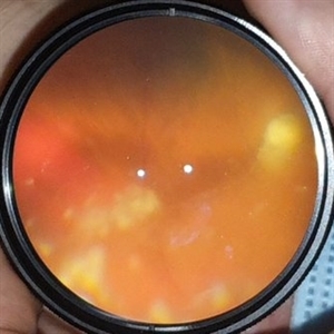
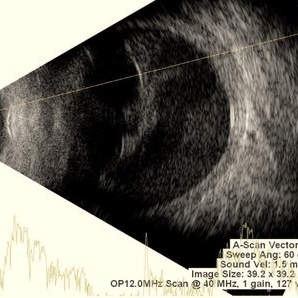 Endophthalmitis
Nov 2 2023 by Anand Temkar
Multiple dot echoes with mild to moderate spikes with free after movements suggestive of vitreous exudates in a case of endophthalmitis.
Photographer: Dr.Anand Temkar- Retina Foundation, Ahmedabad
Condition/keywords: A-scan ultrasound, B scan ultrasound, endophthalmitis
-
 Inflammatory pupillary membrane in patient with endophthalmitis
Jan 28 2023 by Kingston Rodolfo Ureña-Wong, MD, Opht, MSc
Anterior segment photography of a 54-year-old woman with post phacoemulsification endophthalmitis. She did not improve after first intravitreal antibiotics injection and develop an inflammatory pupillary membrane. After two vitrectomies, and a complete three intravitreal injections scheme, we decided to remove the intraocular lens and capsules.
Photographer: Marco Antonio Rubio-Atonal,UNAM, Asociación para evitar la ceguera en México
Imaging device: Zeiss Clarus 700
Condition/keywords: endophthalmitis, pupillary membranes
-
 Endophthalmitis one day after tap and injection.
Nov 19 2022 by Gareth Lema, MD, PhD
A fibrin clot, consolidating one day after tap and injection for post-op endophthalmitis by Staph. aureus.
Photographer: Gareth Lema, MD, PhD, New York Eye and Ear of Mount Sinai
Imaging device: Cell phone with a macro lens and muscle light for illumination.
Condition/keywords: endophthalmitis
-
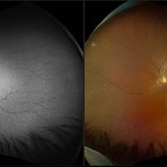
 Central Retinal Artery Occlusion Secondary to Endophthalmitis
Oct 24 2022 by Kelli Nyenhuis
Ultra-widefield fluorescein angiogram of a 64 year old female who developed a Central Retinal Artery Occlusion following acute endophthalmitis. The physician commented that there is no vascular filling with the exception of the papillomacular bundle. The patient's vision was scHM at the time the image was taken.
Photographer: Kelli Nyenhuis
Imaging device: Optos California
Condition/keywords: central retinal artery occlusion (CRAO), endophthalmitis, fluorescein angiogram (FA), left eye, non-perfusion, Optos, ultra-wide field imaging
-
 Fungal Endogenous Endophthalmitis
Oct 18 2022 by Pawel Kolman
60 y.o woman with endogenous endophthalmitis in left eye. She was treated in ICU for few weeks with wide-spectrum antibiotics and i.v steroids because of sever COVID-19 infection. 2 weeks after discharge from ICU, (while taking oral steroids at low dose) she complained about decrased vision in left eye.
Photographer: Pawel Kolman
Imaging device: Volk 20D and Samsung Galaxy S21
Condition/keywords: candida endophthalmitis, COVID-19, endogenous endophthalmitis, fungal endophthalmitis
-
 Fungal Endophthalmitis
Aug 25 2022 by Maxwell J Wingelaar, MD
A 54-year-old male with fungal endophthalmitis
Condition/keywords: fungal endophthalmitis
-
 Post Injection Endophthalmitis
Nov 21 2020 by Suhwan Lee, MD
Stillcut image of eye with post-intravitreal injection endophthalmitis during emergency vitrectomy. The patient received intravitreal injection for neovascular age-related macular degeneration 2 days ago. Image that was taken after anterior chamber irrigation and core vitrectomy showed multiple retinal hemorrhage and whitening of retinal vessels.
Photographer: Suhwan Lee, MD
Imaging device: Stillcut image of surgical video (Lumera 700 )
Condition/keywords: endophthalmitis
-
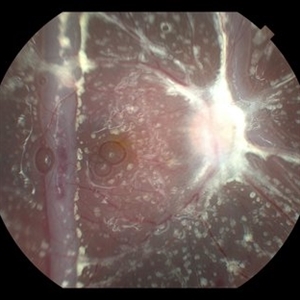
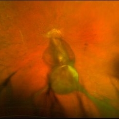
 Bacterial endophthalmitis
Jun 23 2020 by Thirumalesh Mochi Basavaraj, MD
Intraoperative pictures showing bacterial colonies, lab confirmed gram positive cocci(inset).
Imaging device: Leica microsurgery microscope
Condition/keywords: endophthalmitis, intraoperative
-
 Common Artifacts in Macroscopic Ocular Globe Evaluation
May 18 2020 by McGill University Health Centre
This sample was retrieved from a patient with a blind, painful eye. Blind, painful eye may be the end stage of several conditions including glaucoma, retinal detachment, and endophthalmitis, among others. Evisceration specimens are generally submitted in fragments. Different intraocular structures are identifiable: retina, cornea and capsular bag, choroidal tissue, and hematic material.
Condition/keywords: enucleation, evisceration, intraocular structures
-
 Post Endophthalmitis
Mar 12 2020 by Ashley Glace
70-year-old male FA Ultra Wide Field FA at 3:38 of a 70-year-old male with endophthalmitis post vitrectomy to clear the debris.
Photographer: Ashley Glace, Geisinger Ophthalmology, Williamsport Pennsylvania Clinic
Imaging device: Clarus 700
Condition/keywords: endophthalmitis
-
 Candida Endophthalmitis
Jan 26 2020 by Marlon García Roa, MD
Female, 30-years-old with <<< V Pregnancy Currently with 18 weeks gestation. Pathological personal history 1 month prior hospitalization for complicated acute appendicitis + pyelonephritis + severe thrombocytopenia (autoimmune treated with corticosteroids) with septic shock, appendectomy was performed, due to torpid evolution, intensive care unit with placement of central venous catheter treated with intravenous antibiotics is performed, CT scan is performed of thorax, abdomen and pelvis in search of aggregate pathology; finding multiple renal lithiasis that conditions hydronephrosis and reactivation of pyelonephritis, so he continued with antibiotic therapy and underwent endoscopic lithotomy, due to febrile persistence and with a positive blood culture result for candida Albicans, intravenous antifungals (anidulafungin) were started for 1 week, with improvement satisfactory for what was decided his discharge. During hospitalization it was required to transfuse 2 globular packages and platelet plasmapheresis as well as replacement of calcium, phosphorus and potassium. It refers to approximately 3 weeks of visual loss of the left eye associated with myodisopsia. visual acuity 20/100 Vitritis +, with floating vitreous abscess on the posterior pole, round papilla, slightly erased edges, excavation 0.3, macula without foveolar luster, conserved vein artery relationship, with vessels with multiple mineralization areas and superior peripheral lesion of ¼ diameter of papilla as ball of snow applied to retina.
Photographer: MARLON GARCIA ROA, INSTITUTO DE RETINA DEL BAJIO (INDEREB), QUERETARO, MEXICO
Condition/keywords: candida endophthalmitis
-
 Closed Funnel Retinal Detachment
Oct 8 2019 by Olivia Rainey
Ultra-wide field pseudocolor image of a 57-year-old male with a closed funnel retinal detachment with anterior and posterior napkin rings affecting his left eye. Patient presented with klebsiella endophthalmitis in UK, and was in medically induced coma with tracheostomy. He awoke after sedation with loss of vision in both eyes, later developing a retinal detachment in both eyes. Prior inflammation attributable to prephthisical state and chronic funnel retinal detachment. The eye is inoperable and observation is recommended.
Photographer: Olivia Rainey and Amber Poss
Imaging device: Optos
Condition/keywords: blind eye, funnel, hypotony, klebsiella endopthalmitis, left eye, Optos
-
 Chronic Total Retinal Detachment
Oct 8 2019 by Olivia Rainey
Ultra-wide field pseudocolor image of a 57-year-old male with a chronic total retinal detachment affecting his right eye. Patient presented with klebsiella endophthalmitis in UK, and was in medically induced coma with tracheostomy. He awoke after sedation with loss of vision in both eyes, later developing a retinal detachment in both eyes. He had his first repair with gas then with silicone oil. Although he is developing band K and corneal decompensation due to oil in the AC, he has a chronic cicatricial retinal detachment with subretinal and preretinal PVR with large stretch breaks. His vision may worsen with surgery and his eye is hypotenous and if repair is possible, oil will need to be replaced and thus may still migrate to the AC thus would defer intervention until absolutely necessary given risks of vision loss in his only seeing eye. Given this is his only eye, observation is recommended at this time.
Photographer: Olivia Rainey and Amber Poss
Imaging device: Optos
Condition/keywords: chronic retinal detachment, klebsiella endopthalmitis, laser scarring, Optos, proliferative vitreoretinopathy (PVR), retinectomy, silicone oil
-
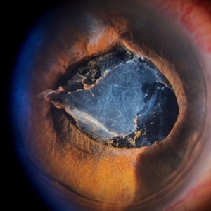
 Fungal Endophthalmitis
Jun 4 2019 by Gary R. Cook, MD, FACS
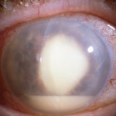
Fungal (Candida) endophthalmitis with hypopyon.
Condition/keywords: candida endophthalmitis, endophthalmitis, fungal endophthalmitis, hypopyon
-
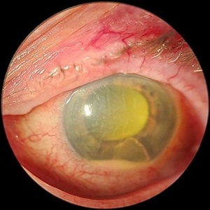
 Septic Endophthalmitis
Apr 12 2019 by Gary R. Cook, MD, FACS
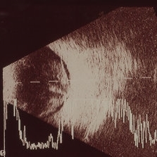
B-Scan ultrasonogram of the vitreous changes in the eye of a 48-year-old male with endogenous endophthalmitis and septic embolus resulting in a BRAO; V.A. = 20/40-2
Imaging device: Ophthscan
Condition/keywords: branch retinal artery occlusion (BRAO), endogenous endophthalmitis, endop, endophthalmitis
-
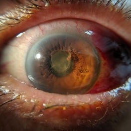
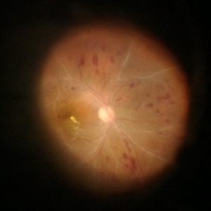
 Endophthalmitis
Apr 1 2019 by Gary R. Cook, MD, FACS
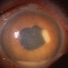
Klebsiella pneumonia endophthalmitis with hypopyon post cataract surgery.
Condition/keywords: endogenous endophthalmitis, endophthalmitis, klebsiella pneumoniae
 Loading…