Search results (75 results)
-
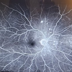
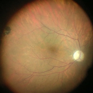
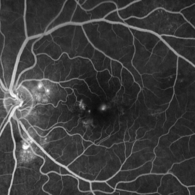 Multifocal CSCR
Apr 10 2025 by Daniela Bogenschutz
FA image from a 65-year-old woman with Multifocal Central Serous Chorioretinopathy. As the dye was working through her veins additional pathology appeared. Monitoring her progression and considering PDT if condition persists.
Photographer: Daniela Bogenschutz, OSC; Retina Consultants of the Carolinas, PA
Condition/keywords: multifocal central serous chorioretinopathy (CSCR)
-
 Sickle Cell Retinopathy
Feb 24 2025 by Kimberly Wakester
Optomap RGB image of an 24-year-old woman with sickle cell retinopathy in both eyes. There is overall progression of the ischemic vessels and vascular drops out compared to previous images completed in 2021. Oral FA was completed and shows possible progression of peripheral non-perfusion but difficult to determine due to drinking FA dye and images not being as bright. On Clinical exam there is no evidence of NV, RD, or RT in either eye. Patient understands the need for continued follow up care and the likely need for PRP laser in both eyes.
Photographer: Kimberly Wakester, COA
Imaging device: Optos California
Condition/keywords: sickle cell retinopathy
-
 Herpetic Corneal Ulcer
Sep 24 2024 by DR Rohit Gupta
Slit lamp photograph of 32 year old male presented with herpetic corneal ulcer on staining with fluorescein dye under cobalt blue filted dendrits can be seen.
Photographer: Dr Rohit gupta
Imaging device: Samsung S21
Condition/keywords: corneal ulcer, dendritic keratitis, herpes dendrite, Herpes simplex infection, Herpes zoster, staining
-
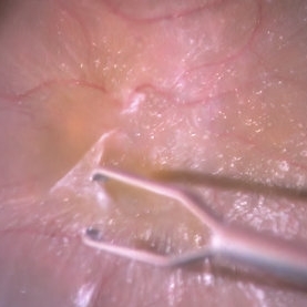 Epiretinal Membrane
Jul 19 2024 by Anjana Mirajkar, MS Ophthalmology
An intra operative still showing removal of the epi -retinal membrane with forceps under high magnification after staining with triamcinolone acetonide dye.
Photographer: Dr. Anjana Mirajkar -Retina Foundation, Ahmedabad
Imaging device: Mirante-Nidek
Condition/keywords: epiretinal membrane removal
-
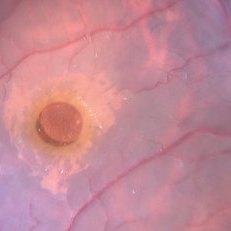 ILM Staining in Case of Macular Hole
Jul 4 2024 by Anjana Mirajkar, MS Ophthalmology
Intra operative still of LE showing ILM staining done with BBG dye in case of macular hole.
Photographer: Dr. Anjana Mirajkar -Retina Foundation, Ahmedabad
Condition/keywords: ILM staining, macular hole
-
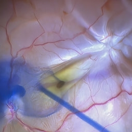 Star Folds in a Chronic Retinal Detachment
Jul 3 2024 by Anjana Mirajkar, MS Ophthalmology
Intra-operative still RE showing a star fold at the parafoveal area causing traction at the macula. Brilliant blue dye being injected to the stain the ILM.
Photographer: Dr. Anjana Mirajkar -Retina Foundation, Ahmedabad
Condition/keywords: brilliant blue staining, proliferative vitreoretinopathy (PVR), star folds
-
 Dye image
Apr 13 2024 by Monica King
Unknown diagnosis. 35yr old female. Blind spots started to form.
Photographer: Dr. Becker, Colorado Eye Institute
Condition/keywords: Unknown diagnosis
-
 Impending STBRVO
Jan 7 2024 by MEENAL SONI
A middle aged female presented to the OPD with diminution of vision in right eye for past 7 days. Fundus examination findings depict supero-temporal AV crossing changes with macular hard exudates and oedema. On FFA we could clearly visualise the artery compressing the vein with leakage of dye in late phase extending into the macular region. On systemic evaluation the patient was found to be hypertensive with deranged lipid profile. She was advised injection anti VEGF for macular oedema and a physician consult for commencing the treatment for systemic condition. Despite a physician reference patient was not started on anti hypertensives and later presented with frank STBRVO with macular oedema after 3 months.
Photographer: Dr. Meenal Soni, VR fellow ASG eye hospital, Jodhpur (Raj)
Imaging device: Visucam
Condition/keywords: Impending BRVO with macular edema
-
Retina Rhexis
Jan 2 2024 by Deepak Bhojwani, MS
THIS SURGICAL VIDEO DEMONSTRATES STANDARD ILM PEELING FOR MACULAR HOLE SURGERIES. THE ILM PEELING IS ASSITED BY BRILLIANT BYE DYE STAINING.
Condition/keywords: ILM peeling, macular hole, Macular surgery
-
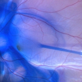
 BBG Dye injection to Stain ILM during Vitrectomy Surgery | Intra-Operative Still
Apr 28 2023 by Veer Singh, MS, FVRS, FMRF, FICO (Retina)
BBG Dye injection to Stain ILM during Vitrectomy Surgery | Intra-Operative Still
Photographer: Dr. Veer Singh
Condition/keywords: brilliant blue staining, ERM, ILM staining
-
Vitrectomy for Macular Hole
Jan 13 2023 by Manish Nagpal, MD, FRCS (UK), FASRS
This is a case of Macular hole for which vitrectomy is being done. After doing core vitrectomy triamcinolone dye is injected to stain the hyaloid. High aspiration is used on cutter to engage the hyaloid and gradually pull it anteriorly. PVD induction is carried out. After this brilliant blue dye is injected to stain the internal limiting membrane. ILM is peeled using a 25 gauge forceps in a tangential manner. After this i use a instrument called the massager which we have developed to gently and atraumatically massage concentrically the edges of the hole. This releases the subtle contaction on the edges of the hole and relaxes the margins. After this air fluid exchange is carried out followed by low vacuum aspiration over the hole. The hole approximates itself gradually as the aspiration dries up the edges.
Condition/keywords: forceps, hyaloid, ILM, macular hole, peeling, staining, video, vitrectomy
-
Vitrectomy for Sub ILM blood over macula
Jan 2 2023 by Manish Nagpal, MD, FRCS (UK), FASRS
This is a case of non resolving ILM hemorrhage over macula. Vitrectomy is carried out and hyaloid is removed after traimcinolone staining. After this brilliant blue dye is injected to stain the ILM. Internal limiting membrane is then removed with a forceps. Once the sub ilm blood is exposed , it easily aspirates with the cutter. The origin is probably from a macroaneurysm and there is a small component of subretinal residual blood noted at the end of the surgery.
Condition/keywords: brilliant blue, hyaloid, internal limiting membrane, macula, microaneurysm, retina, sub ILM blood, sub ILM hemorrhage, triamcinolone, video, vitrectomy
-
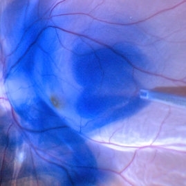
 Brilliant Blue Dye Injection to Stain ILM in a Macular Hole with Retinal Detachment
Feb 4 2022 by Manish Nagpal, MD, FRCS (UK), FASRS
Intraoperative still of a Brilliant blue dye injection being done to stain the ILM.
Photographer: Manish Nagpal, Director, Retina Foundation, Ahmedabad
Imaging device: Sony PMW -10 MD surgical camera
Condition/keywords: full thickness macular hole, macula, retina
-
 Brilliant blue dye injection to stain ILM in a macular hole with retinal detachment
Feb 4 2022 by Manish Nagpal, MD, FRCS (UK), FASRS
Intraoperative still of brilliant blue dye injection in process to initiate ILM peel in a patient who has a retinal detachment with a macular hole
Photographer: Manish Nagpal, Director, Retina Foundation, Ahmedabad
Imaging device: Sony PMW -10 MD surgical camera
Condition/keywords: brilliant blue, ILM flap, macular hole
-
 Brilliant Blue Dye Injected in a Case of Macular Hole to Stain the ILM
Feb 4 2022 by Manish Nagpal, MD, FRCS (UK), FASRS
Intraoperative still of a brilliant blue dye being injected to stain the ILM.
Photographer: Manish Nagpal, Director, Retina Foundation, Ahmedabad
Imaging device: Sony PMW -10 MD surgical camera
Condition/keywords: brilliant blue, ILM flap, ILM staining, macular hole, retina, retina surgery
-
 PVD Induction
Feb 2 2022 by Manish Nagpal, MD, FRCS (UK), FASRS
Intraoperative photo of PVD induction being carried out. Hyaloid has been stained using triamcinolone dye and PVD induction is carried out using high suction on the cutter and engaging the stained hyaloid.
Photographer: Manish Nagpal, Retina Foundation, Ahmedabad, India
Imaging device: Sony PMW -10 MD surgical camera
Condition/keywords: Hyaloid staining, PVD induction, triamcinolone
-
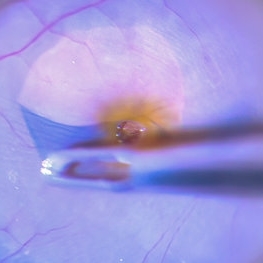 Internal Limiting Membrane Peeling
Jan 10 2022 by Manish Nagpal, MD, FRCS (UK), FASRS
Intraoperative image of internal limiting membrane being peeled using a 25 gauge ILM forceps. Brilliant blue dye has been used to stain the ILM.
Photographer: Manish Nagpal, Director, Retina Foundation, Ahmedabad
Imaging device: Sony PMW -10 MD surgical camera
Condition/keywords: internal limiting membrane (ILM) peeling
-
Epimacular Membrane
Oct 14 2021 by Islam bechakh
A vitrectomy is performed in our 25 G transconjunctival patient after careful decontamination of the cul-de-sacs by washing with povidone-iodine (Betadine®) 5% for 2 minutes. The panoramic system associated with the operating microscope makes it possible to control the traction on the retinal periphery and to facilitate the manipulation of the dye (Brilliant Blue G) during the surgery. The peeling of the membrane is extended to the whole macular area by trying, by a superficial grip begun in the sub-foveolar, to peel only the membrane. The internal limiting is then stained a second time and the total or partial decision is discussed on a case-by-case basis depending on the severity of the retraction and the type of diffuse or cystoid edema.
Photographer: Islam Bechakh
Condition/keywords: Epimacular membrane, vitrectomy
-
 Cilioretinal Artery
Sep 20 2020 by Barton L Blackorby, MD
Early FA just after the choroidal flush. Notice the prominent filling of this cilioretinal artery well in advance of the retinal circulation. This image highlights that the source of blood flow to the cilioretinal artery is from the choroidal circulation as the retinal circulation is just beginning to fill with dye in this image.
Imaging device: Zeiss Clarus
Condition/keywords: cilioretinal artery, fluorescein angiogram (FA)
-
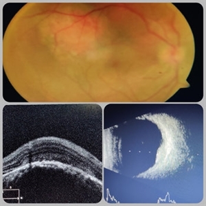 Circumscribed Choroidal Hemangioma
Jul 3 2020 by Dhaivat Shah
A 30-year-old young male presented with drop in vision in right eye since 1 year (6/60). Fundus examination revealed choroidal hemangioma superotemporal to macula. Choroidal hemangioma is an unusual benign vascular tumor of the choroid. It can be circumscribed solitary or diffuse tumor with the later having other systemic associations. Circumscribed choroidal hemangiomas (CCHs) are usually unilateral, unifocal hamartomatous vascular tumor affecting people in second to fourth decade. It appers as round to oval, orangish-red mass in posterior pole with smooth homogenous surface mostly present in macular and peripapillary area. Hyperopic shift is seen in sub-foveal tumors in contrast to para-foveal ones which are usually asymptomatic or present with metamorphopsia or photopsia and diminished vision secondary to exudative retinal detachment. B-scan shows highly reflective tumor without any shadowing or acoustic solidity with high anterior A scan spike. EDI-OCT here depicts a smooth gently sloping choridal mass with compressed choriocapillaries and enlarged medium and large choroidal vessels. Over a period of time structural abnormalities of the outer retina can be visualised. Ancillary testing using Fluorescein Angiography shows lacy hyper-fluorescence during early arterial phase followed by increased hyper-fluorescence due to progressive profuse leakage from pin point foci during arterial and venous phase. Indocyanine green angiography shows lacy diffuse fluorescent tumor in early phase followed by hypo-fluorescent tumor due to dye wash out in late phase. Intrinsic auto-fluorescence is also seen in CCHs from lipofuscin and fresh sub-retinal fluid. Tumor is relatively hyper-intense with respect to vitreous in T1-weighted images in iso-intense in T2-weighted images of MRI. Asymptomatic cases need no treatment, while patients showing vision loss with presence or absence of exudative retinal detachment can be treated with photodynamic therapy which is preferred treatment due to site specific action. Selective occlusion of choroidal neovascularization can be achieved while the neurosensory retinal layers and Bruch membrane are almost unaffected, leaving retinal function intact. Green or rarely red wavelength laser photocoagulation is used to create a chorioretinal adhesion and resolve the SRF. Other treatment modalities include Transpupilary thermotherapy, external beam irradiation, proton beam therapy, brachytherapy and gamma knife.
Photographer: Miss Deepika Nagle
Imaging device: Zeiss
Condition/keywords: B scan ultrasound, choroidal hemangioma, fundus photograph, optical coherence tomography (OCT), photodynamic therapy
-
 ILM staining
Dec 11 2019 by Jennifer R Gallagher, MD
Intra-operative funds photo of the macula after ICG staining with removal of excess dye from the vitreous cavity.
Photographer: Hamzah Khalaf, UT Health San Antonio, University Hospital
Condition/keywords: internal limiting membrane (ILM) peeling, staining, surgical management
-
 Capillary Non-Perfusion
Aug 26 2019 by Narciso F. Atienza, MD, MBA, FASRS, FPCS, FPAO.
FA at 51 sec showing capillary non-perfusion and blocked fluoresence of the inferior macula and infero-temporal area with transit of dye on previously noted infero-temporal branch vein.
Photographer: Narciso F Atienza, Jr. MD, MBA
Imaging device: Topcon TRC
Condition/keywords: capillary nonperfusion
-
 Early Venous Phase
Aug 26 2019 by Narciso F. Atienza, MD, MBA, FASRS, FPCS, FPAO.
Early venous phase shows asymmetrical transit of dye perfusion of the infero-temporal arcade. Infero-temporal arcade shows beginning perfusion. Areas of non perfusion are also more prominent.
Photographer: Narciso F Atienza, Jr. MD, MBA
Imaging device: Topcon TRC
Condition/keywords: capillary nonperfusion, inferotemporal arcade
-
 Neovascular AMD with Active CNV
May 21 2019 by Carolyn Daley
Fluorescein angiogram image of an 86-year-old woman with neovascular AMD. This image was taken 1 minute and 22 seconds into the FA testing, after dye was injected.
Photographer: Carolyn Daley, COA, Retina Specialists of Michigan
Imaging device: Heidelberg Spectralis
Condition/keywords: choroidal neovascularization (CNV), fluorescein angiogram (FA), neovascular age-related macular degeneration (AMD)
 Loading…