 Initializing download.
-
 By Barton L Blackorby, MD
The Retina Institute
Co-author(s): Matthew Cardinale, DO - Uploaded on Sep 20, 2020.
- Last modified by Caroline Bozell on Sep 24, 2020.
- Rating
- Appears in
- Miscellaneous
- Condition/keywords
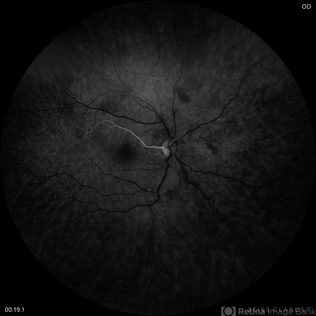
- cilioretinal artery
- Imaging device
- Zeiss Clarus
- Description
- Early FA just after the choroidal flush. Notice the prominent filling of this cilioretinal artery well in advance of the retinal circulation. This image highlights that the source of blood flow to the cilioretinal artery is from the choroidal circulation as the retinal circulation is just beginning to fill with dye in this image.


