Search results (158 results)
-
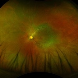
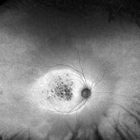
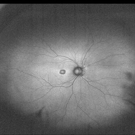
 Rod Cone Dystrophy
Mar 1 2025 by Aditya S Kelkar, MS, FRCS, FASRS,FRCOphth
Fundus Autofluorescence photograph of a 72-year-old woman with a rod cone dystrophy.
Photographer: Optom Rutuja Shelke
Imaging device: OPTOS DAYTONA
Condition/keywords: dystrophy
-
 Robson-Holder Ring
Jul 15 2024 by Arthi Mohankumar , MS,MRCS ED, FICO,FAICO
Robson holder hyper autofluorescent ring in a patient with retinitis pigmentosa.
Photographer: Arthi Mohankumar
Condition/keywords: autofluorescence imaging, retinitis pigmentosa (RP) dystrophy, Rod cone dystrophy
-
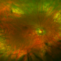
 Cone and Rod Dystrophy with Angioid Streaks
Apr 29 2024 by José Laércio Araújo Filho
Autofluorescence of a 65-year-old patient with rod-cone dystrophy associated with angioid streaks.
Photographer: José Laércio de Araújo Filho, University of São Paulo, Brazil
Imaging device: Optos Daytona P200T / A10600
Condition/keywords: angioid streaks, Cone-Rod Dystrophy
-
 Stargardt Disease
Apr 2 2024 by José Laércio Araújo Filho
Fundus autofluorescence of a 43-year-old woman with a ABCA4 positive macular dystrophy compatible with Stargardt Disease.
Photographer: José Laércio de Araújo Filho, Universidade de São Paulo, São Paulo
Imaging device: Optos Daytona P200T / A10600
Condition/keywords: cone dystrophy, Stargardt disease
-
 Cone Dystrophy
Aug 30 2023 by Vishal Agrawal, MD, FRCS,FACS,FASRS
12 year old male patient presented with photophobia, decrease in vision and Nystagmus. Bulls eye maculopathy gives an appearance of an eye on the fovea on color fundus photo due to nystagmus.
Photographer: Dr Bhagyashree
Imaging device: Clarus 700
Condition/keywords: bull's eye maculopathy, Cone-Rod Dystrophy
-
 Cone-Rod Dystrophy
Jul 20 2023 by Harsh Vardhan Singh, MS
52-year-old male with a advanced stage of cone-rod dystrophy
Photographer: Harsh Vardhan Singh, AIIMS, Guwahati
Imaging device: Zeiss Clarus 700
Condition/keywords: cone dystrophy, Cone-Rod Dystrophy, pigmentary retinal dystrophy, retinal dystrophy
-
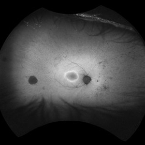
 Retinitis pigmentosa in atypical way
Dec 9 2022 by José Murillo
Fundus autofluorescence of an 78-year-old woman with an history of progressive nightblindness since 10-20 years-old. She has 9 siblings and 7 have the same symptoms, 2 legally blind. We can observe a semi-annular pattern of hipoAF with a at the end of the temporal arcades. The VA was 20/80
Photographer: Dr. José Murillo, Hospital Civil "Fray Antonio Alcalde" Universidad de Guadalajara, Jalisco, México
Condition/keywords: nightblindness, Retina, retinitis pigmentosa (RP) dystrophy, Rod cone dystrophy
-
 Rod Cone dystrophy
Nov 29 2022 by Niloofar Piri, MD
Fundus autofluorescence of the left eye in a 58 yo male with rod cone dystrophy. He presented with night blindness and peripheral vision loss since youth and recent decrease in central vision for the past 10 years. Notice multiple coin shaped hypoautofluorescent pacthes within central 20 degrees which are coalescing centrally. (fundus photo uploaded separately) He has one pathogenic variants of both CEP290 and PRPH2 genes.
Photographer: Sean Kelso, Saint Louis University
Condition/keywords: hereditary retinal degeneration, hereditary retinal dystrophy, rod cone dystrophy
-
 Rod Cone dystrophy
Nov 29 2022 by Niloofar Piri, MD
Fundus photograph of the left eye in a 58 yo male with rod cone dystrophy. He presented with night blindness and peripheral vision loss since youth and recent decrease in central vision for the past 10 years. Notice waxy pallor of the nerve, severe arterial narrowing and chorioretinal atrophy mainly around the arcades as well as posterior pole along with RPE hyperplastic changes and atrophy. RPE atrophy in midperiphery has coin shaped appearance. FAF has characteristic appearance (uploaded separately) He has one pathogenic variants of both CEP290 and PRPH2 genes.
Photographer: Sean Kelso, Saint Louis University
Condition/keywords: hereditary retinal deg, hereditary retinal dystrophy, Rod cone dystrophy
-
 Rod cone dystrophy autofluorescence
Sep 19 2022 by Kenneth Fong
34 year old male with colour blindness and loss of visual field
Condition/keywords: retinal dystrophy
-
 Rod cone dystrophy
Sep 19 2022 by Kenneth Fong
34 year old male with colour blindness and loss of visual field
Condition/keywords: rod cone dystrophy
-
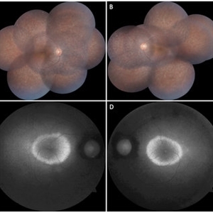
 CERKL-related Cone Rod Dystrophy
Jun 27 2022 by Hanna Choi
37-year-old female with cone-rod dystrophy. Developed photophobia and progressive blurry vision in the third decade. VA 20/40 OD, 20/30 OS. The patient is compound heterozygous for pathogenic mutations in the CERKL gene (Arg465Trp and Arg283*).
Photographer: Kaitlynn Silva, New England Retina Consultants
Imaging device: Ultrawide-field Optos Fundus Photography, Autofluorescence, Fluorescein Angiography
Condition/keywords: cone dystrophy, inherited retinal disease, maculopathy
-
 Cone-Rod Dystrophy
Dec 9 2021 by Filip Kecer
Fundus autofluorescence of a 12-year-old boy with genetically confirmed Cone-rod dystrophy.
Photographer: Filip Kecer
Imaging device: Spectralis, Heidelberg Engineering
Condition/keywords: autofluorescence imaging, cone dystrophy, dystrophy, rod dystrophy, Stargardt disease
-
 Rod/Cone dystrophy
Aug 20 2021 by Jeffrey Barker
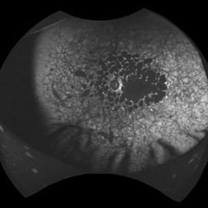
Fluorescein Angiogram Early frame (29 seconds)
Photographer: Jeffrey P. Barker, B.S. Retina Vitreous Surgeons of C.N.Y.
Condition/keywords: cone dystrophy, fluorescein angiogram (FA)
-
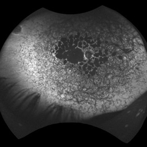
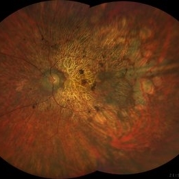
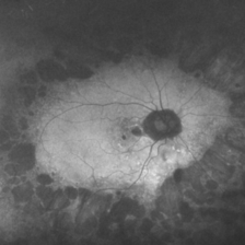
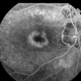
 THE ADDED VALUE OF WIDE-FIELD FUNDUS AUTOFLUORESCENCE IN CONE-ROD DYSTROPHIES
Oct 7 2019 by Mariana Oliveira
This wide-field fundus autofluorescence belongs to a 43 year-old female with CORD and no family history of inherited retinal disease. Her complains were reduced visual acuity and photophobia beginning in early adulthood. Note the central hyperautofluorescence with patchy hypoautofluorescence depicting atrophy of the retinal pigment epithelium in the macular area, an equatorial ring of preserved retinal autofluorescence and peripheral changes portraying rod involvement. The latter would be easily missed without wide-field imaging.
Condition/keywords: cone dystrophy
-
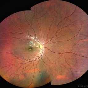
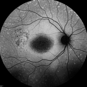
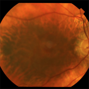
 Cone-Rod Dystrophy
Jun 23 2018 by Hossein Ameri, MD, PhD, FRCSI, MRCOphth
Ultra-wide field fundus photo of a patient with cone-rod dystrophy showing peripheral pigment clumping, mid-peripheral retinal pigment epithelial atrophy, and pigmentary changes in the macula.
Imaging device: Optos
Condition/keywords: cone dystrophy
-
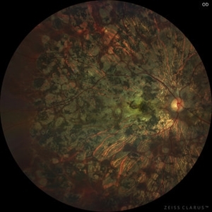
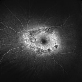
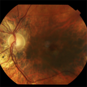
 Cone-Rod Dystrophy
Jun 23 2018 by Hossein Ameri, MD, PhD, FRCSI, MRCOphth
Ultra-wide field autofluorescence of a patient with cone-rod dystrophy showing mid peripheral ring of hypo autofluorescence, as well as autofluorescence changes in the macular area.
Imaging device: Optos
Condition/keywords: cone dystrophy
-
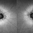
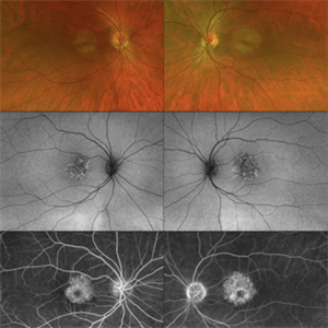
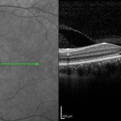
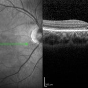 Cone-Rod Dystrophy
Mar 15 2017 by Hamid Ahmadieh, MD
Infrared and OCT images of the right eye of a 16-year-old boy with decreased visual acuity and color vision deficiency due to cone-rod dystrophy.
Photographer: Abazarnezhad , Negah Eye Center, Tehran, Iran
Imaging device: Spectralis OCT
Condition/keywords: cone dystrophy, infrared image, optical coherence tomography (OCT)
-
 Cone-Rod Dystrophy
Mar 15 2017 by Hamid Ahmadieh, MD
Infrared and OCT images of the left eye of a 16-year-old boy with decreased visual acuity and color vision deficiency due to cone-rod dystrophy.
Photographer: Abazarnezhad , Negah Eye Center, Tehran, Iran
Imaging device: Spectralis OCT
Condition/keywords: cone dystrophy, infrared image, optical coherence tomography (OCT)
-
 Cone-Rod Dystrophy
Aug 15 2015 by Thomas A. Ciulla, MD, MBA, FASRS
Fundus image revealing bull's eye maculopathy.
Photographer: Charlotte Harris
Condition/keywords: cone dystrophy
-
 Cone-Rod Dystrophy
Aug 15 2015 by Thomas A. Ciulla, MD, MBA, FASRS
Fundus image revealing bull's eye maculopathy.
Photographer: Charlotte Harris
Condition/keywords: cone dystrophy
-
 Cone-Rod Dystrophy
Aug 15 2015 by Thomas A. Ciulla, MD, MBA, FASRS
Fundus image revealing bull's eye maculopathy.
Photographer: Charlotte Harris
Condition/keywords: cone dystrophy
-
 Cone-Rod Dystrophy
Aug 15 2015 by Thomas A. Ciulla, MD, MBA, FASRS
Fundus image revealing bull's eye maculopathy.
Photographer: Charlotte Harris
Condition/keywords: cone dystrophy
-
 Cone Dystrophy
Mar 22 2015 by Andrea Arriola-Lopez, MD MSc
Fundus autofluorescence of an 31-year-old male with cone dystrophy.
Photographer: Andrea Elizabeth Arriola López, MSc. Asociación para Evitar la Ceguera, I.A.P. México D.F.
Imaging device: OPTOS, Dakota.
Condition/keywords: autofluorescence imaging, cone dystrophy, fundus autofluorescence (FAF), macula, macula lesion
 Loading…