 Initializing download.
-
 By Hossein Ameri, MD, PhD, FRCSI, MRCOphth
USC Roski Eye Institute of Keck School of Medicine, University of Southern California - Uploaded on Jun 23, 2018.
- Last modified by Caroline Bozell on Jun 28, 2018.
- Rating
- Appears in
- Miscellaneous
- Condition/keywords
- cone dystrophy
- Imaging device
-
Fundus camera
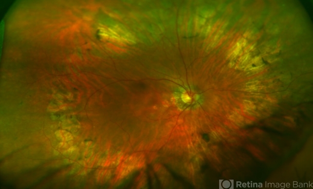
Optos - Description
- Ultra-wide field fundus photo of a patient with cone-rod dystrophy showing peripheral pigment clumping, mid-peripheral retinal pigment epithelial atrophy, and pigmentary changes in the macula.


---thumb.jpg/image-square;max$79,0.ImageHandler "Cone Dystrophy")
---thumb.jpg/image-square;max$79,0.ImageHandler "Cone Dystrophy")
---thumb.jpg/image-square;max$79,0.ImageHandler "Cone Dystrophy")
---thumb.jpg/image-square;max$79,0.ImageHandler "Cone Dystrophy")
---thumb.jpg/image-square;max$79,0.ImageHandler "Cone Dystrophy")
---thumb.jpg/image-square;max$79,0.ImageHandler "Cone Dystrophy")
---thumb.jpg/image-square;max$79,0.ImageHandler "Cone Dystrophy")
