 Initializing download.
-
 By Fabricio Dolores
Instituto mexicano de Oftalmología
Co-author(s): Miguel Vazquez-Membrillo, MD Instituto Mexicano de Oftalmología, Querétaro, México - Uploaded on Mar 13, 2025.
- Last modified by Joshua Friedman on Mar 14, 2025.
- Rating
- Appears in
- Miscellaneous
- Condition/keywords
- Retinal Detachment
- Photographer
- Fabricio Dolores-Villanueva, MD
- Imaging device
-
Fundus camera
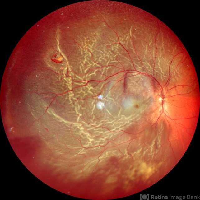
Nidek Mirante - Description
- This color wide-field clinical image depicts the right eye of a female patient who experienced a sudden loss of vision one month earlier. She was initially diagnosed with a vitreous hemorrhage and managed with conservative treatment. Upon presentation to our institute one month later, a superior rhegmatogenous retinal detachment was identified, extending across the 12 o’clock meridian. This was accompanied by an inferior vitreous hemorrhage and a solitary superior retinal lesion located at M11 in the superior triangle of the ora serrata, in alignment with Lincoff's second law.


