Search results (37 results)
-
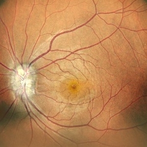
 Optic Nerve Head Drusen With Angiod Streaks in Hyperphosphatemic Familial Tumoral Calcinosis
Aug 8 2024 by Hemanth Murthy, MBBS, MD, FASRS
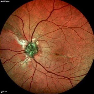
Multicolor image of left eye of 53 year female patient with decreased vision in left eye. Patient gives history of multiple joint swellings with multiple dental procedures due to calcification of the roots. She had type2 MNV demonstrated on OCT and OCTA. Her blood reports showed elevated serum phosphorus (6.4 mg/dl) with normal serum calcium, vitamin D and parathyroid hormone. Her fibroblast growth factor 23 was markedly elevated(>1500RU/ml).
Photographer: Mr Veda Vyas
Condition/keywords: Optic disc drusen and Angiod streaks
-
 Optic Nerve Head Drusen With Angiod Streaks in Hyperphosphatemic Familial Tumoral Calcinosis
Aug 8 2024 by Hemanth Murthy, MBBS, MD, FASRS
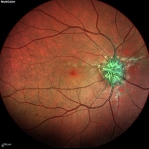
Multicolor image of right eye of 53 year female patient with decreased vision in left eye. Patient gives history of multiple joint swellings with multiple dental procedures due to calcification of the roots. She showed type2 MNV on OCT and OCTA. Her blood reports showed elevated serum phosphorus (6.4 mg/dl) with normal serum calcium, vitamin D and parathyroid hormone. Her fibroblast growth factor 23 was markedly elevated(>1500RU/ml).
Photographer: Mr Veda Vyas
Condition/keywords: Optic disc drusen and Angiod streaks
-
 Optic Nerve Head Drusen With Angiod Streaks in Phosphatemic Familial Tumoral Calcinosis
Aug 8 2024 by Hemanth Murthy, MBBS, MD, FASRS
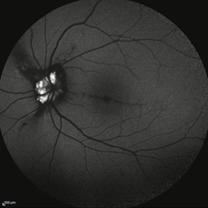
Autofluorescence image of left eye of 53 year female patient with decreased vision in left eye. Patient gives history of multiple joint swellings with multiple dental procedures due to calcification of the roots. She showed type 2 MNV in left eye on OCT and OCTA. Her blood reports showed elevated serum phosphorus (6.4 mg/dl) with normal serum calcium, vitamin D and parathyroid hormone. Her fibroblast growth factor 23 was markedly elevated(>1500RU/ml).
Photographer: Mr Veda Vyas
Condition/keywords: Optic disc drusen and Angiod streaks
-
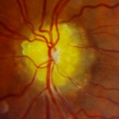
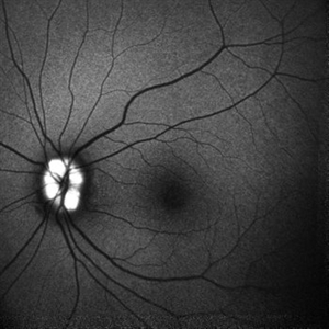
 Autofluorescence in Optic Nerve Head Drusen
May 28 2024 by Nishikant J Borse, MS, FMRF, FASRS
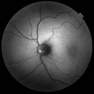
65-year-old female was referred for disc edema. An Autofluorescence Imaging was done which showed the autofluorescence of the optic nerve head drusen.
Photographer: Dr Nishikant Borse , Insight eye Clinic , Mumbai
Imaging device: Topcon Triton
Condition/keywords: Autofluorescence imaging of Optic Disc Drusen
-
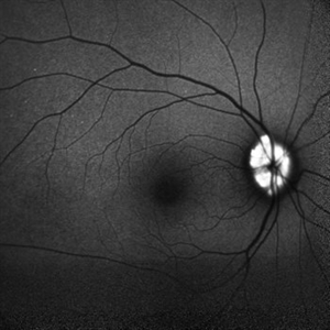
 Autofluorescence in Optic Nerve Head Drusen
May 28 2024 by Nishikant J Borse, MS, FMRF, FASRS
65-year-old female was referred for disc edema. An Autofluorescence Imaging was done which showed the autofluorescence of the optic nerve head drusen.
Photographer: Dr Nishikant Borse , Insight eye Clinic , Mumbai
Imaging device: Topcon Triton
Condition/keywords: Autofluorescence imaging of Optic Disc Drusen
-
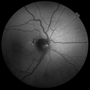
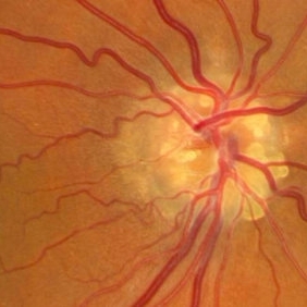
 Optic nerve head drusen
Sep 14 2023 by Ben Serar
Fundus photograph showing a yellowish-white nodule at the superior margin of the optic disc in a case of Optic nerve head drusen.
Condition/keywords: Optic nerve head drusen
-
 Optic nerve head drusen
Sep 14 2023 by Ben Serar
Fundus photograph showing yellowish-white nodules at the optic disc causing blurring of the disc margins in a case of Optic nerve head drusen.
Condition/keywords: Optic nerve head drusen
-
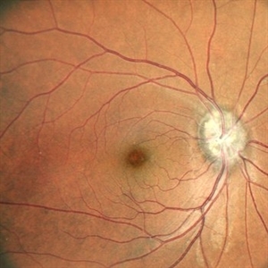 Optic nerve head drusen
Dec 26 2022 by Vaidehi Sathaye
Fundus Photograph of RE of a 32 year old female with Optic nerve head drusen
Photographer: Dr. Vaidehi Sathaye
Imaging device: Mirante
Condition/keywords: drusen of optic disc
-
 Optic nerve head drusen
Dec 26 2022 by Vaidehi Sathaye
Fundus Photograph of LE of a 32 year old female with Optic nerve head drusen
Photographer: Dr. Vaidehi Sathaye
Imaging device: Mirante
Condition/keywords: drusen of optic disc
-
 Optic nerve head drusen
Dec 26 2022 by Vaidehi Sathaye
FAF photograph of LE of a 32 year old female with Optic nerve head drusen
Photographer: Dr. Vaidehi Sathaye
Imaging device: Mirante
Condition/keywords: drusen of optic disc
-
 Optic nerve head drusen
Dec 26 2022 by Vaidehi Sathaye
FAF photograph of RE of a 32 year old female with Optic nerve head drusen
Photographer: Dr. Vaidehi Sathaye
Imaging device: Mirante
Condition/keywords: drusen of optic disc
-
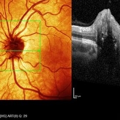
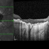
 Right eye SD- OCT-RNFL of optic nerve head drusen showing hypo reflective centre with hyper reflective margins.
Aug 5 2022 by Kavitha Duraipandi, MD DNB FICO FRCS
A 20 year old patient referred to the clinic with blurred disc margins to rule out papilledema.
Photographer: Natalie Fox- Bussell
Condition/keywords: optic nerve drusen, optical coherence tomography (OCT)
-
 Left eye SD- OCT-RNFL of optic nerve head drusen showing hypo reflective centre with hyper reflective margins.
Aug 5 2022 by Kavitha Duraipandi, MD DNB FICO FRCS
A 20 year old patient referred to the clinic with blurred disc margins to rule out papilledema.
Photographer: Natalie Fox- Bussell
Condition/keywords: optic nerve drusen, optical coherence tomography (OCT)
-
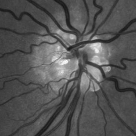
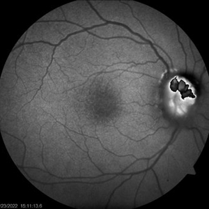 ONH-drusen-OD
Mar 24 2022 by Elite Bor-Shavit, MD
Fundus autofluorescence of a 41-years old patient with combined true papilledema and optic nerve head drusen, treated with Diamox and monitored.
Condition/keywords: optic disc drusen, papilledema
-
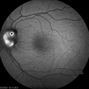 ONH-drusen-OS
Mar 24 2022 by Elite Bor-Shavit, MD
Fundus autofluorescence of a 41-years old patient with combined true papilledema and optic nerve head drusen, treated with Diamox and monitored.
Condition/keywords: optic disc drusen, papilledema
-
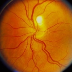
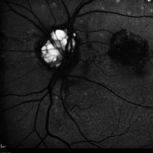 Optic Nerve Head Drusen
Feb 9 2018 by Olivia Rainey
Fundus autofluorescence of a 49-year-old female with optic nerve head drusen affecting her left eye. The patient has pseudoxanthoma elasticum with choroidal neovascularization and has been receiving anti-VEGF treatment for many years.
Photographer: Olivia Rainey
Imaging device: Heidelberg Spectralis
Condition/keywords: 30 degrees, anti-VEGF, choroidal neovascularization (CNV), fundus autofluorescence (FAF), Heidelburg Spectralis, left eye, optic disc, optic nerve drusen, pseudoxanthoma elasticum (PXE)
-
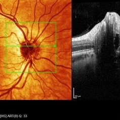
 Optic Nerve Head Drusen with OCT
Feb 2 2018 by Olivia Rainey
Optical coherence tomography with enhanced depth imaging of a 86-year-old male with optic nerve head drusen affecting his right eye. This patient has also been diagnosed with pseudoxanthoma elasticum and macular degeneration.
Photographer: Olivia Rainey
Imaging device: Heidelberg Spectralis
Condition/keywords: enhanced depth imaging, infrared image, macular degeneration, optic disc drusen, optic nerve, optical coherence tomography (OCT), pseudoxanthoma elasticum (PXE)
-
 Optic Nerve Head Drusen With Idiopathic CNV
Feb 17 2017 by Kristen Wagner
22-year-old female fundus photograph of a right eye with Optic Nerve Drusen with Idiopathic CNV.
Photographer: Kristen Wagner, COT, OSC Ophthalmic Photographer, Tennessee Retina, Nashville TN
Condition/keywords: choroidal neovascularization (CNV), drusen of optic disc, optic disc drusen
-
 Optic Nerve Head Drusen
Sep 10 2015 by Mariam A Al-Feky, MD
Left eye 30-year-old obese female patient with BCVA 0.8 OU presenting with severe headache of 1 month duration Ant seg: NAD OU, post. segment: disc edema OU MRI brain is normal ONH drusen is the diagnosis actually, with late staining in FFA (without early hyperfluorescent telangectatic disc capillaries as in cases of papillaedema), well delineated in the ONH map on the Heidelberg machine, and could be detected as a hypereflective material below the nerve fiber layer on the line scan. N.B. Burried ONH drusen don't autofluoresce.
Imaging device: Fundus camera
Condition/keywords: drusen, optic nerve head
-
 Optic Nerve Head Drusen
Sep 10 2015 by Mariam A Al-Feky, MD
Right eye, 30-year-old obese female patient with BCVA 0.8 OU presenting with severe headache of 1 month duration. Ant seg: NAD OU, post. segment: disc edema OU, MRI brain is normal. ONH drusen is the diagnosis actually, with late staining in FFA (without early hyperfluorescent telangectatic disc capillaries as in caes of papillaedema), well delineated in the ONH map on the Heidelberg machine, and could be detected as a hypereflective material below the nerve fiber layer on the line scan. N.B. Burried ONH drusen don't autofluoresce.
Imaging device: Fundus camera
Condition/keywords: optic nerve head
-
 Optic Nerve Head Drusen
Feb 12 2015 by Timothy S Fuller, MD
Fundus autofluorescence image of a 34-year-old woman with striking, asymptomatic optic nerve head drusen.
Photographer: Nice Hesse, Texas Retina Associates
Imaging device: Heidelberg Spectralis
Condition/keywords: drusen of optic disc
-
 Optic Nerve Head Drusen
Feb 12 2015 by Timothy S Fuller, MD
Fundus photograph of a 34-year-old woman with striking, asymptomatic optic nerve head drusen.
Photographer: Nice Hesse, Texas Retina Associates
Condition/keywords: drusen of optic disc
-
 Optic Nerve Head Drusen
Feb 12 2015 by Timothy S Fuller, MD
Fundus autofluorescence image of a 34-year-old woman with striking, asymptomatic nerve head drusen.
Photographer: Nick Hesse, Texas Retina Associates
Imaging device: Heidelberg Spectralis
Condition/keywords: drusen of optic disc
-
 Optic Nerve Head Drusen
Feb 12 2015 by Timothy S Fuller, MD
Fundus photograph of a 34-year-old woman with striking, asymptomatic nerve head drusen.
Photographer: Nick Hesse, Texas Retina Associates
Condition/keywords: drusen of optic disc
-
---thumb.jpg/image-square;max$300,300.ImageHandler) Drusen
Drusen
 Loading…