 Initializing download.
-
 By Mariam A Al-Feky, MD
AIN SHAMS UNIVERSITY - Uploaded on Sep 10, 2015.
- Last modified by Caroline Bozell on Sep 10, 2015.
- Rating
- Appears in
- Miscellaneous
- Condition/keywords
- optic nerve head
- Imaging device
-
Optical coherence tomography system
Fundus camera - Description
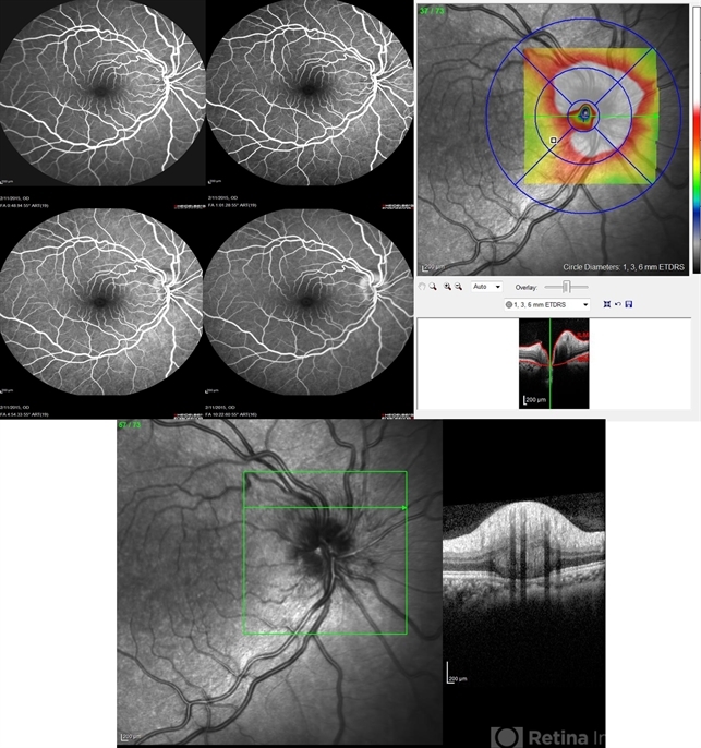
- Right eye, 30-year-old obese female patient with BCVA 0.8 OU presenting with severe headache of 1 month duration. Ant seg: NAD OU, post. segment: disc edema OU, MRI brain is normal. ONH drusen is the diagnosis actually, with late staining in FFA (without early hyperfluorescent telangectatic disc capillaries as in caes of papillaedema), well delineated in the ONH map on the Heidelberg machine, and could be detected as a hypereflective material below the nerve fiber layer on the line scan. N.B. Burried ONH drusen don't autofluoresce.

---thumb.jpg/image-square;max$79,0.ImageHandler "Swollen Optic Nervehead")
---thumb.jpg/image-square;max$79,0.ImageHandler "Swollen Optic Nervehead")
---thumb.jpg/image-square;max$79,0.ImageHandler "Flaked Retina")
---thumb.jpg/image-square;max$79,0.ImageHandler "Drusen")
---thumb.jpg/image-square;max$79,0.ImageHandler "ONH Drusen")
---thumb.jpg/image-square;max$79,0.ImageHandler "Extruded Drusen of ONH")
---thumb.jpg/image-square;max$79,0.ImageHandler "ONH Drusen")
---thumb.jpg/image-square;max$79,0.ImageHandler "Drusen")


