Search results (181 results)
-
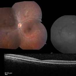 LCA Type 2
Apr 10 2025 by Joshua Friedman
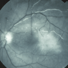
LCA Type 2 (RPE65) showing characteristic hypoautofluorescence and retinal thinning. 8F with best corrected visual acuity of 20/400 (OD) and 20/150 (OS). Small white intraretinal spots and RPE mottling are visible on color fundus photography. Blue light autofluorescence reveals near-complete loss of signal, while OCT demonstrates widespread outer retinal thinning.
Photographer: Stephen Tsang, MD, PhD
Condition/keywords: Leber Congenital Amaurosis
-
 Emulsified Silicone Oil
Apr 3 2025 by Andrew A. Moshfeghi, MD, MBA, FASRS
This is an 87 year- old male with 3.5 year history of retained silicone oil following treatment of late-onset recurrent retinal detachment 18 years following prior primary scleral buckle repair. Robust emulsified silicone oil aggregates are appreciated. Visual acuity is 20/400.
Photographer: Tammy Schoenholz, University of Southern California.
Imaging device: Zeiss Clarus
Condition/keywords: emulsified silicone oil
-
 Ozurdex in AC
Apr 1 2025 by Korey Starkey
90-year-old patient with an Ozurdex implant that migrated into the AC and with the cornea decompensating. Patient recommended for urgent surgery to remove implant. Vision OD at this visit was CF @ 2ft, most recent visit vision is 20/400, PH 20/25.
Photographer: Korey Starkey
Imaging device: Topcon
Condition/keywords: anterior chamber, corneal decompensation, external, external photography, Ozurdex implant, Topcon
-
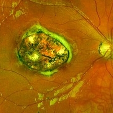
 Choroidal Melanoma 3 Ways
Jan 16 2025 by Virginia Gebhart
RGB/FA/ICG of 76 year old female with a new choroidal melanoma. Pt scheduled for plaque radiation. BCVA 20/400
Photographer: Virginia Gebhart, Retina Consultants of Carolina
Imaging device: Optos California
Condition/keywords: fluorescein angiogram (FA), indocyanine green (ICG) angiography, OPTOS CALIFORNIA RGB
-
 Severe NPDR
Oct 24 2023 by Virginia Gebhart
Fluorescein angiogram of left eye in 60-year-old male with severe non-proliferative diabetic retinopathy with extensive macular edema. Most recent A1c is 11. Vision 20/400. Injection of Eylea given
Photographer: Virginia Gebhart
Imaging device: Topcon
Condition/keywords: diabetic macular edema, Diabetic Retinopathy, fluorescein angiogram (FA), Fluorescein angiography
-
 Methotrexate Bubble following Intravitreal Injection for PVR
Sep 21 2022 by Zach Seim
Ultra-widefield fundus photograph of an 81 year old female with a Methotrexate bubble following an Intravitreal Injection for Proliferative Vitreoretinopathy. Patient has been presenting to the office for two week interval Methotrexate injections in her left eye. The image was taken prior to her eighth injection which revealed a residual Methotrexate bubble in her inferior retinal image. This patient was seeing "lots" of floaters, as well as having visual acuity of cc20/400 cc20/200 PH.
Photographer: Zach Seim
Imaging device: OPTOS California
Condition/keywords: bubble, fundus photograph, fundus photography, intravitreal injection, left eye, methotrexate, nasal retina, Optos, proliferative vitreoretinopathy (PVR), pseudocolor, ultra-wide field imaging
-
 Fundus Photo Montage showing Occlusive Vascultis from Brolucizumab
Jan 21 2022 by Somnath Chakraborty, MD
Right eye of a 62-year-old lady with Inferotemporal Branch Retinal Vein Occlusion, treated with single dose of "off-label" brolucizumab. She developed Occlusive Vasculitis 9 weeks post injection. This is her Fundus Photo Montage at that time, showing evidence of Occlusive Vasculitis with moderate grade vitritis. BCVA OD 20/400.
Photographer: Pulak Roy
Condition/keywords: branch retinal vein occlusion (BRVO), Brolucizumab, occlusive vasculitis, vitritis
-
 Optic Nerve Coloboma
Aug 14 2021 by Narciso F. Atienza, MD, MBA, FASRS, FPCS, FPAO.
19 year old male patient seen on routine examination for refraction. Had blurring of vision on the right eye since childhood. Was initially seen by a general ophthalmologist who diagnosed the patient with glaucoma. Present vision is CF at 3 feet uncorrected, and 20/400 with a refraction of -8.00 -1.50 X 180.
Photographer: Narciso F Atienza, Jr. MD MBA, FASRS, FPCS, FPAO. Legazpi Eye Center
Imaging device: Topcon TRC
Condition/keywords: optic nerve coloboma
-
 Bilateral Calcific Retina Arteriolar Occlusions in a Patient with Metastatic Ovarian Carcinoma
Dec 10 2020 by McGill University Health Centre
47-year-old female with cough and fever. Imaging showed a right pulmonary infiltrate. Transbronchial needle biopsy revealed lymphangitic spread of papillary adenocarcinoma with psammoma bodies. MRI of thyroid, CT of abdomen and pelvis were negative. gynecologic evaluation negative at that time . The patient had bilateral floaters, VA: 20/40 OD and 20/20 OS. Fundus examination showed retinal arteriolar sheathing and a flat choroidal lesion OS and vitritis OD. Fluorescein angiogram showed staining of left superior temporal retinal arterioles and bilateral midperipheral patchy hyperfluorescence at RPE The patient vision in the OD deteriorated to 20/400, and in the OS 20/50. Four months later a new choroidal lesion was diagnosed OS. An abdominal mass consistent with a cystadenoma of the ovary was diagnosed. After a year patient developed systemic metastasis. Autopsy: Metastatic adenocarcinoma to the lung, both adrenals, para-aortic lymph nodes, left hip, right breast, occipital skin, serosal surface of liver, pituitary. In almost all metastatic lesions psammoma bodies were found. Presumptive diagnosis is a primary tumor of the ovary.
Condition/keywords: bilateral, calcification, metastatic adenocarcinoma, retinal arteriolar occlusion
-
 Bilateral Calcific Retina Arteriolar Occlusions in a Patient with Metastatic Ovarian Carcinoma
Dec 10 2020 by McGill University Health Centre
47-year-old female with cough and fever. Imaging showed a right pulmonary infiltrate. Transbronchial needle biopsy revealed lymphangitic spread of papillary adenocarcinoma with psammoma bodies (MRI of thyroid, CT of abdomen and pelvis were negative) gynecologic evaluation negative at that time . The patient had bilateral floaters, VA: 20/40 OD and 20/20 OS. Fundus examination showed retinal arteriolar sheathing and a flat choroidal lesion OS and vitritis OD. Fluorescein angiogram showed staining of left superior temporal retinal arterioles and bilateral midperipheral patchy hyperfluorescence at RPE. The patient vision in the OD deteriorated to 20/400, and in the OS 20/50. Four months later a new choroidal lesion was diagnosed OS. An abdominal mass consistent with a cystadenoma of the ovary was diagnosed. After a year patient developed systemic metastasis. Autopsy: Metastatic adenocarcinoma to the lung, both adrenals, para-aortic lymph nodes, left hip, right breast, occipital skin, serosal surface of liver, pituitary. In almost all metastatic lesions psammoma bodies were found. Presumptive diagnosis is a primary tumor of the ovary.
Imaging device: Fluoroscein angiogram
Condition/keywords: bilateral, calcification, metastatic adenocarcinoma, retinal arteriolar occlusion
-
 Bilateral Calcific Retina Arteriolar Occlusions in a Patient with Metastatic Ovarian Carcinoma
Dec 10 2020 by McGill University Health Centre
47-year-old female with cough and fever. Imaging showed a right pulmonary infiltrate. Transbronchial needle biopsy revealed lymphangitic spread of papillary adenocarcinoma with psammoma bodies (MRI of thyroid, CT of abdomen and pelvis were negative) gynecologic evaluation negative at that time . The patient had bilateral floaters, VA: 20/40 OD and 20/20 OS. Fundus examination showed retinal arteriolar sheathing and a flat choroidal lesion OS and vitritis OD. Fluorescein angiogram showed staining of left superior temporal retinal arterioles and bilateral midperipheral patchy hyperfluorescence at RPE The patient vision in the OD deteriorated to 20/400, and in the OS 20/50. Four months later a new choroidal lesion was diagnosed OS. An abdominal mass consistent with a cystadenoma of the ovary was diagnosed. After a year patient developed systemic metastasis. Autopsy: Metastatic adenocarcinoma to the lung, both adrenals, para-aortic lymph nodes, left hip, right breast, occipital skin, serosal surface of liver, pituitary. In almost all metastatic lesions psammoma bodies were found. Presumptive diagnosis is a primary tumor of the ovary. Histopathologic examination of both eyes disclosed : Bilateral metastatic adenocarcinoma to the vitreous with partially calcified proliferation along internal limiting membrane, OS. Metastatic adenocarcinoma to choroid, OS. Bilateral optic atrophy secondary to retinal arteriolar occlusion with calcification.
Condition/keywords: bilateral, calcification, histopathology, metastatic adenocarcinoma, pathology, retinal arteriolar occlusion
-
 Bilateral Calcific Retina Arteriolar Occlusions in a Patient with Metastatic Ovarian Carcinoma
Dec 10 2020 by McGill University Health Centre
47-year-old female with cough and fever. Imaging showed a right pulmonary infiltrate. Transbronchial needle biopsy revealed lymphangitic spread of papillary adenocarcinoma with psammoma bodies (MRI of thyroid, CT of abdomen and pelvis were negative) gynecologic evaluation negative at that time Patient had bilateral floaters, VA: 20/40 OD and 20/20 OS. Fundus examination showed retinal arteriolar sheathing and a flat choroidal lesion OS and vitritis OD. Fluorescein angiogram showed staining of left superior temporal retinal arterioles and bilateral midperipheral patchy hyperfluorescence at RPE The patient vision in the OD deteriorated to 20/400, and in the OS 20/50. Four months later a new choroidal lesion was diagnosed OS. An abdominal mass consistent with a cystadenoma of the ovary was diagnosed. After a year patient developed systemic metastasis. Autopsy: Metastatic adenocarcinoma to the lung, both adrenals, para-aortic lymph nodes, left hip, right breast, occipital skin, serosal surface of liver, pituitary. In almost all metastatic lesions psammoma bodies were found. Presumptive diagnosis is a primary tumor of the ovary. Histopathologic examination of both eyes disclosed : Bilateral metastatic adenocarcinoma to the vitreous with partially calcified proliferation along internal limiting membrane, OS. Metastatic adenocarcinoma to choroid, OS. Bilateral optic atrophy secondary to retinal arteriolar occlusion with calcification.
Condition/keywords: bilateral, calcification, histopathology, metastatic adenocarcinoma, pathology, retinal arteriolar occlusion
-
 Bilateral Calcific Retina Arteriolar Occlusions in a Patient with Metastatic Ovarian Carcinoma
Dec 10 2020 by McGill University Health Centre
47-year-old female with cough and fever. Imaging showed a right pulmonary infiltrate. Transbronchial needle biopsy revealed lymphangitic spread of papillary adenocarcinoma with psammoma bodies (MRI of thyroid, CT of abdomen and pelvis were negative) gynecologic evaluation negative at that time . The patient had bilateral floaters, VA: 20/40 OD and 20/20 OS. Fundus examination showed retinal arteriolar sheathing and a flat choroidal lesion OS and vitritis OD. Fluorescein angiogram showed staining of left superior temporal retinal arterioles and bilateral midperipheral patchy hyperfluorescence at RPE. The patient vision in the OD deteriorated to 20/400, and in the OS 20/50. Four months later a new choroidal lesion was diagnosed OS. An abdominal mass consistent with a cystadenoma of the ovary was diagnosed. After a year patient developed systemic metastasis. Autopsy: Metastatic adenocarcinoma to the lung, both adrenals, para-aortic lymph nodes, left hip, right breast, occipital skin, serosal surface of liver, pituitary. In almost all metastatic lesions psammoma bodies were found. Presumptive diagnosis is a primary tumor of the ovary. Histopathologic examination of both eyes disclosed : Bilateral metastatic adenocarcinoma to the vitreous with partially calcified proliferation along internal limiting membrane, OS. Metastatic adenocarcinoma to choroid, OS. Bilateral optic atrophy secondary to retinal arteriolar occlusion with calcification.
Condition/keywords: bilateral, calcification, histopathology, metastatic adenocarcinoma, pathology, retinal arteriolar occlusion
-
 Bilateral Calcific Retina Arteriolar Occlusions in a Patient with Metastatic Ovarian Carcinoma
Dec 10 2020 by McGill University Health Centre
47-year-old female with cough and fever. Imaging showed a right pulmonary infiltrate. Transbronchial needle biopsy revealed lymphangitic spread of papillary adenocarcinoma with psammoma bodies (MRI of thyroid, CT of abdomen and pelvis were negative) gynecologic evaluation negative at that time . The patient had bilateral floaters, VA: 20/40 OD and 20/20 OS. Fundus examination showed retinal arteriolar sheathing and a flat choroidal lesion OS and vitritis OD. Fluorescein angiogram showed staining of left superior temporal retinal arterioles and bilateral midperipheral patchy hyperfluorescence at RPE The patient vision in the OD deteriorated to 20/400, and in the OS 20/50. Four months later a new choroidal lesion was diagnosed OS. An abdominal mass consistent with a cystadenoma of the ovary was diagnosed. After a year patient developed systemic metastasis. Autopsy: Metastatic adenocarcinoma to the lung, both adrenals, para-aortic lymph nodes, left hip, right breast, occipital skin, serosal surface of liver, pituitary. In almost all metastatic lesions psammoma bodies were found. Presumptive diagnosis is a primary tumor of the ovary. Histopathologic examination of both eyes disclosed : Bilateral metastatic adenocarcinoma to the vitreous with partially calcified proliferation along internal limiting membrane, OS. Metastatic adenocarcinoma to choroid, OS. Bilateral optic atrophy secondary to retinal arteriolar occlusion with calcification.
Condition/keywords: bilateral, calcification, histopathology, metastatic adenocarcinoma, pathology, retinal arteriolar occlusion
-
 Bilateral Calcific Retina Arteriolar Occlusions in a Patient with Metastatic Ovarian Carcinoma
Dec 10 2020 by McGill University Health Centre
47-year-old female with cough and fever. Imaging showed a right pulmonary infiltrate. Transbronchial needle biopsy revealed lymphangitic spread of papillary adenocarcinoma with psammoma bodies (MRI of thyroid, CT of abdomen and pelvis were negative) gynecologic evaluation negative at that time . The patient had bilateral floaters, VA: 20/40 OD and 20/20 OS. Fundus examination showed retinal arteriolar sheathing and a flat choroidal lesion OS and vitritis OD. Fluorescein angiogram showed staining of left superior temporal retinal arterioles and bilateral midperipheral patchy hyperfluorescence at RPE. The patient vision in the OD deteriorated to 20/400, and in the OS 20/50. Four months later a new choroidal lesion was diagnosed OS. An abdominal mass consistent with a cystadenoma of the ovary was diagnosed. After a year patient developed systemic metastasis. Autopsy: Metastatic adenocarcinoma to the lung, both adrenals, para-aortic lymph nodes, left hip, right breast, occipital skin, serosal surface of liver, pituitary. In almost all metastatic lesions psammoma bodies were found. Presumptive diagnosis is a primary tumor of the ovary. Histopathologic examination of both eyes disclosed : Bilateral metastatic adenocarcinoma to the vitreous with partially calcified proliferation along internal limiting membrane, OS. Metastatic adenocarcinoma to choroid, OS. Bilateral optic atrophy secondary to retinal arteriolar occlusion with calcification.
Condition/keywords: bilateral, calcification, histopathology, metastatic adenocarcinoma, pathology, retinal arteriolar occlusion
-
 Congenital Toxoplasmosis
Dec 18 2019 by Yoshihiro Yonekawa, MD, FASRS
Widefield fundus image of a teenage girl's right eye with an inactive congenital toxoplasmosis macular lesion. Her vision is 20/400 in this eye.
Photographer: Netanya Lerner, COA, Wills Eye Hospital/Mid Atlantic Retina
Imaging device: Optos California
Condition/keywords: congenital toxoplasmosis, pediatric retina
-
 Tractional Retinal Detachment with an Epiretinal Membrane
Nov 5 2019 by Nichole Lewis
69-year-old female with a tractional retinal detachment, mutiple tears and epiretinal membrane. VA 20/400
Photographer: Nichole Lewis
Imaging device: Optos
Condition/keywords: epiretinal membrane (ERM), retinal tear, tractional retinal detachment
-
 02123-20190508-171643-Fluorescein-R-001
Aug 26 2019 by Narciso F. Atienza, MD, MBA, FASRS, FPCS, FPAO.
47-year-old female who came in with blurring of vision of the right eye of 2 weeks duration. She is hypertensive with poor control, taking Amlodipine irregularly. Denies any cardiac problem non-diabetic. Vision upon presentation was 20/400 (OD), 20/20 (OS) . Early arterial phase shows beginning asymmetrical perfusion of the supero-temporal arcade supplying the macula. Infero-temporal arcade shows no perfusion.
Photographer: Narciso F Atienza, Jr. MD, MBA
Imaging device: Topcon TRC
Condition/keywords: asymmetrical perfusion, inferotemporal arcade, superotemporal arcade
-
 Retinal Ischemia, Edema, and Hemorrhages on the Infero-Temporal Macula
Aug 26 2019 by Narciso F. Atienza, MD, MBA, FASRS, FPCS, FPAO.
47-year-old female who came in with blurring of vision of the right eye of 2 weeks duration. She is hypertensive with poor control, taking Amlodipine irregularly. Denies any cardiac problem non-diabetic. Vision upon presentation was 20/400 (OD), 20/20 (OS) colored fundus photo of the right eye showing areas of retinal ischemia, edema and hemorrhages on the infero-temporal macula extending to the arcade.
Photographer: Narciso F Atienza, Jr. MD, MBA
Imaging device: Topcon TRC
Condition/keywords: edema, hemorrhage, inferotemporal arcade, retinal ischemia
-
 Tractional Retinal Detachment
Jun 27 2019 by Nichole Lewis
69-year-old female with an epiretinal membrane and tractional retinal detachment. VA 20/400.
Photographer: Nichole Lewis
Imaging device: Optos
Condition/keywords: epiretinal membrane (ERM), fibrosis, retinoschisis, tractional retinal detachment
-
 Ruptured Macroaneurysm
May 22 2019 by Nichole Lewis
FA of a 91-year-old woman with a ruptured macroaneurysm, intraretinal hemorrhage and subretinal hemorrhage. VA 20/400.
Photographer: Nichole Lewis
Condition/keywords: intraretinal hemorrhage, ruptured macroaneurysm, subretinal hemorrhage
 Loading…