Search results (6 results)
-
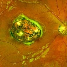
 Methotrexate Bubble following Intravitreal Injection for PVR
Sep 21 2022 by Zach Seim
Ultra-widefield fundus photograph of an 81 year old female with a Methotrexate bubble following an Intravitreal Injection for Proliferative Vitreoretinopathy. Patient has been presenting to the office for two week interval Methotrexate injections in her left eye. The image was taken prior to her eighth injection which revealed a residual Methotrexate bubble in her inferior retinal image. This patient was seeing "lots" of floaters, as well as having visual acuity of cc20/400 cc20/200 PH.
Photographer: Zach Seim
Imaging device: OPTOS California
Condition/keywords: bubble, fundus photograph, fundus photography, intravitreal injection, left eye, methotrexate, nasal retina, Optos, proliferative vitreoretinopathy (PVR), pseudocolor, ultra-wide field imaging
-
 Congenital Toxoplasmosis
Dec 18 2019 by Yoshihiro Yonekawa, MD, FASRS
Widefield fundus image of a teenage girl's right eye with an inactive congenital toxoplasmosis macular lesion. Her vision is 20/400 in this eye.
Photographer: Netanya Lerner, COA, Wills Eye Hospital/Mid Atlantic Retina
Imaging device: Optos California
Condition/keywords: congenital toxoplasmosis, pediatric retina
-
 Retinal Ischemia, Edema, and Hemorrhages on the Infero-Temporal Macula
Aug 26 2019 by Narciso F. Atienza, MD, MBA, FASRS, FPCS, FPAO.
47-year-old female who came in with blurring of vision of the right eye of 2 weeks duration. She is hypertensive with poor control, taking Amlodipine irregularly. Denies any cardiac problem non-diabetic. Vision upon presentation was 20/400 (OD), 20/20 (OS) colored fundus photo of the right eye showing areas of retinal ischemia, edema and hemorrhages on the infero-temporal macula extending to the arcade.
Photographer: Narciso F Atienza, Jr. MD, MBA
Imaging device: Topcon TRC
Condition/keywords: edema, hemorrhage, inferotemporal arcade, retinal ischemia
-
 Ruptured Macroaneurysm
May 22 2019 by Nichole Lewis
FA of a 91-year-old woman with a ruptured macroaneurysm, intraretinal hemorrhage and subretinal hemorrhage. VA 20/400.
Photographer: Nichole Lewis
Condition/keywords: intraretinal hemorrhage, ruptured macroaneurysm, subretinal hemorrhage
-
 Optos Picture With Speculum: Dislocated Natural Lens
Oct 9 2018 by John S. King, MD
55-year-old white female with history of pathologic myopia+, lattice (laser), SB OU (1990s), and dislocated natural lenses OU that had been watched for years. In the fellow eye she developed phacolytic glaucoma and a PPV, PPL was performed. Plan for both eyes are monitoring. I wanted to get a good picture of her lens today with the optos machine, as the other pics had artifact from the lower lid. It worked out well to use a speculum in the left eye. Vision cc is 20/400 J1+ OD and 20/40 J2 OS; aphakic OU; vitreous clear OD; dislocated lens OS (see pic); retinas attached.
Photographer: Maisee Yang
Imaging device: Optos California
Condition/keywords: dislocated crystalline lens, pathologic myopia, scleral buckle, staphyloma
-
 RAMA
RAMA
Jun 20 2016 by John S. King, MD
RAMA with 2 w co decreased vision; htn, afib using anticoag; light laser applied; 20/400.
Condition/keywords: ruptured macroaneurysm
 Loading…