-
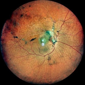
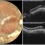
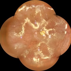
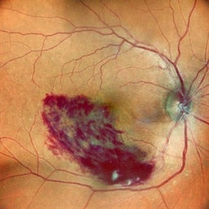
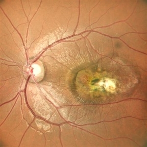
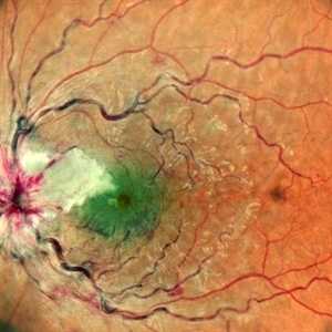
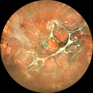
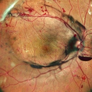
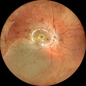
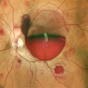
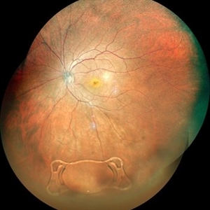
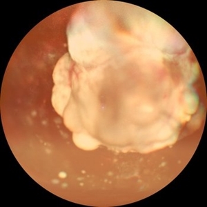
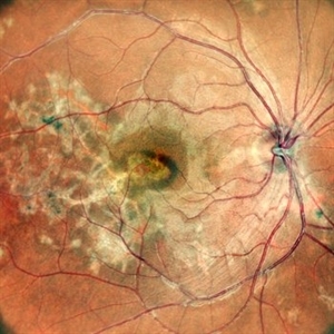
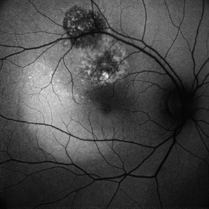
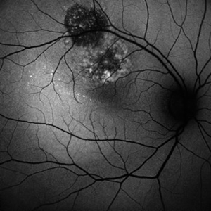
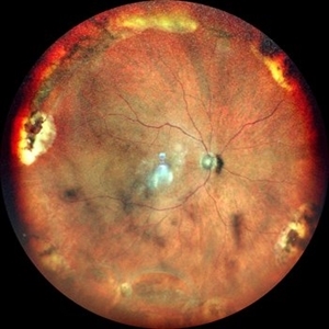
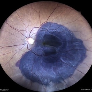
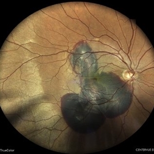
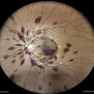
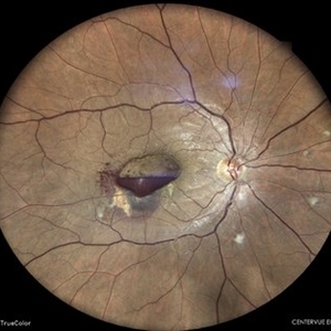
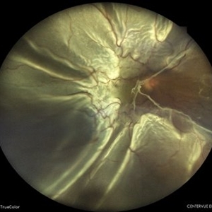
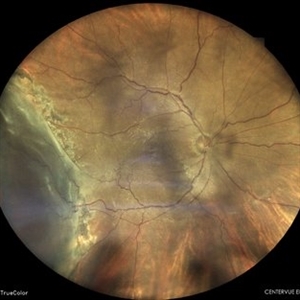
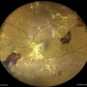
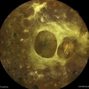
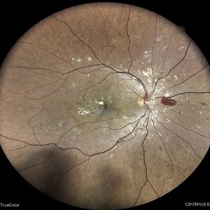
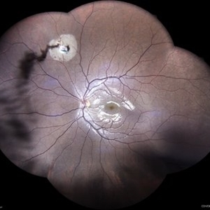
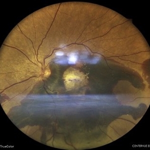
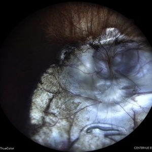
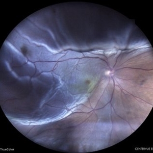
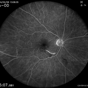
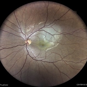
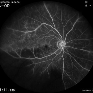
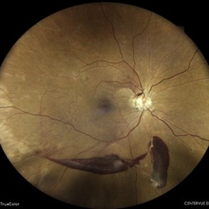
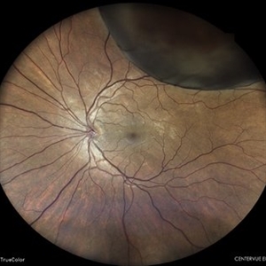
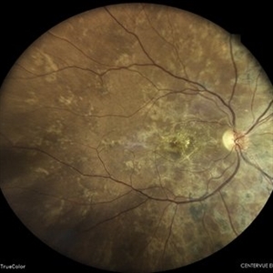
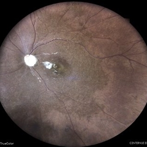
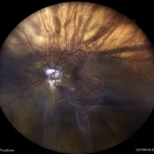
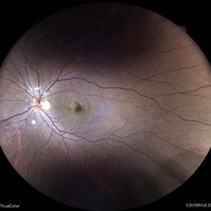
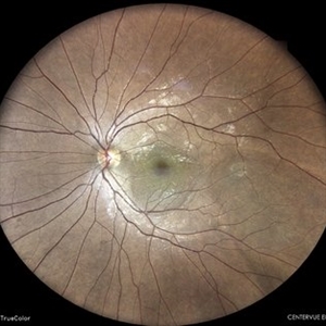
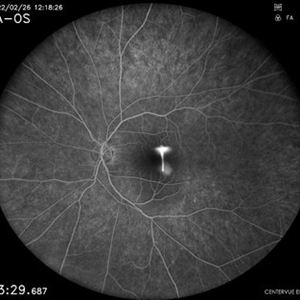
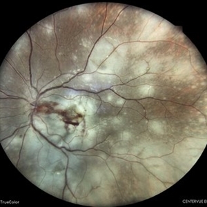
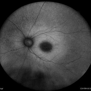
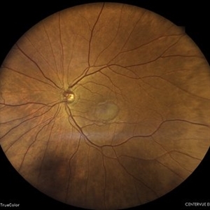
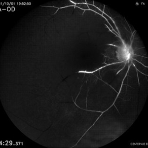
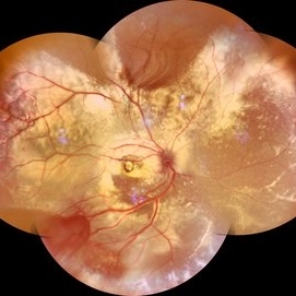 Coats' Disease Montage
Feb 5 2021 by Akansha Sharma
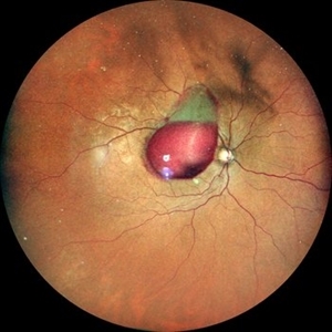
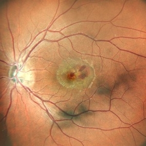
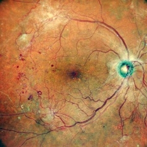
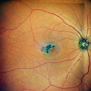
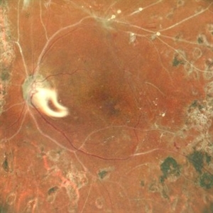
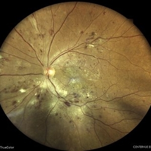
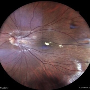
Fundus photograph of a 5-year-old male child who presented with unilateral diminution of vision since one month.
Photographer: Dr. Nivesh Gupta, M.S., Retina Foundation, Ahmedabad
Condition/keywords: angiomatosis retinae, Coats' disease, exudative detachment, subretinal exudates
-
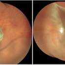
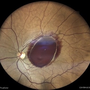
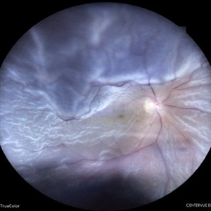
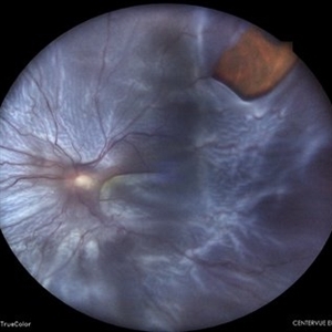
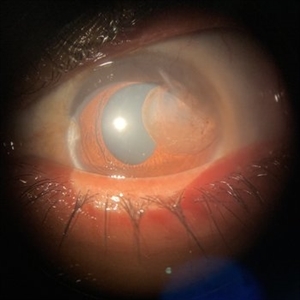 Traumatic Iris Cyst
Feb 24 2021 by Akansha Sharma
Diffuse illumination slit lamp image of a 30-year-old female patient with a traumatic iris cyst in left eye.
Photographer: Dr. Akansha Sharma - Retina Foundation, Ahmedabad
Condition/keywords: cyst, iris, trauma
-
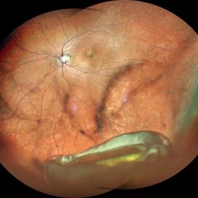
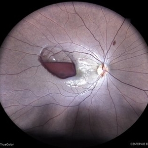
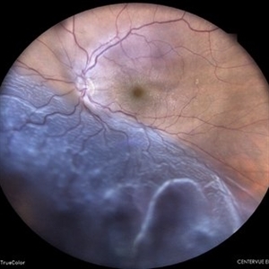
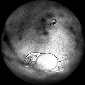 IOL in Vitreous Cavity Right Eye
May 22 2021 by Akansha Sharma
Fundus infra-red image of a 47-year-old male with IOL in vitreous cavity right eye.
Photographer: Dr. Akansha Sharma-Retina Foundation, Ahmedabad
Condition/keywords: intraocular lens (IOL)
-
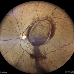
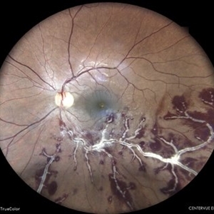
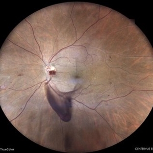
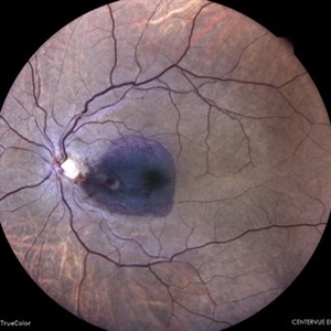
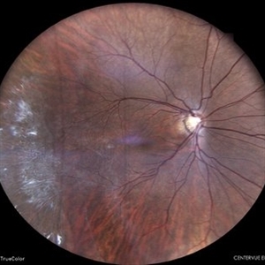
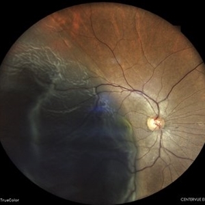
 Old Supero-Temporal Branch Retinal Vein Occlusion with Macular Hole Right Eye Fundus
May 23 2021 by Akansha Sharma
Fundus photograph of a 36-year-old female presented with supero-temporal branch retinal vein occlusion with macular hole in right eye.
Photographer: Dr. Akansha Sharma-Retina Foundation
Condition/keywords: branch retinal vein occlusion (BRVO), macular hole
-
 Old Supero-Temporal Branch Retinal Vein Occlusion with Macular Hole
May 23 2021 by Akansha Sharma
Fundus photograph of a 36-year-old female with supero-temporal branch retinal vein occlusion with macular hole in right eye.
Photographer: Dr. Akansha Sharma- Retina Foundation, Ahmedabad
Condition/keywords: branch retinal vein occlusion (BRVO), macular hole
-
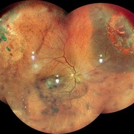
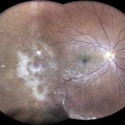
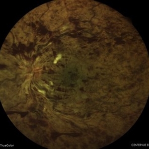
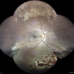
 Familial Exudative Vitreo-retinopathy
Jul 6 2021 by Akansha Sharma
Color photo montage of 21-year-old male with familial exudative vitreoretinopathy in an amblyopic eye.
Photographer: Dr. Akansha Sharma-Retina Foundation, Ahmedabad
Condition/keywords: familial exudative vitreoretinopathy (FEVR), retinal ischemia
-
 Familial Exudative Vitreo-retinopathy
Jul 6 2021 by Akansha Sharma
Infra-red capture of 5-year-old male with familial exudative vitro-retinopathy with disc pallor.
Photographer: Dr. Akansha Sharma-Retina Foundation, Ahmedabad
Condition/keywords: familial exudative vitreoretinopathy (FEVR), retinal ischemia
-
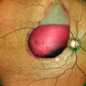
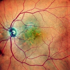
 Von Hippel-Lindau Syndrome
Jul 12 2021 by Akansha Sharma
Color photo montage of a 19-year-old female with Von-Hippel-Lindau syndrome.
Photographer: Dr. Akansha Sharma-Retina Foundation, Ahmedabad
Condition/keywords: angioma, Von Hippel-Lindau
-
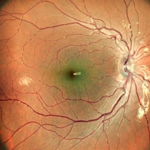
 Macular Pseudo-Hole in a Buckled Eye
Sep 10 2021 by Akansha Sharma
Fundus Photograph and Optical Coherence Tomography of left eye showing a macular pseudo-hole in a buckled eye.
Photographer: Dr. Akansha Sharma-Retina Foundation, Ahmedabad
Condition/keywords: macular hole, macular pseudohole, scleral buckle
-
 Giant Retinal Tear with Retina Detachment, Both Eyes
Sep 15 2021 by Akansha Sharma
Fundus photograph of a 13 year-old female with giant retinal tear and retinal detachment in both eyes.
Photographer: Dr. Akansha Sharma-Retina Foundation, Ahmedabad
Condition/keywords: giant retinal tear, proliferative vitreoretinopathy (PVR)
-
 IOL Drop
Dec 20 2021 by Akansha Sharma
Wide-Field Fundus Photograph of a 66-year-old female patient with a dropped IOL and Capsule.
Photographer: Dr. Akansha Sharma-Retina Foundation, Ahmedabad
Condition/keywords: dislocated posterior chamber intraocular lens (PCIOL), dropped capsular IOL bag complex
-
 Pre-Retinal Hemorrhage
Jan 26 2022 by Akansha Sharma
Wide field fundus photograph of a 36 year-old male with a macular pre-retinal hemorrhage.
Photographer: Dr. Akansha Sharma
Condition/keywords: color wide field, macular pre-retinal hemorrhage, ultra-wide field imaging
-
 Pre-retinal Hemorrhage
Jan 26 2022 by Akansha Sharma
Wide-field fundus photograph of a 36 year-old male with a macular pre-retinal hemorrhage.
Photographer: Dr. Akansha Sharma - Retina Foundation, Ahmedabad
Condition/keywords: color wide field, macular pre-retinal hemorrhage
-
 Status Post Scleral Buckling
Jan 26 2022 by Akansha Sharma
Wide field fundus photograph of a 55-year-old male with a scleral buckle in situ post scleral buckling performed 15 years back.
Photographer: Dr. Akansha Sharma - Retina Foundation, Ahmedabad
Condition/keywords: color wide field, encircling scleral buckle
-
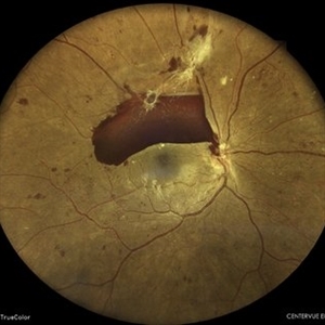
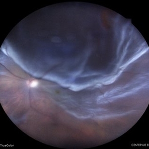
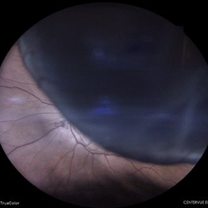
 Retinal Detachment
Feb 2 2022 by Akansha Sharma
Montage of right eye of a 31-year-old male with a sub-retinal fluid pocket around a horseshoe tear, status post laser barrage, status post blunt trauma.
Photographer: Dr. Akansha Sharma-Retina Foundation, Ahmedabad
Condition/keywords: subretinal fluid
-
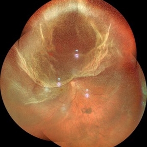
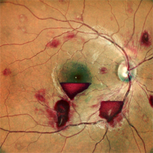
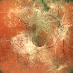
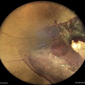
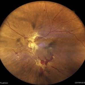
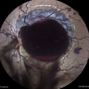
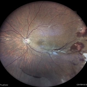
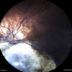
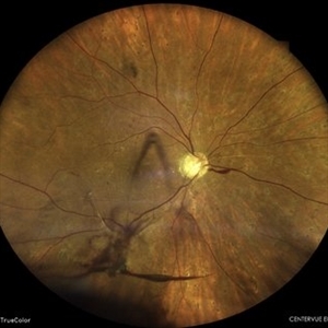
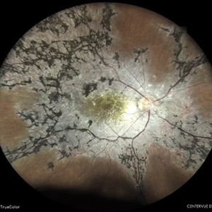
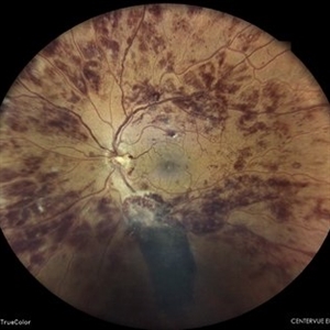
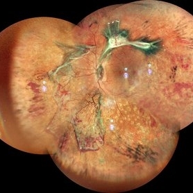 TRACTIONAL RETINAL DETACHMENT IN A CASE OF VASCULITIS
Mar 14 2022 by Akansha Sharma
MONTAGE OF A 27 YEAR OLD MALE WITH TREACTIONAL RETINAL DETACHMENT IN A CASE OF VASCULITIS
Photographer: Dr. Akansha Sharma-Retina Foundation, Ahmedabad
Condition/keywords: pan-retinal photocoagulation (PRP), tractional retinal detachment, VASCULITIS
-
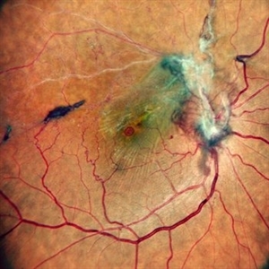
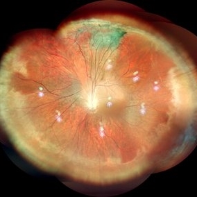
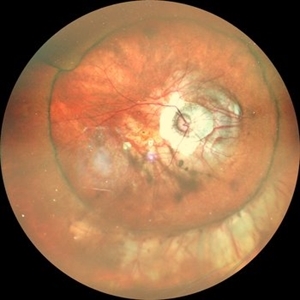
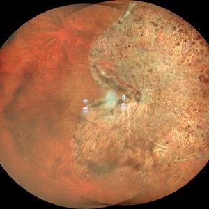
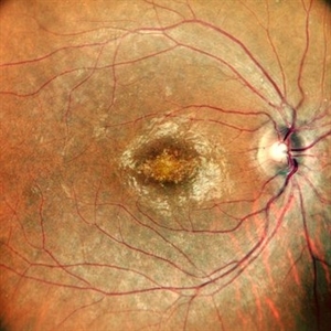
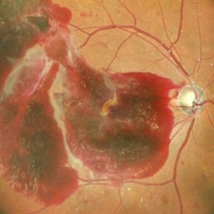
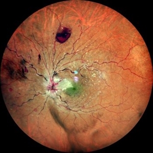
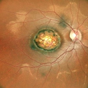
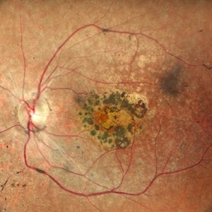
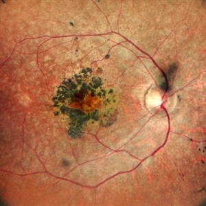
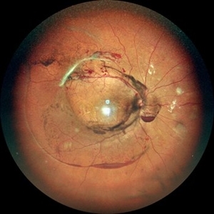
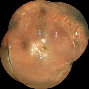
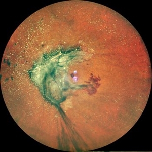
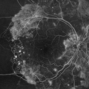
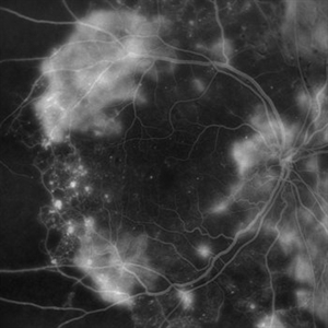
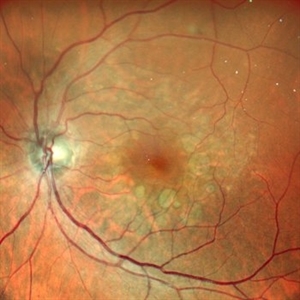
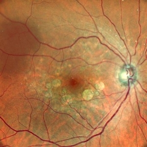
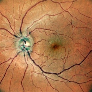
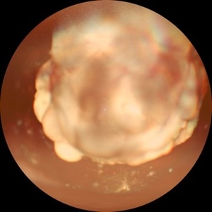
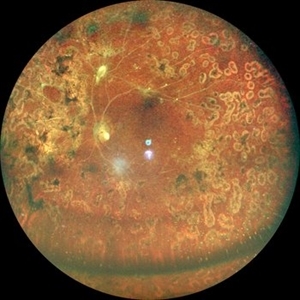
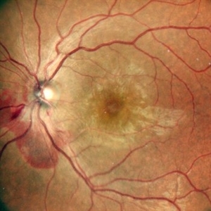
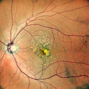
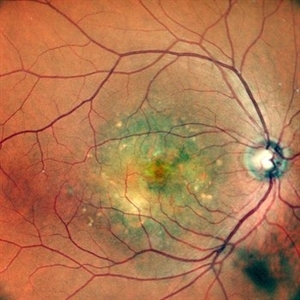
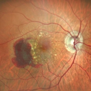
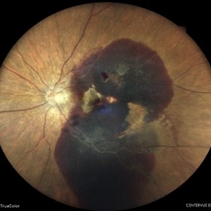
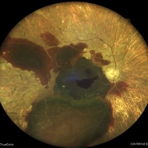
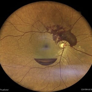
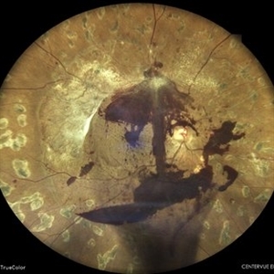
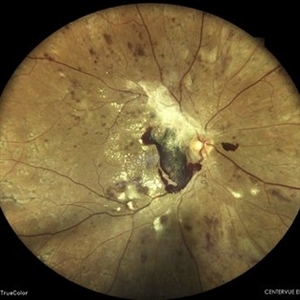
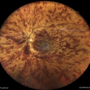
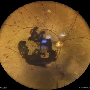
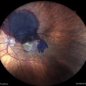
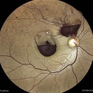
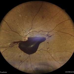
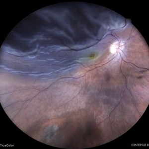
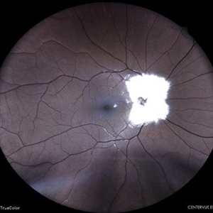
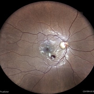
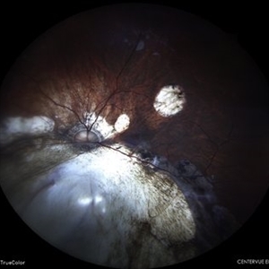
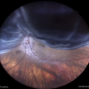
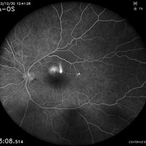
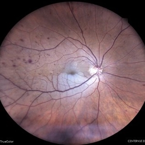
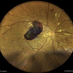
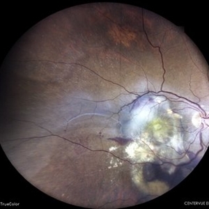
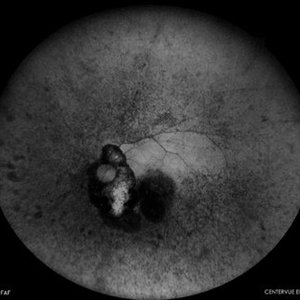
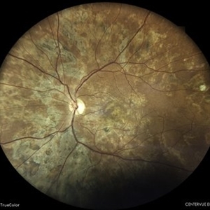
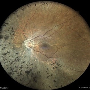
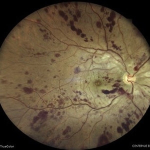
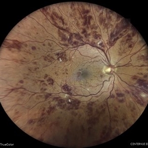
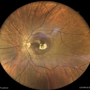
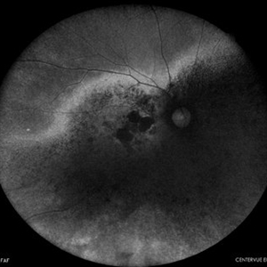
 COATS' DISEASE
Mar 14 2022 by Akansha Sharma
MONTAGE OF A 11 YEAR OLD MALE WITH COATS' DISEASE
Photographer: Dr. Akansha Sharma-Retina Foundation, Ahmedabad
Condition/keywords: Coats' disease, exudates, telangiectatic vessels
-
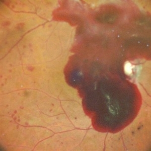
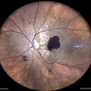
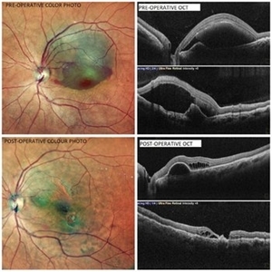 Subretinal Bleed
Jul 12 2022 by Akansha Sharma
A 60 YEAR OLD FEMALE PRESENTED WITH COUNTING FINGERS VISION FROM A SUB-RETINAL HEMORRHAGE AT THE MACULA. OCT SHOWS VARIABLE SUB -RETINAL FLUID. PARS PLANA VITRECTOMY WITH DRAINAGE OF THE SUB-RETINAL BLOOD WAS PERFORMED. POST-OPERATIVE OCT SHOWS NO SUB-RETINAL FLUID WITH VARIABLE OUTER RETINAL CYSTIC CHANGES AND VISUAL ACUITY IMPROVING TO 20/120.
Photographer: Dr. Akansha Sharma-Retina Foundation, Ahmedabad
Condition/keywords: subretinal hemorrhage, subretinal blood
-
 Subretinal Bleed
Jul 12 2022 by Akansha Sharma
73 year old diabetic and hypertensive female presented with sub-retinal hemorrhage for which she was operated with pars-plana vitrectomy with intra-vitreal anti-VEGF
Photographer: Dr. Akansha Sharma-Retina Foundation, Ahmedabad
Condition/keywords: subretinal hemorrhage, subretinal blood
-
 Hypotonous Maculopathy
Jul 12 2022 by Akansha Sharma
FUNDUS PHOTOGRAPH OF A 37 YEAR OLD MALE WITH HISTORY OF BLUNT TRAUMA WITH TENNIS BALL PRESENTING WITH HYPOTONOUS MACULOPATHY
Photographer: Dr. Akansha Sharma-Retina Foundation, Ahmedabad
Condition/keywords: hypotonous retinopathy
-
 SUB RETINAL HEMORRHAGE
Oct 11 2022 by Akansha Sharma
COLUR FUNDUS PHOTOGRAPH OF A 28 YEAR OLD MALE WITH SUB RETINAL NEOVASCULAR MEMBRANE
Photographer: Dr. Akansha Sharma-Retina Foundation, Ahmedabad
Condition/keywords: choroidal neovascularization (CNV), subretinal hemorrhage
-
 STATUS POST PAN-RETINAL PHOTOCOAGULATION
Oct 11 2022 by Akansha Sharma
COLOUR FUNDUS PHOTO OF A 71 YEAR OLD MALE WITH SCARRING POST PAN-RETINAL PHOTOCOAGULATION
Photographer: Dr. Akansha Sharma-Retina Foundation, Ahmedabad
Condition/keywords: laser photocoagulation, pan-retinal photocoagulation (PRP)
-
 CENTRAL RETINAL VEIN WITH CENTRAL RETINAL ARTERY OCCLUSION
Oct 11 2022 by Akansha Sharma
OCT OF 47 YEAR OLD MALE WITH CENTRAL RETINAL VEIN WITH CENTRAL RETINAL ARTERY OCCLUSION
Photographer: Dr. Akansha Sharma-Retina Foundation, Ahmedabad
Condition/keywords: central retinal artery occlusion (CRAO), central retinal vein occlusion (CRVO)
-
 BRANCH RETINAL VEIN OCCLUSION
Oct 11 2022 by Akansha Sharma
COLOUR FUNDUS PHOTOGRAPH OF A 59 YEAR OLD FEMALE WITH BRACH RETINAL VEIN OCCLUSION
Photographer: Dr. Akansha Sharma-Retina Foundation, Ahmedabad
Condition/keywords: BRVO
-
 MACULAR DYSTROPHY
Oct 11 2022 by Akansha Sharma
COLOUR FUNDUS PHOTOGRAPH OF 20 YEAR OLD MALE WITH MACULAR DYSTROPHY
Photographer: Dr. Akansha Sharma-Retina Foundation, Ahmedabad
Condition/keywords: Macular Dystrophy
-
 SUB-HYALOID HEMORRHAGE
Oct 11 2022 by Akansha Sharma
FUNDUS PHOTOGRAPH OF 63 YEAR OLD MALE WITH SUB-HYALOID HEMORRHAGE IN A CASE OF PROLIFERATIVE DIABETIC RETINOPATHY
Photographer: Dr. Akansha Sharma-Retina Foundation, Ahmedabad
Condition/keywords: proliferative diabetic retinopathy (PDR), SHH
-
 SIDEROSIS BULBI
Oct 11 2022 by Akansha Sharma
FUNDUS PHOTOGRAPH OF A 42 YEAR OLD MALE WITH IRON FOREIGN BODY IN FOR 13 YEARS WHICH LEAD TO SIDEROSIS BULBI
Photographer: Dr. Akansha Sharma-Retina Foundation, Ahmedabad
Condition/keywords: siderosis
-
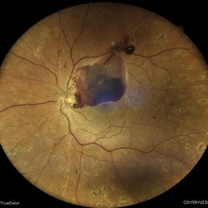
 SCARRED CHOROIDAL NEOVASCULAR MEMBRANE
Oct 15 2022 by Akansha Sharma
FUNDUS PHOTOGRAPH OF AN 18 YEAR OLD MALE WITH A SCARRED CHOROIDAL NEOVASCULAR MEMBRANE
Photographer: Dr. Akansha Sharma-Retina Foundation, Ahmedabad
Condition/keywords: choroidal neovascularization (CNV), CNVM
-
 SUB-HYALOID HEMORRHAGE
Oct 15 2022 by Akansha Sharma
COLOUR FUNDUS PHOTOGRAPH OF A 46 YEAR OLD DIABETIC MALE WITH SUBHYALOID HEMORRHAGE IN A CASE OF PROLIFERATIVE DIABETIC RETINOPATHY
Photographer: Dr. Akansha Sharma-Retina Foundation, Ahmedabad
Condition/keywords: subhyaloid hemorrhage
-
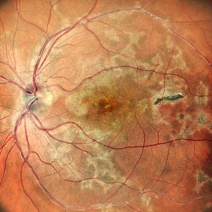
 HEREDITARY MACULAR DEGENERATION
Oct 15 2022 by Akansha Sharma
COLOUR FUNDUS PHOTOGRAPH OF A 43 YEAR OLD MALE PATIENT WITH HEREDITARY MACULAR DEGENERATION
Photographer: Dr. Akansha Sharma-Retina Foundation, Ahmedabad
Condition/keywords: hereditary retinal degeneration
-
 HEREDITARY MACULAR DEGENERATION
Oct 15 2022 by Akansha Sharma
COLOUR FUNDUS PHOTOGRAPH OF A 43 YEAR OLD MALE PATIENT WITH HEREDITARY MACULAR DEGENERATION
Photographer: Dr. Akansha Sharma-Retina Foundation, Ahmedabad
Condition/keywords: hereditary retinal degeneration
-
 ANAEMIC RETINOPATHY
Oct 15 2022 by Akansha Sharma
COLOUR FUNDUS PHOTOGRAPH OF A 24 YEAR OLD FEMALE WITH ANAEMIC RETINOPATHY AND MACULAR EDEMA
Photographer: Dr. Akansha Sharma-Retina Foundation, Ahmedabad
Condition/keywords: anaemic retinopathy
-
 ANAEMIC RETINOPATHY
Oct 15 2022 by Akansha Sharma
COLOUR FUNDUS PHOTOGRAPH OF A 24 YEAR OLD FEMALE WITH ANAEMIC RETINOPATHY AND MACULAR EDEMA
Photographer: Dr. Akansha Sharma-Retina Foundation, Ahmedabad
Condition/keywords: anaemic retinopathy
-
 ANAEMIC RETINOPATHY
Oct 15 2022 by Akansha Sharma
COLOUR FUNDUS PHOTOGRAPH OF A 24 YEAR OLD FEMALE WITH ANAEMIC RETINOPATHY AND MACULAR EDEMA
Photographer: Dr. Akansha Sharma-Retina Foundation, Ahmedabad
Condition/keywords: anaemic retinopathy
-
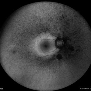
 MACULAR COLOBOMA
Oct 15 2022 by Akansha Sharma
COLOUR FUNDUS PHOTOGRAPH OF A 32 YEAR OLD MALE WITH MACULAR COLOBOMA
Photographer: Dr. Akansha Sharma-Retina Foundation, Ahmedabad
Condition/keywords: coloboma of macula
-
 MACULAR COLOBOMA
Oct 15 2022 by Akansha Sharma
COLOUR FUNDUS PHOTOGRAPH OF A 32 YEAR OLD MALE WITH MACULAR COLOBOMA
Photographer: Dr. Akansha Sharma-Retina Foundation, Ahmedabad
Condition/keywords: coloboma of macula
-
 TRACTIONAL RETINAL DETACHMENT
Oct 15 2022 by Akansha Sharma
COLOUR FUNDUS PHOTOGRAPH OF A 71 YEAR OLD MALE WITH A TRACTIONAL RETINAL DETACHMENT
Photographer: Dr. Akansha Sharma-Retina Foundation, Ahmedabad
Condition/keywords: tractional retinal detachment, TRD
-
 RETINITIS PIGMENTOSA
Oct 15 2022 by Akansha Sharma
COLOUR FUNDUS PHOTOGRAPH OF A 30 YEAR OLD MALE WITH RETINITIS PIGMENTOSA
Photographer: Dr. Akansha Sharma-Retina Foundation, Ahmedabad
Condition/keywords: retinitis pigmentosa (RP) dystrophy, RP variant
-
 RETINITIS PIGMENTOSA
Oct 15 2022 by Akansha Sharma
COLOUR FUNDUS PHOTOGRAPH OF A 30 YEAR OLD MALE WITH RETINITIS PIGMENTOSA
Photographer: Dr. Akansha Sharma-Retina Foundation, Ahmedabad
Condition/keywords: retinitis pigmentosa (RP) dystrophy, RP variant
-
 SUBHYALOID HEMORRHAGE
Oct 15 2022 by Akansha Sharma
COLOUR FUNDUS PHOTOGRAPH OF A 23 YEAR OLD TYPE-1 DIABETIC WITH SUBHYALOID HEMORRHAGE IN A CASE OF PROLIFERATIVE DIABETIC RETINOPATHY
Photographer: Dr. Akansha Sharma-Retina Foundation, Ahmedabad
Condition/keywords: SHH
-
 SUB-HYALOID HEMORRHAGE
Oct 15 2022 by Akansha Sharma
COLOUR FUNDUS PHOTOGRAPH OF A 23 YEAR OLD TYPE-1 DIABETIC WITH SUBHYALOID HEMORRHAGE IN A CASE OF PROLIFERATIVE DIABETIC RETINOPATHY
Photographer: Dr. Akansha Sharma-Retina Foundation, Ahmedabad
Condition/keywords: SHH
-
 RHEGMATOGENOUS RETINAL DETACHMENT
Oct 19 2022 by Akansha Sharma
FUNDUS PHOTOGRAPH OF A 40 YEAR OLD MALE WITH RHEGMATOGENOUS RETINAL DETACHMENT
Photographer: Dr. Akansha Sharma-Retina Foundation, Ahmedabad
Condition/keywords: rhegmatogenous retinal detachment
-
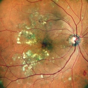 CIRCINATE RETINOPATHY
Oct 19 2022 by Akansha Sharma
COLOUR FUNDUS PHOTOGRAPH OF A 51 YEAR OLD MALE WITH DIABETIC MACULOPATHY
Photographer: Dr. Akansha Sharma-Retina Foundation, Ahmedabad
Condition/keywords: circinate retinopathy, diabetic macular edema, diabetic maculopathy
-
 RHEGMATOGENOUS RETINAL DETACHMENT
Oct 19 2022 by Akansha Sharma
WIDE-FIELD COLOUR FUNDUS PHOTOGRAPH OF AN 8 YEAR OLD MALE CHILD WITH RHEGMATOGENOUS RETINAL DETACHMENT
Photographer: Dr. Akansha Sharma-Retina Foundation, Ahmedabad
Condition/keywords: Retinal Detachment, rhegmatogenous retinal detachment
-
 AMBLYOPIA
Oct 19 2022 by Akansha Sharma
COLOUR FUNDUS PHOTOGRAPH OF A 12 YEAR OLD MALE CHILD WITH HIGH REFRACTIVE ERROR WITH AMBLYOPIA
Photographer: Dr. Akansha Sharma-Retina Foundation, Ahmedabad
Condition/keywords: amblyopia
-
 SILICONE OIL FILLED VITRECTOMISED EYE STATUS POST PARS PLANA VITRECTOMY WITH SILICONE OIL INFUSION IN A CASE OF RHEGMATOGENOUS RETINAL DETACHMENT
Oct 19 2022 by Akansha Sharma
COLOUR FUNDUS MONTAGE OF A 11 YEAR OLD MALE WITH SILICONE OIL FILLED VITRECTOMISED EYE STATUS POST PARS PLANA VITRECTOMY WITH SILICONE OIL INFUSION IN A CASE OF RHEGMATOGENOUS RETINAL DETACHMENT
Photographer: Dr. Akansha Sharma-Retina Foundation, Ahmedabad
Condition/keywords: retina surgery, rhegmatogenous retinal detachment, silicone oil
-
 SUB HYALOID HEMORRHAGE
Oct 20 2022 by Akansha Sharma
COLOUR FUNDUS PHOTOGRAPH OF A 23 YEAR OLD MALE WITH SUB HYALOID HEMORRHAGE IN A CASE OF ANAEMIC RETINOPATHY
Photographer: Dr. Akansha Sharma-Retina Foundation, Ahmedabad
Condition/keywords: anaemic retinopathy, preretinal hemorrhage, subhyaloid hemorrhage
-
 SUB HYALOID HEMORRHAGE
Oct 20 2022 by Akansha Sharma
COLOUR FUNDUS PHOTO OF A 23 YEAR OLD MALE WITH SUBHYALOID HEMORRHAGE IN A CASE OF ANAEMIC RETINOPATHY
Photographer: Dr. Akansha Sharma-Retina Foundation, Ahmedabad
Condition/keywords: anaemic retinopathy, preretinal hemorrhage, subhyaloid hemorrhage
-
 SUBHYALOID HEMORRHAGE
Oct 20 2022 by Akansha Sharma
AUTOFLUORESCENCE IMAGE OF A 23 YEAR OLD MALE WITH SUBHYALOID HEMORRHAGE IN A CASE OF ANAEMIC RETINOPATHY
Photographer: Dr. Akansha Sharma-Retina Foundation, Ahmedabad
Condition/keywords: anaemic retinopathy, preretinal hemorrhage, subhyaloid hemorrhage
-
 SUB-HYALOID HEMORRHAGE
Oct 20 2022 by Akansha Sharma
RETRO IMAGE OF A 23 YEAR OLD MALE WITH SUBHYALOID HEMORRHAGE IN A CASE OF ANAEMIC RETINOPATHY
Photographer: Dr. Akansha Sharma-Retina Foundation, Ahmedabad
Condition/keywords: anaemic retinopathy, preretinal hemorrhage, subhyaloid hemorrhage
-
 TRACTIONAL RETINAL DETACHMENT
Oct 31 2022 by Akansha Sharma
COLOUR FUNDUS WIDE-FIELD PHOTO OF A 50 YEAR OLD DIABETIC MALE WITH TRACTIONAL RETINAL DETACHMENT, SUB-HYALOID HEMORRHAGR AND ASTEROID HYALOSIS IN A CASE OF PROLIFERATIVE DIABETIC RETINOPATHY
Photographer: Dr. Akansha Sharma-Retina Foundation, Ahmedabad
Condition/keywords: asteroid hyalosis, florid type PDR, proliferative diabetic retinopathy (PDR), SHH, subhyaloid hemorrhage, TRD
-
 IOL DROP
Oct 31 2022 by Akansha Sharma
COLOUR FUNDUS MONTAGE OF A 73 YEAR OLD MALE WITH IOL IN VITREOUS CAVITY
Photographer: Dr. Akansha Sharma-Retina Foundation, Ahmedabad
Condition/keywords: dislocated intraocular lens (IOL), dislocated posterior chamber intraocular lens (PCIOL)
-
 IOL DROP
Oct 31 2022 by Akansha Sharma
RETRO IMAGE OF A 73 YEAR OLD MALE WITH IOL IN VITREOUS CAVITY
Photographer: Dr. Akansha Sharma-Retina Foundation, Ahmedabad
Condition/keywords: dislocated intraocular lens (IOL), dislocated posterior chamber intraocular lens (PCIOL)
-
 PROLIFERATIVE DIABETIC RETINOPATHY
Oct 31 2022 by Akansha Sharma
EARLY PHASE FLUORESCEIN ANGIOGRAPHY OF A 50 YEAR OLD MALE WITH PROLIFERATIVE DIABETIC RETINOPATHY
Photographer: Dr. Akansha Sharma-Retina Foundation, Ahmedabad
Condition/keywords: florid type PDR, proliferative diabetic retinopathy (PDR)
-
 PROLIFERATIVE DIABETIC RETINOPATHY
Oct 31 2022 by Akansha Sharma
LATE PHASE FLUORESCEIN ANGIOGRAPHY OF A 50 YEAR OLD MALE WITH PROLIFERATIVE DIABETIC RETINOPATHY
Photographer: Dr. Akansha Sharma-Retina Foundation, Ahmedabad
Condition/keywords: florid type PDR, proliferative diabetic retinopathy (PDR)
-
 PROLIFERATIVE DIABETIC RETINOPATHY
Oct 31 2022 by Akansha Sharma
COLOUR FUNDUS PHOTOGRAPH OF A 50 YEAR OLD MALE WITH PROLIFERATIVE DIABETIC RETINOPATHY
Photographer: Dr. Akansha Sharma-Retina Foundation, Ahmedabad
Condition/keywords: florid type PDR, proliferative diabetic retinopathy (PDR)
-
 HEREDITARY MACULAR DEGENERATION WITH ETHAMBUTOL TOXICITY
Nov 1 2022 by Akansha Sharma
COLOUR FUNDUS PHOTOGRAPH OF A 45 YEAR OLD MALE WITH HEREDITARY MACULAR DEGENERATION WITH ETHAMBUTOL TOXICITY
Photographer: Dr. Akansha Sharma-Retina Foundation, Ahmedabad
Condition/keywords: drug toxicity, hereditary retinal degeneration, toxic optic neuropathy
-
 JUXTAFOVEAL TELANGIECTASIS
Nov 1 2022 by Akansha Sharma
COLOUR FUNDUS PHOTOGRAPH OF 72 YEAR OLD FEMALE WITH JUXTAFOVEAL TELANGIECTASIS
Photographer: Dr. Akansha Sharma-Retina Foundation, Ahmedabad
Condition/keywords: juxtafoveal telangiectasis, JXT
-
 JUXTAFOVEAL TELANGIECTASIS
Nov 1 2022 by Akansha Sharma
COLOUR FUNDUS PHOTOGRAPH OF 72 YEAR OLD FEMALE WITH JUXTAFOVEAL TELANGIECTASIS
Photographer: Dr. Akansha Sharma-Retina Foundation, Ahmedabad
Condition/keywords: juxtafoveal telangiectasis, JXT
-
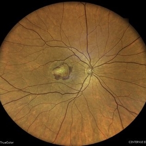
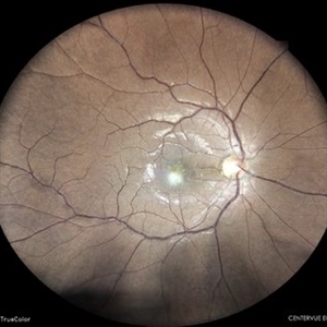
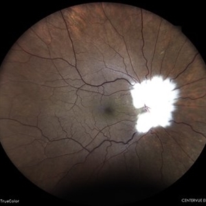
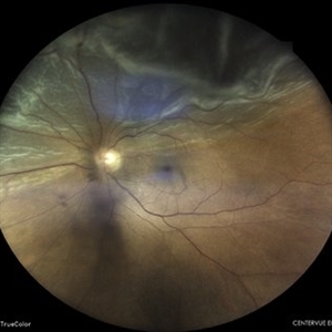
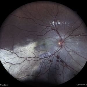
 DISC EDEMA
Nov 1 2022 by Akansha Sharma
COLOUR FUNDUS PHOTOGRAPH OF A 33 YEAR OLD FEMALE WITH DISC EDEMA IN LEFT EYE
Photographer: Dr. Akansha Sharma-Retina Foundation, Ahmedabad
Condition/keywords: disc edema
-
 TRACTIONAL RETINAL DETACHMENT
Nov 1 2022 by Akansha Sharma
COLOUR FUNDUS PHOTOGRAPH OF A 62 YEAR OLD FEMALE WITH TRACTIONAL RETINAL DETACHMENT
Photographer: Dr. Akansha Sharma-Retina Foundation, Ahmedabad
Condition/keywords: tractional retinal detachment, TRD
-
 TRACTIONAL RETINAL DETACHMENT
Nov 1 2022 by Akansha Sharma
COLOUR FUNDUS PHOTOGRAPH OF A 62 YEAR OLD FEMALE WITH TRACTIONAL RETINAL DETACHMENT
Photographer: Dr. Akansha Sharma-Retina Foundation, Ahmedabad
Condition/keywords: tractional retinal detachment, TRD
-
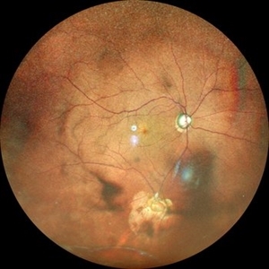
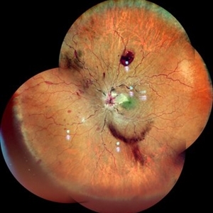
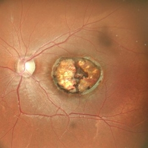
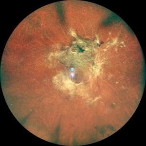
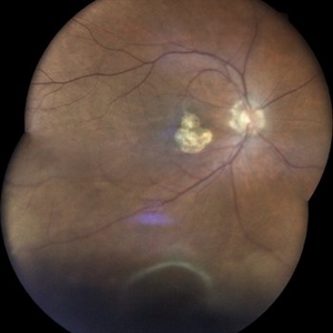
 Retinoblastoma
Nov 6 2022 by Akansha Sharma
Wide-field color fundus photograph of a 2-month old female with retinoblastoma.
Photographer: Dr. Akansha Sharma-Retina Foundation, Ahmedabad
Condition/keywords: RB gene mutation, retinoblastoma
-
 Retinoblastoma
Nov 6 2022 by Akansha Sharma
WIDE-FIELD COLOUR FUNDUS PHOTOGRAPH OF A 2 MONTH OLD FEMALE WITH RETINOBLASTOMA
Photographer: Dr. Akansha Sharma-Retina Foundation, Ahmedabad
Condition/keywords: RB gene mutation, retinoblastoma
-
 SUB-RETINAL NEOVASCULAR MEMBRANE
Nov 21 2022 by Akansha Sharma
COLOUR FUNDUS PHOTO OF A 79 YEAR OLD MALE PATIENT WITH SUBRETINAL NEOVASCULAR MEMBRANE
Photographer: Dr. Akansha Sharma-Retina Foundation, Ahmedabad
Condition/keywords: choroidal neovascular membrane (CNVM), CNVM, subretinal neovascularization (SRNV)
-
 TRACTIONAL RETINAL DETACHMENT
Nov 21 2022 by Akansha Sharma
WIDE-FIELD COLOUR FUNDUS PHOTOGRAPH OF A 46 YEAR OLD MALE WITH TRACTIONAL RETINAL DETACHMENT IN A CASE OF PROLIFERATIVE DIABETIC RETINOPATHY
Photographer: Dr. Akansha Sharma-Retina Foundation, Ahmedabad
Condition/keywords: proliferative diabetic retinopathy (PDR), tractional retinal detachment, TRD
-
 SILICONE OIL FILLED VITRECTOMISED EYE IN A CASE OF TRACTIONAL RETINAL DETACHMENT
Nov 21 2022 by Akansha Sharma
WIDE-FIELD COLOUR FUNDUS PHOTOGRAPH OF A 46 YEAR OLD MALE WITH SILICONE OIL FILLED VITRECTOMISED EYE OPERATED FOR TRACTIONAL RETINAL DETACHMENT IN A CASE OF PROLIFERATIVE DIABETIC RETINOPATHY
Photographer: Dr. Akansha Sharma-Retina Foundation, Ahmedabad
Condition/keywords: proliferative diabetic retinopathy (PDR), tractional retinal detachment, TRD
-
 TRACTIONAL RETINAL DETACHMENT
Nov 21 2022 by Akansha Sharma
WIDE-FIELD COLOUR FUNDUS PHOTOGRAPH OF A 46 YEAR OLD MALE WITH TRACTIONAL RETINAL DETACHMENT IN A CASE OF PROLIFERATIVE DIABETIC RETINOPATHY
Photographer: Dr. Akansha Sharma-Retina Foundation, Ahmedabad
Condition/keywords: proliferative diabetic retinopathy (PDR), tractional retinal detachment, TRD
-
 HEALED MULTIFOCAL CHOROIDITIS
Nov 21 2022 by Akansha Sharma
COLOUR FUNDUS PHOTO OF A 32 YEAR OLD FEMALE WITH HEALED MULTIFOCAL CHOROIDITIS
Photographer: Dr. Akansha Sharma-Retina Foundation, Ahmedabad
Condition/keywords: multifocal choroiditis
-
 HEALED MULTIFOCAL CHOROIDITIS
Nov 21 2022 by Akansha Sharma
COLOUR FUNDUS PHOTO OF A 32 YEAR OLD FEMALE WITH HEALED MULTIFOCAL CHOROIDITIS
Photographer: Dr. Akansha Sharma-Retina Foundation, Ahmedabad
Condition/keywords: multifocal choroiditis
-
 BERLIN'S EDEMA
Nov 21 2022 by Akansha Sharma
COLOUR FUNDUS PHOTOGRAPH OF A 35 YEAR OLD MALE WITH BERLIN'S EDEMA STATUS POST FIRE-CRACKER INJURY
Photographer: Dr. Akansha Sharma-Retina Foundation, Ahmedabad
Condition/keywords: Berlin's edema, commotio retinae, firework injury
-
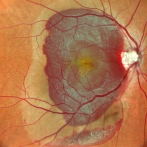
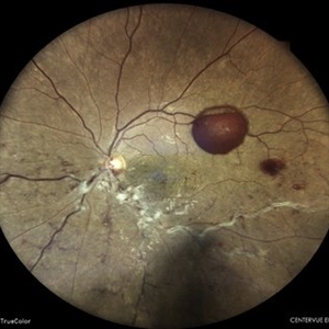
 CHOROIDAL HEMANGIOMA
Nov 21 2022 by Akansha Sharma
COLOU FUNDUS IMAGE OF A 32 YEAR OLD MALE WITH CHOROIDAL HEMANGIOMA WITH SUB RETINAL FLUID
Photographer: Dr. Akansha Sharma-Retina Foundation, Ahmedabad
Condition/keywords: choroidal hemangioma, subretinal fluid
-
 CHOROIDAL HEMANGIOMA
Nov 21 2022 by Akansha Sharma
RETRO IMAGE OF A 32 YEAR OLD MALE WITH CHOROIDAL HEMANGIOMA WITH SUB RETINAL FLUID
Photographer: Dr. Akansha Sharma-Retina Foundation, Ahmedabad
Condition/keywords: choroidal hemangioma, subretinal fluid
-
 CHOROIDAL HEMANGIOMA
Nov 21 2022 by Akansha Sharma
BLUE-AUTOFLUORESCENCE IMAGE OF A 32 YEAR OLD MALE WITH CHOROIDAL HEMANGIOMA WITH SUB RETINAL FLUID
Photographer: Dr. Akansha Sharma-Retina Foundation, Ahmedabad
Condition/keywords: choroidal hemangioma, subretinal fluid
-
 CHOROIDAL HEMANGIOMA
Nov 21 2022 by Akansha Sharma
GREEN-AUTOFLUORESCENCE IMAGE OF A 32 YEAR OLD MALE WITH CHOROIDAL HEMANGIOMA WITH SUB RETINAL FLUID
Photographer: Dr. Akansha Sharma-Retina Foundation, Ahmedabad
Condition/keywords: choroidal hemangioma, subretinal fluid
-
 DRY ARMD
Nov 21 2022 by Akansha Sharma
COLOUR FUNDUS PHOTOGRAPH OF A 57 YEAR OLD MALE WITH DRY AGE RELATED MACULAR DEGENERATION
Photographer: Dr. Akansha Sharma-Retina Foundation, Ahmedabad
Condition/keywords: drusen, dry age-related macular degeneration (dry AMD)
-
 SUB-RETINAL NEOVASCULAR MEMBRANE
Nov 21 2022 by Akansha Sharma
COLOUR FUNDUS PHOTOGRAPH OF A 57 YEAR OLD MALE WITH SUBRETINAL NEOVASCULAR MEMBRANE
Photographer: Dr. Akansha Sharma-Retina Foundation, Ahmedabad
Condition/keywords: choroidal neovascularization (CNV), CNVM, subretinal neovascularization (SRNV)
-
 RETINAL BREAKS
Nov 21 2022 by Akansha Sharma
COLOUR FUNDUS PHOTOGRAPH OF A 60 YEAR OLD MALE PATIENT WITH RETINAL BREAK STATUS POST SCLERAL BUCKLING 25 YEARS AGO
Photographer: Dr. Akansha Sharma-Retina Foundation, Ahmedabad
Condition/keywords: retinal break, scleral buckle
-
 WET AGE RELATED MACULAR DEGENERATION
Nov 21 2022 by Akansha Sharma
COLOUR FUNDUS PHOTOGRAPH OF A 71 YEAR OLD MALE WITH SUBRETINAL BLEED IN A CASE OF WET AGE RELATED MACULAR DEGENERATION
Photographer: Dr. Akansha Sharma-Retina Foundation, Ahmedabad
Condition/keywords: CNVM, subretinal neovascularization (SRNV), wet age-related macular degeneration (wet AMD)
-
 RESOLVING CENTRAL RETINAL VEIN OCCLUSION
Nov 21 2022 by Akansha Sharma
COLOUR FUNDUS PHOTOGRAPH OF A 49 YEAR OLD MALE WITH A RESOLVING CENTRAL RETINAL VEIN OCCLUSION WITH MACULAR EDEMA
Photographer: Dr. Akansha Sharma-Retina Foundation, Ahmedabad
Condition/keywords: central retinal vein occlusion (CRVO)
-
 Subretinal Hemorrhage
Feb 28 2023 by Akansha Sharma
Color fundus photograph of an 84-year old male with subretinal hemorrhage associated with areas of scarring.
Photographer: Dr. Urmil Shah, Dr. Denish Patel, Dr. Akansha Sharma, Bharati Eye Hospital, Ahmedabad, Gujarat
Condition/keywords: choroidal neovascularization (CNV), subretinal hemorrhage
-
 SUB-RETINAL HEMORRHAGE
Feb 28 2023 by Akansha Sharma
COLOUR FUNDUS PHOTOGRAPH OF AN 84 YEAR OLD MALE WITH SUB RETINAL BLEED
Photographer: Dr. Urmil Shah, Dr. Denish Patel, Dr. Akansha Sharma, Bharati Eye Hospital, Ahmedabad, Gujarat
Condition/keywords: choroidal neovascular membrane (CNVM), subretinal hemorrhage
-
 SUB HYALOID HEMORRHAGE
Feb 28 2023 by Akansha Sharma
COLOUR FUNDUS PHOTOGRAPH OF A 55 YEAR OLD FEMALE WITH SUB HYALOID HEMORRHAGE
Photographer: Dr. Urmil Shah, Dr. Denish Patel, Dr. Akansha Sharma, Bharati Eye Hospital, Ahmedabad, Gujarat
Condition/keywords: SHH
-
 SUB-RETINAL HEMORRHAGE WITH FOVEOLAR DETACHMENT
Feb 28 2023 by Akansha Sharma
COLOUR FUNDUS PHOTO OF A 62 YEAR OLD FEMALE WITH SUB-RETINAL BLEED OPERATED WITH BIMANUAL VITRECTOMY WITH SUBRETINAL TPA INJECTION WITH SILICONE OIL INFUSION
Photographer: Dr. Akansha Sharma, Dr. Urmil Shah, Dr. Denish Patel, Eye Hospital, Ahmedabad, Gujarat
Condition/keywords: subretinal hemorrhage
-
 ISCHAEMIC CENTRAL RETINAL VEIN OCCLUSION
Feb 28 2023 by Akansha Sharma
COLOUR FUNDUS PHOTOGRAPH OF A 54 YEAR OLD FEMALE WITH ISCHAEMIC CENTRAL RETINAL VEIN OCCLUSION WITH MACULAR EDEMA
Photographer: Dr. Urmil Shah, Dr. Denish Patel, Dr. Akansha Sharma, Bharati Eye Hospital, Ahmedabad, Gujarat
Condition/keywords: central retinal vein occlusion (CRVO), ischemic CRVO
-
 SUB-RETINAL HEMORRHAGE WITH CHOROIDAL RUPTURE
Feb 28 2023 by Akansha Sharma
COLOUR FUNDUS PHOTOGRAPH OF A 45 YEAR OLD MALE WITH SUBRETINAL HEMORRHAGE WITH CHOROIDAL RUPTURE
Photographer: Dr. Urmil Shah, Dr. Denish Patel, Dr. Akansha Sharma, Bharati Eye Hospital, Ahmedabad, Gujarat
Condition/keywords: subretinal hemorrhage
-
 SUB HYALOID HEMORRHAGE
Feb 28 2023 by Akansha Sharma
COLOUR FUNDUS PHOTOGRAPH OF A 40 YEAR OLD MALE WITH SUB HYALOID HEMORRHAGE
Photographer: Dr. Urmil Shah, Dr. Denish Patel, Dr. Akansha Sharma, Bharati Eye Hospital, Ahmedabad, Gujarat
Condition/keywords: subhyaloid hemorrhage
-
 SUB HYALOID HEMORRHAGE POST YAG LASER HYALOIDOTOMY
Feb 28 2023 by Akansha Sharma
COLOUR FUNDUS PHOTOGRAPH OF A 40 YEAR OLD MALE WITH SUB HYALOID HEMORRHAGE POST YAG LASER HYALOIDOTOMY
Photographer: Dr. Urmil Shah, Dr. Denish Patel, Dr. Akansha Sharma, Bharati Eye Hospital, Ahmedabad, Gujarat
Condition/keywords: SUBHYALOID HEMORRHAGE, YAG HYALOIDOTOMY
-
 SUB-RETINAL HEMORRHAGE WITH RETINITIS PIGMENTOSA
Feb 28 2023 by Akansha Sharma
COLOUR FUNDUS PHOTOGRAPH OF A 77 YEAR OLD MALE WITH SUBRETINAL HEMORRHAGE IN A CASE OF RETINITIS PIGMENTOSA
Photographer: Dr. Urmil Shah, Dr. Denish Patel, Dr. Akansha Sharma, Bharati Eye Hospital, Ahmedabad, Gujarat
Condition/keywords: retinitis pigmentosa, subretinal hemorrhage
-
 Anaemic Retinopathy
May 8 2023 by Akansha Sharma
Colour fundus photograph of a 38 year old male with anaemic retinoathy
Photographer: Dr. Urmil Shah, Dr. Denish Patel, Dr. Akansha Sharma, Bharati Eye Clinic, Ahmedabad, Gujarat
Condition/keywords: anaemic retinopathy, flame shaped retinal hemorrhage, subhyaloid hemorrhage
-
 Subretinal Neovascular Membrane
May 8 2023 by Akansha Sharma
Colour fundus photograph of a 60 year old female with subretinal blood suggestive of subretinal neovascular membrane
Photographer: Dr. Urmil Shah, Dr. Denish Patel, Dr. Akansha Sharma, Bharati Eye Clinic, Ahmedabad, Gujarat
Condition/keywords: CNVM, SRNVM, subretinal blood
-
 Familial Exudative Vitreo-Retinopathy
May 8 2023 by Akansha Sharma
Colour fundus photograph of a 28 year old female with familial exudative vitreoretinopathy
Photographer: Dr. Urmil Shah, Dr. Denish Patel, Dr. Akansha Sharma, Bharati Eye Clinic, Ahmedabad, Gujarat
Condition/keywords: familial exudative vitreoretinopathy (FEVR)
-
 Cytomegalovirus Retinitis
May 8 2023 by Akansha Sharma
Colour fundus photograph of a 37 year old male with cytomegalovirus retinitis with macular edema
Photographer: Dr. Urmil Shah, Dr. Denish Patel, Dr. Akansha Sharma, Bharati Eye Clinic, Ahmedabad, Gujarat
Condition/keywords: CMV retinitis
-
 Subhyaloid with Subretinal Bleed
May 8 2023 by Akansha Sharma
Colour fundus photograph of a 38 year old male with subhyaloid bleed with subretinal bleed
Photographer: Dr. Urmil Shah, Dr. Denish Patel, Dr. Akansha Sharma, Bharati Eye Clinic, Ahmedabad, Gujarat
Condition/keywords: SHH, subretinal neovascularization (SRNV)
-
 Proliferative Diabetic Retinopathy
May 8 2023 by Akansha Sharma
Colour fundus photograph of a 53 year old male with fibrovascular proliferations and subhyaloid hemorrhage in a case of proliferative diabetic retinopathy
Photographer: Dr. Urmil Shah, Dr. Denish Patel, Dr. Akansha Sharma, Bharati Eye Clinic, Ahmedabad, Gujarat
Condition/keywords: proliferative diabetic retinopathy (PDR)
-
 Subhyaloid Hemorrhage
May 8 2023 by Akansha Sharma
Colour fundus photograph of 62 year old hypertensive male with subhyaloid hemorrhage
Photographer: Dr. Urmil Shah, Dr. Denish Patel, Dr. Akansha Sharma, Bharati Eye Clinic, Ahmedabad, Gujarat
Condition/keywords: SHH, subhyaloid blood, subhyaloid hemorrhage
-
 Subretinal Neovascular Membrane
May 8 2023 by Akansha Sharma
Colour fundus photograph of a 81 year old female with subretinal neovascular memebrane
Photographer: Dr. Urmil Shah, Dr. Denish Patel, Dr. Akansha Sharma, Bharati Eye Clinic, Ahmedabad, Gujarat
Condition/keywords: CNVM, subretinal neovascularization (SRNV)
-
 Retinal Detachment
May 8 2023 by Akansha Sharma
Colour fundus photograph of a 52 year old female with retinal detachment
Photographer: Dr. Urmil Shah, Dr. Denish Patel, Dr. Akansha Sharma, Bharati Eye Clinic, Ahmedabad, Gujarat
Condition/keywords: RD, Retinal Detachment
-
 Lasered Profilerative Diabetic Retinopathy
May 8 2023 by Akansha Sharma
COLOUR FUNDUS PHOTOGRAPH OF A 42 YEAR OLD MALE WITH SUBHYALOID HEMORRHAGE IN A CASE OF LASERED PROLIFERATIVE DIABETIC RETINOPATHY
Photographer: Dr. Urmil Shah, Dr. Denish Patel, Dr. Akansha Sharma, Bharati Eye Clinic, Ahmedabad, Gujarat
Condition/keywords: PDR, proliferative diabetic retinopathy (PDR), subhyaloid hemorrhage
-
 Atypical Retinitis Pigmentosa
May 9 2023 by Akansha Sharma
Autofluorescence image of a 52 year old female with atypical retinitis pigmentosa
Photographer: Dr. Urmil Shah, Dr. Denish Patel, Dr. Akansha Sharma
Condition/keywords: RP variant
-
 Atypical Retinitis Pigmentosa
May 9 2023 by Akansha Sharma
Colour fundus photo of a 52 year old female with atypical retinitis pigmentosa
Photographer: Dr. Urmil Shah, Dr. Denish Patel, Dr. Akansha Sharma
Condition/keywords: RP variant
-
 Tractional retinal detachment with subhyaloid hemorrhage in a case of proliferative diabetic retinopathy
May 9 2023 by Akansha Sharma
COLOUR FUNDUS PHOTO OF A 42 YEAR OLD MALE WITH TRACTIONAL RETINAL DETACHMENT WITH SUBHYALOID HEMORRHAGE IN A CASE OF PROLIFERATIVE DIABETIC RETINOPATHY
Photographer: Dr. Urmil Shah, Dr. Denish Patel, Dr. Akansha Sharma
Condition/keywords: proliferative diabetic retinopathy (PDR), subhyaloid hemorrhage, tractional retinal detachment
-
 Ocular Toxoplasmosis
May 9 2023 by Akansha Sharma
Color fundus photograph of a 33 year old male with ocular toxoplasmosis
Photographer: Dr. Urmil Shah, Dr. Denish Patel, Dr. Akansha Sharma
Condition/keywords: toxoplasmosis
-
 Subretinal Neovascular Membrane
May 10 2023 by Akansha Sharma
COLOUR FUNDUS PHOTOGRAPH OF A 26 YEAR OLD MALE WITH SUBRETINAL BLEED WITH SUBRETINAL HYPERREFLECTIVE MATERIAL SUGGESTIVE OF SUBRETINAL NEOVASCULAR MEMBRANE
Photographer: Dr. Denish Patel, Dr. Akansha Sharma, Dr. Urmil Shah, Bharati Eye Hospital
Condition/keywords: CNVM, SRNVM, subretinal hemorrhage
-
 CENTRAL SEROUS RETINOPATHY
May 31 2023 by Akansha Sharma
COLOUR FUNDUS PHOTOGRAPH OF A 33 YEAR OLD MALE WITH CENTRAL SEROUS RETINOPATHY
Photographer: Dr. Denish Patel, Dr. Akansha Sharma, Dr. Urmil Shah, Bharati Eye Hospital
Condition/keywords: central serous retinopathy (CSR)
-
 RETINAL DETACHMENT STATUS POST BARRAGE LASER FOR HORSE SHOE TEAR
May 31 2023 by Akansha Sharma
COLOUR FUNDUS PHOTOGRAPH OF A 58 YEAR OLD MALE PATIENT WITH RETINAL DETACHMENT STATUS POST BARRAGE LASER FOR HORSE SHOE TEAR
Photographer: Dr. Urmil Shah, Dr. Akansha Sharma, Dr. Denish Patel,
Condition/keywords: barrier laser, Retinal Detachment
-
 ISCHAEMIC CENTRAL RETINAL VEIN OCCLUSION
May 31 2023 by Akansha Sharma
COLOUR FUNDUS PHOTOGRAPH OF A 65 YEAR OLD FEMALE WITH ISCHAEMIC CENTRAL RETINAL VEIN OCCLUSION
Photographer: Dr. Urmil Shah, Dr. Akansha Sharma, Dr. Denish Patel
Condition/keywords: central retinal vein occlusion (CRVO), ischemic CRVO
-
 SUBHYALOID HEMORRHAGE IN A CASE OF VALSALVA RETINOPATHY
May 31 2023 by Akansha Sharma
COLOUR FUNDUS PHOTOGRAPH OF A 20 YEAR OLD MALE WITH SUBHYALOID HEMORRHAGE IN A CASE OF VALSALVA RETINOPATHY
Photographer: Dr. Urmil Shah, Dr. Akansha Sharma, Dr. Denish Patel
Condition/keywords: valsalva retinopathy
-
 ISCHAEMIC CENTRAL RETINAL VEIN OCCLUSION
May 31 2023 by Akansha Sharma
COLOUR FUNDUS PHOTOGRAPH OF A 60 YEAR OLD HYPERTENSIVE MALE WITH ISCHAEMIC CENTRAL RETINAL VEIN OCCLUSION
Photographer: Dr. Urmil Shah, Dr. Akansha Sharma, Dr. Denish Patel
Condition/keywords: central retinal vein occlusion (CRVO), ischemic CRVO
-
 Tractional retinal detachment in a case of proliferative diabetic retinopathy
May 31 2023 by Akansha Sharma
COLOUR FUNDUS PHOTOGRAPH OF A 54 YEAR OLD MALE WITH TRACTIONAL RETINAL DETACHMENT IN A CASE OF PROLIFERATIVE DIABETIC RETINOPATHY
Photographer: Dr. Urmil Shah, Dr. Akansha Sharma, Dr. Denish Patel
Condition/keywords: tractional retinal detachment, TRD
-
 PROLIFERATIVE DIABETIC RETINOPATHY
May 31 2023 by Akansha Sharma
colour fundus photograph of a 56 year old male with fibrovascular proliferations and subhyaloid hemorrhage in a case of proliferative diabetic retinopathy
Photographer: Dr. Urmil Shah, Dr. Akansha Sharma, Dr. Denish Patel
Condition/keywords: PDR, proliferative diabetic retinopathy (PDR)
-
 MYELINATED NERVE FIBRES
May 31 2023 by Akansha Sharma
COLOUR FUNDUS PHOTOGRAPH OF A 32 YEAR OLD FEMALE WITH MYELINATED NERVE FIBRES
Photographer: Dr. Urmil Shah, Dr. Akansha Sharma, Dr. Denish Patel
Condition/keywords: myelinated nerve fibers
-
 EPIRETINAL MEMBRANE
Jun 6 2023 by Akansha Sharma
COLOUR FUNDUS PHOTOGRAPH OF A 40 YAER OLD MALE WITH EPIRETINAL MEMBRANE FORMATION
Photographer: Dr. Akansha Sharma, Dr. Denish Patel, Dr. Urmil Shah,
Condition/keywords: epiretinal membrane (ERM), ERM
-
 NEOVASCULARIZATION OVER DISC IN A CASE OF PROLIFERATIVE DIABETIC RETINOPATHY
Jun 6 2023 by Akansha Sharma
COLOUR FUNDUS PHOTOGRAPH OF A 54 YEAR OLD MALE WITH NEOVASCULARIZATION OVER DISC WITH VITREOUS HEMORRHAGE IN A CASE OF PROLIFERATIVE DIABETIC RETINOPATHY
Photographer: Dr. Akansha Sharma, Dr. Denish Patel, Dr. Urmil Shah
Condition/keywords: neovascularization of the disc (NVD), proliferative diabetic retinopathy (PDR), vitreous hemorrhage
-
 Tractional retinal detachment in a case of proliferative diabetic retinopathy
Jun 6 2023 by Akansha Sharma
COLOUR FUNDUS PHOTOGRAPH OF A 54 YEAR OLD MALE WITH TRACTIONAL RETINAL DETACHMENT IN A CASE OF LASERED PROLIFERATIVE DIABETIC RETINOPATHY
Photographer: Dr. Urmil Shah, Dr. Akansha Sharma, Dr. Denish Patel
Condition/keywords: PDR, proliferative diabetic retinopathy (PDR), tractional retinal detachment, TRD
-
 SUBHYALOID HEMORRHAGE IN A CASE OF PROLIFERATIVE DIABETIC RETINOPATHY
Jun 6 2023 by Akansha Sharma
COLOUR FUNDUS PHOTOGRAPH OF A 46 YEAR OLD MALE WITH SUBHYALOID HEMORRHAGE IN A CASE OF PROLIFERATIVE DIABETIC RETINOPATHY
Photographer: Dr. Urmil Shah, Dr. Akansha Sharma, Dr. Denish Patel
Condition/keywords: PDR, proliferative diabetic retinopathy (PDR), SHH, subhyaloid hemorrhage
-
 SCARRED CHOROIDAL NEOVACULAR MEMBRANE
Jun 6 2023 by Akansha Sharma
COLOUR FUNDUS PHOTOGRAPH OF A 82 YEAR OLD MALE WITH SCARRED CHOROIDAL NEOVASCULAR MEMBRANE
Photographer: Dr. Urmil Shah, Dr. Akansha Sharma, Dr. Denish Patel
Condition/keywords: choroidal neovascular membrane (CNVM), wet age-related macular degeneration (wet AMD)
-
 Tractional retinal detachment in a case of proliferative diabetic retinopathy
Jun 6 2023 by Akansha Sharma
COLOUR FUNDUS PHOTOGRAPH OF 65 YEAR OLD MALE WITH TRACTIONAL RETINAL DETACHMENT IN A CASE OF PROLIFERATIVE DIABETIC RETINOPATHY
Photographer: Dr. Urmil Shah, Dr. Akansha Sharma, Dr. Denish Patel
Condition/keywords: PDR, proliferative diabetic retinopathy (PDR), tractional retinal detachment, TRD
-
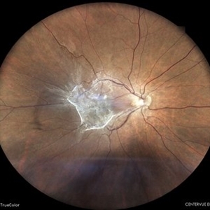
 DISC EDEMA WITH MACULAR EDEMA
Jun 6 2023 by Akansha Sharma
COLOUR FUNDUS PHOTOGRAPH OF 43 YEAR OLD MALE WITH DISC EDEMA AND MACULAR EDEMA
Photographer: Dr. Urmil Shah, Dr. Denish Patel, Dr. Akansha Sharma
Condition/keywords: cystoid macular edema (CME), disc edema
-
 PROLIFERATIVE DIABETIC RETINOPATHY
Jun 6 2023 by Akansha Sharma
COLOUR FUNDUS PHOTOGRAPH OF A 24 YEAR OLD DIABETIC MALE WITH CHANGES OF PROLIFERATIVE DIABETIC RETINOPATHY
Photographer: Dr. Urmil Shah, Dr. Denish Patel, Dr. Akansha Sharma
Condition/keywords: PDR, proliferative diabetic retinopathy (PDR)
-
 IDIOPATHIC POLYPOIDAL CHOROIDAL VASCULOPATHY
Jun 6 2023 by Akansha Sharma
COLOUR FUNDUS PHOTOGRAPH OF AN 80 YEAR OLD MALE PATIENT WITH IDIOPATHIC POLYPOIDAL CHOROIDAL VASCULOPATHY
Photographer: Dr. Urmil Shah, Dr. Denish Patel, Dr. Akansha Sharma
Condition/keywords: idiopathic polypoidal choroidal vasculopathy, polypoidal choroidal vasculopathy (PCV)
-
 VALSALVA RETINOPATHY
Jun 6 2023 by Akansha Sharma
COLOUR FUNDUS PHOTOGRAPH OF A 21 YEAR OLD MALE WITH VALSALVA RETINOPATHY
Photographer: Dr. Urmil Shah, Dr. Denish Patel, Dr. Akansha Sharma, Bharati Eye Clinic, Ahmedabad, Gujarat
Condition/keywords: SUB RETINAL HEMORRHAGE, valsalva retinopathy
-
 RETINAL ARTERIAL MACROANEURYSM
Jun 6 2023 by Akansha Sharma
COLOUR FUNDUS PHOTOGRAPH OF A 60 YEAR OLD HYPERTENSIVE MALE WITH RETINAL ARTERIAL MACROANEURYSM LEADING TO SUBRETINAL BLEED
Photographer: Dr. Urmil Shah, Dr. Denish Patel, Dr. Akansha Sharma, BHARATI EYE CLINIC, AHMEDABAD, GUJARAT
Condition/keywords: retinal arterial macroaneurysm
-
 PROLIFERATIVE DIABETIC RETINOPATHY
Jun 6 2023 by Akansha Sharma
COLOUR FUNDUS PHOTOGRAPH OF A 42 YEAR OLD DIABETIC MALE WITH PROLIFERATIVE DIABETIC RETINOPATHY AND MACULAR EDEMA
Photographer: Dr. Denish Patel, Dr. Akansha Sharma, Dr. Urmil Shah, Bharati Eye Hospital, Ahmedabad, Gujarat
Condition/keywords: diabetes, diabetic macular edema, macular edema, PDR, proliferative diabetic retinopathy (PDR)
-
 SUBHYALOID HEMORRHAGE IN A CASE OF VALSALVA RETINOPATHY
Jun 6 2023 by Akansha Sharma
colour fundus photograph of a 40 year old female with subhyaloid hemorrhage in a case of valsalva retinopathy
Photographer: Dr. Denish Patel, Dr. Akansha Sharma, Dr. Urmil Shah, Bharati Eye Hospital, Ahmedabad, Gujarat
Condition/keywords: SHH, subhyaloid hemorrhage
-
 CHRONIC CENTRAL SEROUS RETINOPATHY
Jun 20 2023 by Akansha Sharma
COLOUR FUNDUS PHOTGRAPH OF A 49 YEAR OLD MALE WITH CHRONIC CENTRAL SEROUS RETINOPATHY WITH PIGMENT EPITHELIAL DETACHMENT
Photographer: Dr. Urmil Shah, Dr. Denish Patel, Dr. Akansha Sharma, BHARATI EYE CLINIC, AHMEDABAD, GUJARAT
Condition/keywords: chronic central serous chorioretinopathy (CSCR)
-
 Berlin's Edema with Retinal Tear with Vitreous Hemorrhage Status post Blunt Trauma
Aug 19 2023 by Akansha Sharma
Colour fundus photo of a 22 year old male with retinal tear with vitreous hemorrhage with berlin's edema status post blunt trauma with a cricket ball
Photographer: Dr. Akansha Sharma, Dr. Urmil Shah, Dr. Denish Patel, Bharati Eye Hospital
Condition/keywords: Berlin's edema, blunt trauma, retinal tear
-
 Intra-ocular Foreign Body with Vitreous Hemorrhage
Aug 19 2023 by Akansha Sharma
Colour fundus photograph of a 30 year old male with impacted intra-ocular foreign body with vitreous hemorrhage status post penetrating trauma while tailoring
Photographer: Dr. Akansha Sharma, Dr. Urmil Shah, Dr. Denish Patel, Bharati Eye Hospital, Ahmedabad, Gujarat
Condition/keywords: intraocular foreign body, penetrating trauma, vitreous hemorrhage
-
 Rhegmatogenous Retina Detachment
Aug 19 2023 by Akansha Sharma
COLOUR FUNDUS PHOTOGRAPH OF A 65 YEAR OLD MALE WITH RHEGMATOGENOUS RETINAL DETACHMENT
Photographer: Dr. Akansha Sharma, Dr. Urmil Shah, Dr. Denish Patel, Bharati Eye Hospital, Ahmedabad, Gujarat
Condition/keywords: Retinal Detachment, rhegmatogenous retinal detachment, rrd
-
 Rhegmatogenous Retina Detachment
Aug 19 2023 by Akansha Sharma
Colour fundus photograph of a 64 year old male with rhegmatogenous retinal detachment
Photographer: Dr. Akansha Sharma, Bharati Eye Hospital, Ahmedabad, Gujarat
Condition/keywords: Retinal Detachment, rhegmatogenous retinal detachment, rrd
-
 Familial Exudative Vitreo-Retinopathy
Jan 30 2024 by Akansha Sharma
Colour fundus photograph of a 19 year old male with both eyes familial exudative vitreo-retinopathy. Right eye shows peripheral avasular areas.
Photographer: Dr. Akansha Sharma, Bharati Eye Hospital
Condition/keywords: familial exudative vitreoretinopathy (FEVR), REGRESSED ROP
-
 Familial Exudative Vitreo-Retinopathy
Jan 30 2024 by Akansha Sharma
Colour fundus photograph of a 19 year old male with both eyes familial exudative vitreo-retinopathy. Left eye shows aggregation of hard exudates over the fovea.
Photographer: Dr. Akansha Sharma, Bharati Eye Hospital
Condition/keywords: familial exudative vitreoretinopathy (FEVR), REGRESSED ROP
-
 Subhyaloid Hemorrhage in a Case of Proliferative Diabetic Retinopathy
Jan 30 2024 by Akansha Sharma
Colour fundus photo of a 52 year old male with subhyaloid hemorrhage in a case of proliferative diabetic retinopathy
Photographer: Dr. Akansha Sharma, Bharati Eye Hospital
Condition/keywords: florid type PDR, proliferative diabetic retinopathy (PDR), SHH
-
 Vasculitis
Jan 30 2024 by Akansha Sharma
Color fundus photograph of a 34 year old seronegative male patient with vasculitis presenting with macular edema associated with a macroaneurysm.
Photographer: Dr. Akansha Sharma, Bharati Eye Hospital
Condition/keywords: macroaneurysm, vasculitis
-
 Sub-total Retinal Detachment
Jan 30 2024 by Akansha Sharma
Color fundus photograph of a 53 year old male patient with sub-total retinal detachment.
Photographer: Dr. Akansha Sharma, Bharati Eye Hospital
Condition/keywords: RD, Retinal Detachment, sub-total retinal detachment
-
 Sub Retinal Hemorrhage
Jan 30 2024 by Akansha Sharma
Color fundus photograph of a 62 year old female patient with sub retinal bleed in a case of polypoidal choroidal vasculopathy.
Photographer: Dr. Akansha Sharma, Bharati Eye Hospital
Condition/keywords: polypoidal choroidal vasculopathy (PCV), subretinal hemorrhage, subretinal blood
-
 Superior Retinal Detachment
Jan 30 2024 by Akansha Sharma
Color fundus photograph of a 72 year old female with superior retinal detachment with macula off.
Photographer: Dr. Akansha Sharma, Bharati Eye Hospital
Condition/keywords: RD, Retinal Detachment
-
 Epiretinal Membrane
Jan 30 2024 by Akansha Sharma
Color fundus photograph of a 65 year old hypertensive male with an epiretinal membrane in a case of old branch retinal vein occlusion with a subhyaloid hemorrhage seen inferiorly.
Photographer: Dr. Akansha Sharma, Bharati Eye Hospital
Condition/keywords: epiretinal membrane (ERM), ERM
-
 Subhyaloid Hemorrhage From a Macroaneurysm
Jan 30 2024 by Akansha Sharma
Color fundus photograph of a 58 year old hypertensive and diabetic female patient with lasered proliferative diabetic retinopathy developing a subhyaloid hemorrhage from a macroaneurysm.
Photographer: Dr. Akansha Sharma, Bharati Eye Hospital
Condition/keywords: florid type PDR, macroaneurysm, proliferative diabetic retinopathy (PDR), SHH
-
 Myelinated Nerve Fibres
Jan 30 2024 by Akansha Sharma
Color fundus photograph of a 15 year old male with myelinated nerve fibres all around the disc in the right eye.
Photographer: Dr. Akansha Sharma, Bharati Eye Hospital
Condition/keywords: MNF, myelinated nerve fibers
-
 Superior Retinal Detachment
Jan 30 2024 by Akansha Sharma
Color fundus photograph of a 62 year old male patient with superior retinal detachment.
Photographer: Dr. Akansha Sharma, Bharati Eye Hospital
Condition/keywords: RD
-
 Berlin's Edema Status Post Blunt Trauma
Jan 30 2024 by Akansha Sharma
Color fundus photograph of a 17 year old male patient who developed berlin's edema post blunt trauma from a cricket ball.
Photographer: Dr. Akansha Sharma, Bharati Eye Hospital
Condition/keywords: Berlin's edema, blunt trauma
-
 Retinitis
Jan 30 2024 by Akansha Sharma
Color fundus photograph of a 32 year old male patient presenting with retinitis post fever.
Photographer: Dr. Akansha Sharma, Bharati Eye Hospital
Condition/keywords: cotton wool exudates, retinitis
-
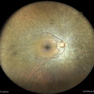
 IOL Drop in a Case of Iridofundal Coloboma
Feb 7 2024 by Akansha Sharma
Color fundus photograph of a 43 year old male with IOL drop in a case of iridofundal coloboma.
Photographer: Dr. Akansha Sharma, Bharati Eye Hospital
Condition/keywords: chorioretinal coloboma, IOL drop
-
 Status Post Retinal Detachment Surgery in a Case of Iridofundal Coloboma
Feb 7 2024 by Akansha Sharma
Color fundus photograph of a 43 year old male post retinal detachment surgery in a case of iridofundal coloboma.
Photographer: Dr. Akansha Sharma, Bharati Eye Hospital
Condition/keywords: chorioretinal coloboma, RD
-
 Retinal Detachment With Irido-fundal Coloboma
Feb 7 2024 by Akansha Sharma
Color fundus photograph of a 43 year old male with retinal detachment in a case of iridofundal coloboma.
Photographer: Dr. Akansha Sharma, Bharati Eye Hospital
Condition/keywords: chorioretinal coloboma, RD
-
 Scarred Choroidal Neovacular Membrane With Large Inferior Horse Shoe Tear
Feb 7 2024 by Akansha Sharma
Color fundus photograph of a 73 year old male with scarred choroidal neovascular membrane with large horse shoe tear inferiorly.
Photographer: Dr. Akansha Sharma, Bharati Eye Hospital
Condition/keywords: atrophic scar, choroidal neovascular membrane (CNVM), CNVM
-
 Retinal Detachment
Feb 7 2024 by Akansha Sharma
Color fundus photograph of a 62 year old male with retinal detachment.
Photographer: Dr. Akansha Sharma, Bharati Eye Hospital
Condition/keywords: RD
-
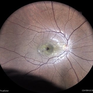
 Retinal Detachment With Disc Edema
Mar 6 2024 by Akansha Sharma
Color fundus photograph of a 60 year old female with retinal detachment associated with disc edema.
Photographer: Dr. Akansha Sharma, Bharati Eye Hospital
Condition/keywords: disc edema, RD, Retinal Detachment
-
 Branch Retinal Artery Occlusion
Mar 6 2024 by Akansha Sharma
Color fundus photograph of a 65 year old male with sudden dimness of vision presenting with a branch retinal artery occlusion.
Photographer: Dr. Akansha Sharma, Bharati Eye Hospital
Condition/keywords: branch retinal artery occlusion, BRAO
-
 Branch Retinal Artery Occlusion
Mar 6 2024 by Akansha Sharma
Fluorescein angiography of a 65 year old male with absence of filling in the retinal artery close to the arcade in the early phase.
Photographer: Dr. Akansha Sharma, Bharati Eye Hospital
Condition/keywords: BRAO
-
 Branch Retinal Artery Occlusion
Mar 6 2024 by Akansha Sharma
Fluorescein angiography of a 65 year old male showing delayed filling of the retinal artery in the late phase.
Photographer: Dr. Akansha Sharma, Bharati Eye Hospital
Condition/keywords: branch retinal artery occlusion
-
 Central Serous Retinopathy
Mar 6 2024 by Akansha Sharma
Color fundus photograph of a 34 year old male with central serous retinopathy.
Photographer: Dr. Akansha Sharma, Bharati Eye Hospital
Condition/keywords: Central Serous Chorioretinopathy (CSR), central serous retinopathy (CSR)
-
 Central Serous Retinopathy
Mar 6 2024 by Akansha Sharma
Fluorescein angiography of a 34 year old male with smoke stack leak in the late phase in a case of central serous retinopathy.
Photographer: Dr. Akansha Sharma, Bharati Eye Hospital
Condition/keywords: Central Serous Chorioretinopathy (CSR), central serous retinopathy (CSR)
-
 Branch Retinal Artery Occlusion
Mar 6 2024 by Akansha Sharma
Color fundus photograph of a 67 year old male with branch retinal artery occlusion.
Photographer: Dr. Akansha Sharma, Bharati Eye Hospital
Condition/keywords: branch retinal artery occlusion, BRAO
-
 Branch Retinal Artery Occlusion
Mar 6 2024 by Akansha Sharma
Fluorescein angiography of a 67 year old male with capillary non perfusion areas in early phase in a case of branch retinal artery occlusion.
Photographer: Dr. Akansha Sharma, Bharati Eye Hospital
Condition/keywords: branch retinal artery occlusion, BRAO
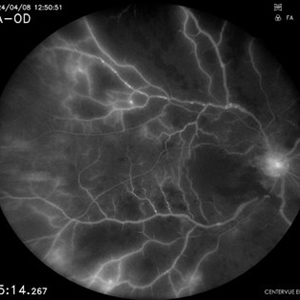
-
 Subhyaloid Hemorrhage in a Case of Proliferative Diabetic Retinopathy
Mar 26 2024 by Akansha Sharma
Color fundus photograph of a 44 year old diabetic male with subhyaloid hemorrhage in a case of proliferative diabetic retinopathy.
Photographer: Dr. Akansha Sharma, Bharati Eye Hospital
Condition/keywords: PDR, proliferative diabetic retinopathy (PDR), SHH, subhyaloid hemorrhage
-
 Retinal Detachment
Mar 26 2024 by Akansha Sharma
Color fundus photograph of a 62 year old male with left eye retinal detachment with superotemporal horse shoe tear.
Photographer: Dr. Akansha Sharma, Bharati Eye Hospital
Condition/keywords: RD
-
 Subretinal Neovascular Membrane
Mar 26 2024 by Akansha Sharma
Color fundus photograph of a 65 year old female patient with subretinal bleed suggestive of subretinal neovascular membrane.
Photographer: Dr. Akansha Sharma, Bharati Eye Hospital
Condition/keywords: CNVM, SRNVM, subretinal neovascularization (SRNV), wet age-related macular degeneration (wet AMD)
-
 Subhyaloid Hemorrhage in a Case of Proliferative Diabetic Retinopathy
Mar 26 2024 by Akansha Sharma
Color fundus photograph of a 56 year old male patient with subhyaloid hemorrhage in a case of proliferative diabetic retinopathy.
Photographer: Dr. Akansha Sharma, Bharati Eye Hospital
Condition/keywords: PDR, proliferative diabetic retinopathy (PDR), SHH
-
 Best Disease
Mar 26 2024 by Akansha Sharma
Color fundus photograph of a 58 year old male with best disease.
Photographer: Dr. Akansha Sharma, Bharati Eye Hospital
Condition/keywords: Best disease
-
 Drusen
Mar 26 2024 by Akansha Sharma
Autofluorescence photograph of a 52 year old female patient with dry age related macular degeneration.
Photographer: Dr. Akansha Sharma, Bharati Eye Hospital
Condition/keywords: age-related macular degeneration (AMD), drusen, dry age-related macular degeneration (dry AMD)
-
 Idiopathic Polypoidal Choroidal Vasculopathy
Mar 26 2024 by Akansha Sharma
Color fundus photograph of a 61 year old treatment naive female patient with scarring at fovea with surrounding subretinal bleed suggestive of idiopathic polypoidal choroidal vasculopathy.
Photographer: Dr. Akansha Sharma, Bharati Eye Hospital
Condition/keywords: polypoidal choroidal vasculopathy (PCV)
-
 Choroidal Melanoma
Mar 26 2024 by Akansha Sharma
Color fundus photograph of a 50 year old male patient with choroidal melanoma.
Photographer: Dr. Akansha Sharma, Bharati Eye Hospital
-
 Retinal Detachment
Mar 26 2024 by Akansha Sharma
Color fundus photograph of a 54 year old female with right eye retinal detachment.
Photographer: Dr. Akansha Sharma, Bharati Eye Hospital
Condition/keywords: RD
-
 Retinitis Pigmentosa
Mar 26 2024 by Akansha Sharma
Autofluorescence photograph of a 68 year old female patient with retinitis pigmentosa with macular thinning.
Photographer: Dr. Akansha Sharma, Bharati Eye Hospital
Condition/keywords: Retinitis Pigmentosa, RP
-
 Subhyaloid Hemorrhage in a Case of Proliferative Diabetic Retinopathy
Apr 8 2024 by Akansha Sharma
Color fundus photograph of a 46 year old diabetic female with subhyaloid hemorrhage in a case of proliferative diabetic retinopathy.
Photographer: Dr. Akansha Sharma, Bharati Eye Hospital
Condition/keywords: PDR, proliferative diabetic retinopathy (PDR), SHH, subhyaloid hemorrhage
-
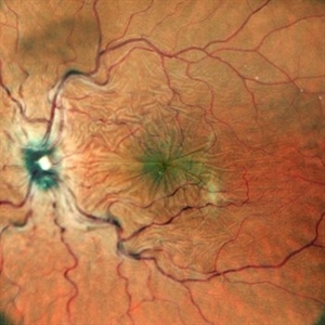
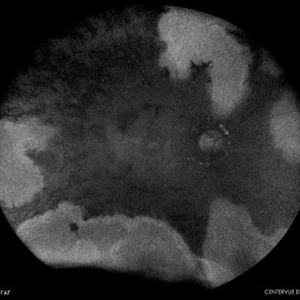
 Old Healed Serpiginous Like Choroiditis
Apr 8 2024 by Akansha Sharma
Color fundus photograph of a 40 year old male patient with old healed serpiginous like choroiditis.
Photographer: Dr. Akansha Sharma, Bharati Eye Hospital
Condition/keywords: serpiginous like choroiditis, Tuberculosis
-
 Old Healed Serpiginous Like Choroiditis With Disc Pallor
Apr 8 2024 by Akansha Sharma
Color fundus photograph of a 40 year old male patient with old healed serpiginous like choroiditis with disc pallor.
Photographer: Dr. Akansha Sharma, Bharati Eye Hospital
Condition/keywords: Tuberculosis
-
 Subhyaloid Hemorrhage With Vitreous Hemorrhage in a Case of Proliferative Diabetic Retinopathy
Apr 8 2024 by Akansha Sharma
Color fundus photograph of a 48 year old diabetic male patient with subhyaloid hemorrhage with disperse vitreous hemorrhage with disc pallor in a case of proliferative diabetic retinopathy.
Photographer: Dr. Akansha Sharma, Bharati Eye Hospital
Condition/keywords: PDR, proliferative diabetic retinopathy (PDR), tractional retinal detachment, TRD
-
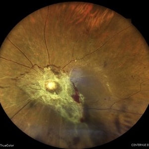 Table Top Tractional Retinal Detachment
Apr 8 2024 by Akansha Sharma
Color fundus photograph of a 48 year old diabetic male patient with a table top tractional retinal detachment in a case of proliferative diabetic retinopathy.
Photographer: Dr. Akansha Sharma, Bharati Eye Hospital
Condition/keywords: PDR, proliferative diabetic retinopathy (PDR), tractional retinal detachment, TRD
-
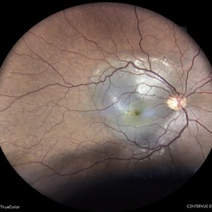
 Disc Pallor With Retinal Atrophy Status Post Ischaemic Vascular Event
Apr 8 2024 by Akansha Sharma
Color fundus photograph of a 22 year old female with disc pallor with retinal atrophy status post ischaemic vascular event.
Photographer: Dr. Akansha Sharma, Bharati Eye Hospital
Condition/keywords: inner retinal thinning, optic disc pallor
-
 Retinal Detachment
Apr 8 2024 by Akansha Sharma
Color fundus photograph of a 65 year old male with retinal detachment and superotemporal horse shoe tear.
Photographer: Dr. Akansha Sharma, Bharati Eye Hospital
Condition/keywords: RD
-
 Retinitis Pigmentosa
Apr 8 2024 by Akansha Sharma
Color fundus photograph of a 45 year old male with disc pallor and peripheral bony spicules suggestive of retinitis pigmentosa.
Photographer: Dr. Akansha Sharma, Bharati Eye Hospital
Condition/keywords: Retinitis Pigmentosa, RP
-
 Combined Central Retinal Artery and Vein Occlusion
Apr 8 2024 by Akansha Sharma
Fundus fluorescein angiography of a 63 year old male with combined central retinal artery and vein occlusion with carotid artery stenosis and infarct in the brain demonstrating late filling.
Photographer: Dr. Akansha Sharma, Bharati Eye Hospital
Condition/keywords: central retinal artery occlusion (CRAO), central retinal vein occlusion (CRVO), CRAO
-
 Combined Central Retinal Artery and Vein Occlusion
Apr 8 2024 by Akansha Sharma
Color fundus photograph of a 63 year old male with combined central retinal artery and vein occlusion with carotid artery stenosis and infarct in the brain.
Photographer: Dr. Akansha Sharma, Bharati Eye Hospital
Condition/keywords: central retinal artery occlusion (CRAO), central retinal vein occlusion (CRVO), CRAO
-
 Retinitis Pigmentosa
Apr 9 2024 by Akansha Sharma
Color fundus photograph of a 30 year old male with retinitis pigmentosa.
Photographer: Dr. Akansha Sharma, Bharati Eye Hospital
Condition/keywords: retinitis pigmentosa, RP, RP variant
-
 Retinitis Pigmentosa
Apr 9 2024 by Akansha Sharma
Autofluorescence image of a 30 year old male with retinitis pigmentosa.
Photographer: Dr. Akansha Sharma, Bharati Eye Hospital
Condition/keywords: retinitis pigmentosa, RP, RP variant
-
 Retinal Detachment
Apr 9 2024 by Akansha Sharma
Color fundus photograph of a 57 year old female with retinal detachment.
Photographer: Dr. Akansha Sharma, Bharati Eye Hospital
Condition/keywords: RD, Retinal Detachment
-
 Angiographic Diabetic Macular Edema in a Case of Proliferative Diabetic Retinopathy
Apr 9 2024 by Akansha Sharma
Fundus fluorescein angiographic image of 62 year old male demonstrating angiographic diabetic macular edema in a case of proliferative diabetic retinopathy.
Photographer: Dr. Akansha Sharma, Bharati Eye Hospital
Condition/keywords: clinically significant macular edema (CSME), diabetic blindness, diabetic macular edema, proliferative diabetic retinopathy (PDR)
-
 Retinal Arterial Macroaneurysm
Apr 9 2024 by Akansha Sharma
Color fundus photograph of a 68 year old female patient with retinal arterial macroaneurysm with subretinal bleed.
Photographer: Dr. Akansha Sharma, Bharati Eye Hospital
Condition/keywords: macroaneurysm, subretinal hemorrhage
-
 Status Post Prophylactic Barrage Laser for Inferior Retinal Detachment
Apr 9 2024 by Akansha Sharma
Color fundus photograph of a 19 year old male with an inferior retinal detachment holding well years after prophylactic barrage laser in a one eyed patient.
Photographer: Dr. Akansha Sharma, Bharati Eye Hospital
Condition/keywords: BARRAGE LASER, RD
-
 Myopic Subretinal Neovascular Membrane
Apr 9 2024 by Akansha Sharma
Color fundus photograph of a 23 year old female with subretinal bleed in a case of high myopia.
Photographer: Dr. Akansha Sharma, Bharati Eye Hospital
Condition/keywords: myopic choroidal neovascularization (CNV), SRNVM, subretinal hemorrhage
-
 Retinal Detachment
Apr 9 2024 by Akansha Sharma
Color fundus photograph of a 67 year old male patient with retinal detachment involving macula.
Photographer: Dr. Akansha Sharma, Bharati Eye Hospital
Condition/keywords: RD, Retinal Detachment
-
 Central Retinal Vein Occlusion
Apr 9 2024 by Akansha Sharma
Color fundus photograph of a 73 year old hypertensive male with central retinal vein occlusion.
Photographer: Dr. Akansha Sharma, Bharati Eye Hospital
Condition/keywords: central retinal vein occlusion (CRVO), ischemic CRVO
-
 Central Retinal Vein Occlusion
Apr 9 2024 by Akansha Sharma
Color fundus photograph of a 73 year old hypertensive male with central retinal vein occlusion.
Photographer: Dr. Akansha Sharma, Bharati Eye Hospital
Condition/keywords: central retinal vein occlusion (CRVO), ischemic CRVO
-
 Central Serous Retinopathy
Apr 9 2024 by Akansha Sharma
Color fundus photograph of a 35 year old male with central serous retinopathy.
Photographer: Dr. Akansha Sharma, Bharati Eye Hospital
Condition/keywords: Central Serous Chorioretinopathy (CSR), central serous retinopathy (CSR)
-
 Central Serous Retinopathy
Apr 9 2024 by Akansha Sharma
Fundus fluorescein angiography of a 35 year old male with central serous retinopathy demonstrating leaks.
Photographer: Dr. Akansha Sharma, Bharati Eye Hospital
Condition/keywords: Central Serous Chorioretinopathy (CSR), central serous retinopathy (CSR)
-
 Idiopathic Polypoidal Choroidal Vasculopathy
Apr 9 2024 by Akansha Sharma
Color fundus photograph of a 74 year old female with subretinal bleed in a case of polypoidal choroidal vasculopathy.
Photographer: Dr. Akansha Sharma, Bharati Eye Hospital
Condition/keywords: PCV, polypoidal choroidal vasculopathy (PCV), wet age-related macular degeneration (wet AMD)
-
 Central Serous Retinopathy
Apr 9 2024 by Akansha Sharma
Color fundus photograph of a 39 year old male patient with smoke stack pattern of central serous retinopathy.
Photographer: Dr. Akansha Sharma, Bharati Eye Hospital
Condition/keywords: Central Serous Chorioretinopathy (CSR), central serous retinopathy (CSR)
-
 Central Serous Retinopathy
Apr 9 2024 by Akansha Sharma
Fundus fluorescein angiography of a 39 year old male patient with smoke stack pattern of central serous retinopathy.
Photographer: Dr. Akansha Sharma, Bharati Eye Hospital
Condition/keywords: Central Serous Chorioretinopathy (CSR), central serous retinopathy (CSR)
-
 Retinitis Pigmentosa
Apr 9 2024 by Akansha Sharma
Autofluorescence image of a 35 year old male with retinitis pigmentosa.
Photographer: Dr. Akansha Sharma, Bharati Eye Hospital
Condition/keywords: retinitis pigmentosa, RP, RP variant
-
 Retinitis Pigmentosa
Apr 9 2024 by Akansha Sharma
Color fundus photograph of a 35 year old male with retinitis pigmentosa.
Photographer: Dr. Akansha Sharma, Bharati Eye Hospital
Condition/keywords: retinitis pigmentosa, RP, RP variant
-
 Multifocal Chorioretinitis
Apr 9 2024 by Akansha Sharma
Color fundus photograph of a 34 year old male patient with multifocal chorioretinitis.
Photographer: Dr. Akansha Sharma, Bharati Eye Hospital
Condition/keywords: chorioretinal inflammations, chorioretinitis
-
 Multifocal Chorioretinitis
Apr 9 2024 by Akansha Sharma
Color fundus photograph of a 34 year old male patient with multifocal chorioretinitis with subretinal bleed.
Photographer: Dr. Akansha Sharma, Bharati Eye Hospital
Condition/keywords: chorioretinal inflammations, chorioretinitis, subretinal hemorrhage
-
 Retinitis Pigmentosa
Apr 9 2024 by Akansha Sharma
Autofluorescence image of a 57 year old male with retinitis pigmentosa.
Photographer: Dr. Akansha Sharma, Bharati Eye Hospital
Condition/keywords: retinitis pigmentosa, RP
-
 Retinitis Pigmentosa
Apr 9 2024 by Akansha Sharma
Color fundus photograph of a 57 year old male with retinitis pigmentosa.
Photographer: Dr. Akansha Sharma, Bharati Eye Hospital
Condition/keywords: retinitis pigmentosa, RP
-
 Stargardts' Disease
Apr 9 2024 by Akansha Sharma
Color fundus photograph of a 28 year old male with stargardts' disease.
Photographer: Dr. Akansha Sharma, Bharati Eye Hospital
Condition/keywords: Stargardt disease
-
 Stargardts' Disease
Apr 9 2024 by Akansha Sharma
Autofluorescence image of a 28 year old male with stargardts' disease.
Photographer: Dr. Akansha Sharma, Bharati Eye Hospital
Condition/keywords: Stargardt disease
-
 Subretinal Neovascular Membrane with PED
Apr 17 2024 by Akansha Sharma
Color fundus photograph of a 72 year old male with ped along with subretinal bleed around it.
Photographer: Dr. Akansha Sharma, Bharati Eye Hospital
Condition/keywords: CNVM, PED, SRNVM, subretinal neovascularization (SRNV), wet age-related macular degeneration (wet AMD)
-
 Retinitis Pigmentosa
Apr 17 2024 by Akansha Sharma
Color fundus photograph of a 42 year old male with bony spicules and waxy pallor disc suggestive of retinitis pigmentosa.
Photographer: Dr. Akansha Sharma, Bharati Eye Hospital
Condition/keywords: retinitis pigmentosa, RP
-
 Retinitis Pigmentosa
Apr 17 2024 by Akansha Sharma
Color fundus photograph of a 42 year old male with bony spicules and waxy pallor disc suggestive of retinitis pigmentosa.
Photographer: Dr. Akansha Sharma, Bharati Eye Hospital
Condition/keywords: retinitis pigmentosa, RP
-
 Central Retinal Artery Occlusion
Apr 17 2024 by Akansha Sharma
Fluorescein angiography of a 48 year old male with central retinal artery occlusion.
Photographer: Dr. Akansha Sharma, Bharati Eye Hospital
Condition/keywords: central retinal artery occlusion (CRAO), CRAO
-
 Central Retinal Artery Occlusion
Apr 17 2024 by Akansha Sharma
Color fundus photograph of a 48 year old male with central retinal artery occlusion.
Photographer: Dr. Akansha Sharma, Bharati Eye Hospital
Condition/keywords: central retinal artery occlusion (CRAO), CRAO
-
 Disc Edema
Apr 17 2024 by Akansha Sharma
Color fundus photograph of a 35 year old female with disc edema.
Photographer: Dr. Akansha Sharma, Bharati Eye Hospital
Condition/keywords: Disc Edema, optic disc edema
-
 Retinal Detachment
Apr 17 2024 by Akansha Sharma
Color fundus photograph of a 42 year old male with retinal detachment.
Photographer: Dr. Akansha Sharma, Bharati Eye Hospital
Condition/keywords: RD, Retinal Detachment
-
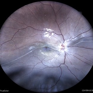 Old Retinal Detachment
Apr 17 2024 by Akansha Sharma
Color fundus photograph of a 13 year old female with old retinal detachment at presentation as is demonstrated by a demarcation line.
Photographer: Dr. Akansha Sharma, Bharati Eye Hospital
Condition/keywords: delimited old retinal detachment, demarcation line, RD, Retinal Detachment
-
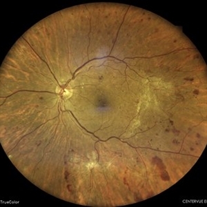 Proliferative Diabetic Retinopathy
Apr 17 2024 by Akansha Sharma
Color fundus photograph of a 68 year old diabetic and hypertensive male with proliferative diabetic retinopathy.
Photographer: Dr. Akansha Sharma, Bharati Eye Hospital
Condition/keywords: PDR, proliferative diabetic retinopathy (PDR)
-
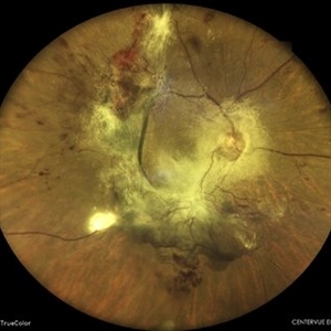 Table Top Tractional Retinal Detachment
Apr 17 2024 by Akansha Sharma
Color fundus photograph of a 68 year old diabetic and hypertensive male with a table top tractional retinal detachment.
Photographer: Dr. Akansha Sharma, Bharati Eye Hospital
Condition/keywords: PDR, proliferative diabetic retinopathy (PDR), tractional retinal detachment, TRD
-
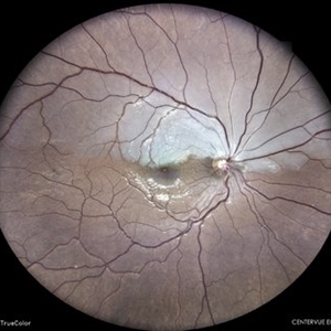 Hemi Central Retinal Artery Occlusion
Apr 17 2024 by Akansha Sharma
Color fundus photograph of a 30 year old female with hemi central retinal artery occlusion.
Photographer: Dr. Akansha Sharma, Bharati Eye Hospital
Condition/keywords: central retinal artery occlusion (CRAO), CRAO, Hemi-Central Retinal Artery Occlusion (CRAO)
-
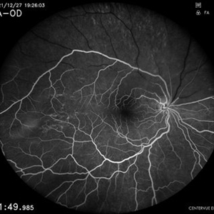 Hemi Central Retinal Artery Occlusion
Apr 17 2024 by Akansha Sharma
Early phase fluorecein angiogram of a 30 year old female with perfusion lacking along the vessels of the superior arcade.
Photographer: Dr. Akansha Sharma, Bharati Eye Hospital
Condition/keywords: central retinal artery occlusion (CRAO), CRAO, Hemi-Central Retinal Artery Occlusion (CRAO)
-
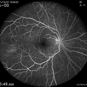 Hemi Central Retinal Artery Occlusion
Apr 17 2024 by Akansha Sharma
Late phase fluorescein angiogram of a 30 year old female with complete perfusion.
Photographer: Dr. Akansha Sharma, Bharati Eye Hospital
Condition/keywords: central retinal artery occlusion (CRAO), CRAO, Hemi-Central Retinal Artery Occlusion (CRAO)
-
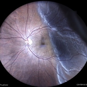 Retinal Detachment
Apr 17 2024 by Akansha Sharma
Color fundus photograph of a 61 year old male with retinal detachment.
Photographer: Dr. Akansha Sharma, Bharati Eye Hospital
Condition/keywords: RD, Retinal Detachment
-
 Valsalva Retinopathy
Apr 19 2024 by Akansha Sharma
Color fundus photograph of a 24 year old male with valsalva retinopathy.
Photographer: Dr. Akansha Sharma, Bharati Eye Hospital
Condition/keywords: valsalva retinopathy
-
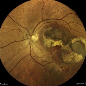 Polypoidal Choroidal Vasculopathy
Apr 19 2024 by Akansha Sharma
Color fundus photograph of a 63 year old female with polypoidal choroidal vasculopathy.
Photographer: Dr. Akansha Sharma, Bharati Eye Hospital
Condition/keywords: PCV, polypoidal choroidal vasculopathy (PCV)
-
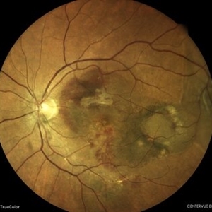 Retinal Detachment Secondary to a Resolving Polypoidal Choroidal Vasculopathy
Apr 19 2024 by Akansha Sharma
Color fundus photograph of a 63 year old female with a retinal detachment secondary to a resolving polypoidal choroidal vasculopathy.
Photographer: Dr. Akansha Sharma, Bharati Eye Hospital
Condition/keywords: PCV, polypoidal choroidal vasculopathy (PCV), RD, Retinal Detachment
-
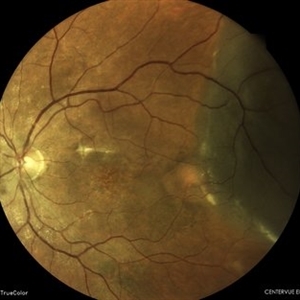 Retinal Detachment Secondary to Polypoidal Choroidal Vasculopathy
Apr 19 2024 by Akansha Sharma
Color fundus photograph of a 63 year old female with a retinal detachment secondary to polypoidal choroidal vasculopathy.
Photographer: Dr. Akansha Sharma, Bharati Eye Hospital
Condition/keywords: PCV, polypoidal choroidal vasculopathy (PCV), RD, Retinal Detachment
-
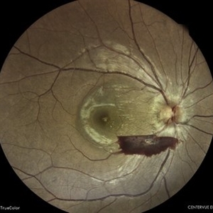 Pre-Retinal Hemorrhage With Disc Edema
Apr 19 2024 by Akansha Sharma
Color fundus photograph of a 39 year old female with disc edema along with a pre-retinal hemorrhage.
Photographer: Dr. Akansha Sharma, Bharati Eye Hospital
Condition/keywords: disc edema, preretinal hemorrhage
-
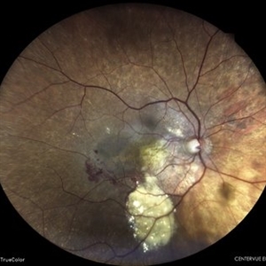 Polypoidal Choroidal Vasculopathy
Apr 19 2024 by Akansha Sharma
Color fundus photograph of a 84 year old female with subretinal hemorrhage in a case of polypoidal choroidal vasculopathy.
Photographer: Dr. Akansha Sharma, Bharati Eye Hospital
Condition/keywords: PCV, polypoidal choroidal vasculopathy (PCV)
-
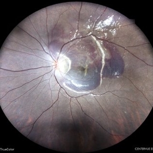 Choroidal Rupture With Traumatic Subretinal Hemorrhage
Apr 19 2024 by Akansha Sharma
Color fundus photograph of a 11 year old female with subretinal bleed and choroidal rupture status post trauma.
Photographer: Dr. Akansha Sharma, Bharati Eye Hospital
Condition/keywords: Choroidal Rupture, subretinal hemorrhage, subretinal blood
-
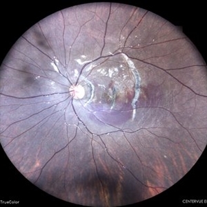 Choroidal Rupture With Traumatic Subretinal Hemorrhage
Apr 19 2024 by Akansha Sharma
Color fundus photograph of a 11 year old female with subretinal bleed and choroidal rupture status post trauma.
Photographer: Dr. Akansha Sharma, Bharati Eye Hospital
Condition/keywords: Choroidal Rupture, subretinal hemorrhage, subretinal blood
-
 Subretinal Neovascular Membrane
Apr 19 2024 by Akansha Sharma
Color fundus photograph of a 74 year old female with subretinal neovascular membrane with scarring below the fovea.
Photographer: Dr. Akansha Sharma, Bharati Eye Hospital
Condition/keywords: SRNVM, subretinal neovascularization (SRNV)
-
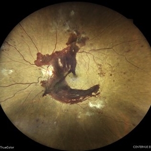 Subhyaloid Hemorrhage With Vascular Proliferations in a Case of Proliferative Diabetic Retinopathy
Apr 19 2024 by Akansha Sharma
Color fundus photograph of a 60 year old male with subhyaloid hemorrhage with vascular proliferations in a case of proliferative diabetic retinopathy,
Photographer: Dr. Akansha Sharma, Bharati Eye Hospital
Condition/keywords: proliferative diabetic retinopathy (PDR), SHH, subhyaloid blood, subhyaloid hemorrhage, TRACTION
-
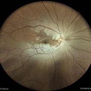 Central Retinal Artery Occlusion
May 7 2024 by Akansha Sharma
Color fundus photograph of a 40 year old female with central retinal artery occlusion.
Photographer: Dr. Akansha Sharma, Bharati Eye Hospital
Condition/keywords: central retinal artery occlusion (CRAO), CRAO
-
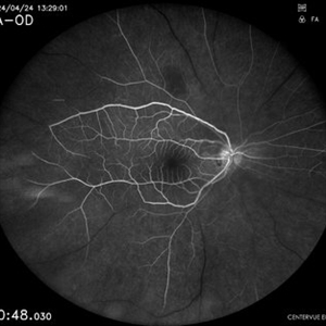 Central Retinal Artery Occlusion
May 7 2024 by Akansha Sharma
Early phase fluorescein angiography of a 40 year old female with central retinal artery occlusion.
Photographer: Dr. Akansha Sharma, Bharati Eye Hospital
Condition/keywords: central retinal artery occlusion (CRAO), CRAO
-
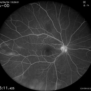 Central Retinal Artery Occlusion
May 7 2024 by Akansha Sharma
Late phase fluorescein angiography of a 40 year old female with central retinal artery occlusion.
Photographer: Dr. Akansha Sharma, Bharati Eye Hospital
Condition/keywords: central retinal artery occlusion (CRAO), CRAO
-
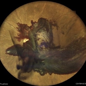 Subhyaloid Hemorrhage in a Case of Proliferative Diabetic Retinopathy
May 7 2024 by Akansha Sharma
Color fundus photograph of a 45 year old male with subhyaloid hemorrhage in a case of proliferative diabetic retinopathy.
Photographer: Dr. Akansha Sharma, Bharati Eye Hospital
Condition/keywords: florid type PDR, proliferative diabetic retinopathy (PDR), SHH, subhyaloid hemorrhage
-
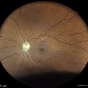 Disc Pallor Status Post NAION
May 7 2024 by Akansha Sharma
Color fundus photograph of a 48 year old male with disc pallor status post NAION.
Photographer: Dr. Akansha Sharma, Bharati Eye Hospital
Condition/keywords: NAION, optic disc pallor
-
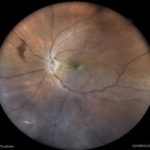 Retinal Detachment
May 7 2024 by Akansha Sharma
Color fundus photograph of a 65 year old female with retinal detachment with inferonasal horse shoe tear.
Photographer: Dr. Akansha Sharma, Bharati Eye Hospital
Condition/keywords: RD, Retinal Detachment
-
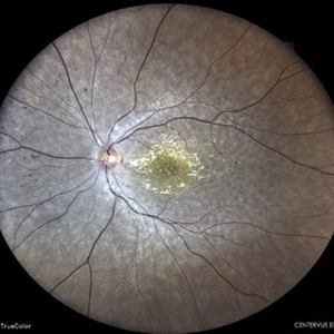 Atypical Retinitis Pigmentosa
May 7 2024 by Akansha Sharma
Color fundus photograph of a 23 year old female with atypical retinitis pigmentosa.
Photographer: Dr. Akansha Sharma, Bharati Eye Hospital
Condition/keywords: retinitis pigmentosa, RP
-
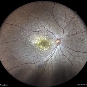 Atypical Retinitis Pigmentosa
May 7 2024 by Akansha Sharma
Color fundus photograph of a 23 year old female with atypical retinitis pigmentosa.
Photographer: Dr. Akansha Sharma, Bharati Eye Hospital
Condition/keywords: retinitis pigmentosa, RP
-
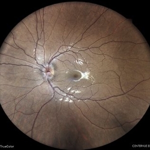 Unilateral Disc Edema
May 7 2024 by Akansha Sharma
Color fundus photograph of a 27 year old male with unilateral optic disc edema.
Photographer: Dr. Akansha Sharma, Bharati Eye Hospital
Condition/keywords: disc edema, optic disc edema
-
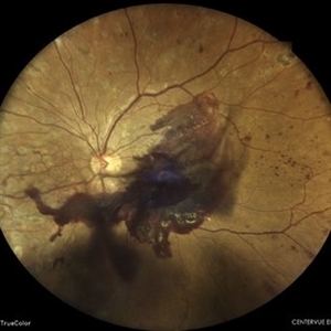 Subhyaloid Hemorrhage With Vitreous Hemorrhage in a Case of Proliferative Diabetic Retinopathy
May 7 2024 by Akansha Sharma
Color fundus photograph of a 39 year old female with vitreous hemorrhage and subhyaloid hemorrhage in proliferative diabetic retinopathy.
Photographer: Dr. Akansha Sharma, Bharati Eye Hospital
Condition/keywords: PDR, proliferative diabetic retinopathy (PDR), SHH, subhyaloid hemorrhage, vitreous hemorrhage
-
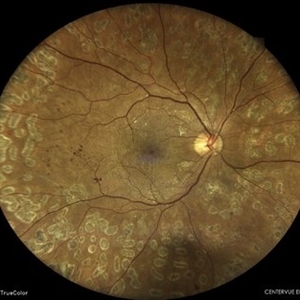 Status Post Pars Plana Vitrectomy in a Case of Vitreous Hemorrhage in Proliferative Diabetic Retinopathy
May 7 2024 by Akansha Sharma
Color fundus photograph of a 39 year old female with stable retina status post pars plana vitrectomy in a case of vitreous hemorrhage in proliferative diabetic retinopathy.
Photographer: Dr. Akansha Sharma, Bharati Eye Hospital
Condition/keywords: PDR, POST SURGERY, proliferative diabetic retinopathy (PDR)
-
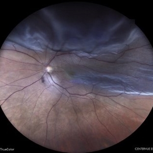 Retinal Detachment
May 7 2024 by Akansha Sharma
Color fundus photograph of a 60 year old male with retinal detachment.
Photographer: Dr. Akansha Sharma, Bharati Eye Hospital
Condition/keywords: RD, Retinal Detachment
-
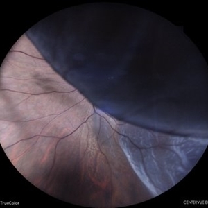 Retinal Detachment
May 7 2024 by Akansha Sharma
Color fundus photograph of a 50 year old male with retinal detachment.
Photographer: Dr. Akansha Sharma, Bharati Eye Hospital
Condition/keywords: RD, Retinal Detachment
-
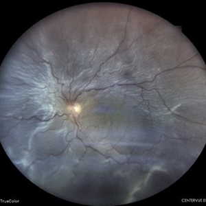 Retinal Detachment Status Post Trauma
May 7 2024 by Akansha Sharma
Color fundus photograph of a 47 year old male with total retinal detachment.
Photographer: Dr. Akansha Sharma, Bharati Eye Hospital
Condition/keywords: RD, Retinal Detachment
-
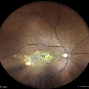 Scarred Choroidal Neovacular Membrane
May 7 2024 by Akansha Sharma
Color fundus photograph of a 75 year old male with scarred choroidal neovascular membrane.
Photographer: Dr. Akansha Sharma, Bharati Eye Hospital
Condition/keywords: choroidal neovascular membrane (CNVM), CNVM
-
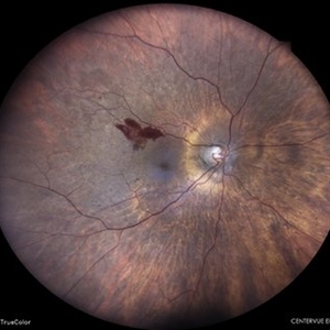 Neovascularization Elsewhere in a Case of Proliferative Diabetic Retinopathy
May 7 2024 by Akansha Sharma
Color fundus photograph of a 65 year old female with neovascularization elsewhere in a case of proliferative diabetic retinopathy.
Photographer: Dr. Akansha Sharma, Bharati Eye Hospital
Condition/keywords: NVE, PDR, proliferative diabetic retinopathy (PDR)
-
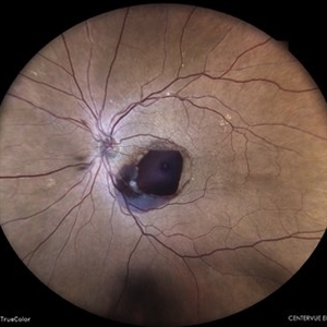 Retinal Macroaneurysm
May 7 2024 by Akansha Sharma
Color fundus photograph of a 74 year old female with retinal artery macroaneurysm.
Photographer: Dr. Akansha Sharma, Bharati Eye Hospital
Condition/keywords: macroaneurysm, RAM
-
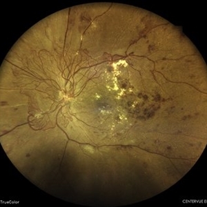 Proliferative Diabetic Retinopathy With Clinically Significant Macular Edema
May 7 2024 by Akansha Sharma
Color fundus photograph of a 41 year old female with proliferative diabetic retinopathy with clinically significant macular edema.
Photographer: Dr. Akansha Sharma, Bharati Eye Hospital
Condition/keywords: clinically significant macular edema (CSME), DME, PDR, proliferative diabetic retinopathy (PDR)
-
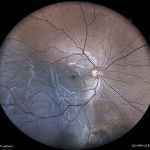 Retinal Detachment
May 27 2024 by Akansha Sharma
Color fundus photograph of a 30 year old male with retinal detachment status post blunt trauma.
Photographer: Dr. Akansha Sharma, Bharati Eye Hospital
Condition/keywords: RD, Retinal Detachment
-
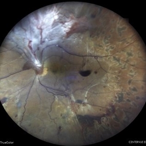 Subhyaloid Hemorrhage With Tractional Retinal Detachment in a Case of Lasered Proliferative Diabetic Retinopathy
May 27 2024 by Akansha Sharma
Color fundus photograph of a 52 year old diabetic and hypertensive male with subhyaloid hemorrhage with tractional retinal detachment in a case of lasered proliferative diabetic retinopathy.
Photographer: Dr. Akansha Sharma, Bharati Eye Hospital
Condition/keywords: PDR, proliferative diabetic retinopathy (PDR), SHH, subhyaloid hemorrhage, tractional retinal detachment, TRD
-
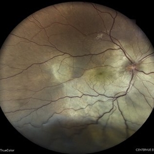 Exudative Retinal Detachment
May 27 2024 by Akansha Sharma
Color fundus photograph of a 38 year old male with breast carcinoma leading to intraocular metastasis as represented by an exudative retinal detachment.
Photographer: Dr. Akansha Sharma, Bharati Eye Hospital
Condition/keywords: Disc Edema, exudative detachment, macular pucker, METASTATSIS
-
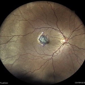 Subretinal Hemorrhage Status Post Blunt Trauma
May 27 2024 by Akansha Sharma
Color fundus photograph of a 41 year old male with subretinal bleed status post blunt trauma.
Photographer: Dr. Akansha Sharma, Bharati Eye Hospital
Condition/keywords: subretinal hemorrhage, trauma
-
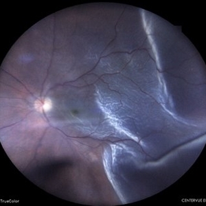 Retinal Detachment
May 27 2024 by Akansha Sharma
Color fundus photograph of a 64 year old male with retinal detachment.
Photographer: Dr. Akansha Sharma, Bharati Eye Hospital
Condition/keywords: RD, Retinal Detachment
-
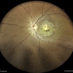 Hereditary Macular Degeneration
May 27 2024 by Akansha Sharma
Color fundus photograph of a 42 year old female with hereditary macular degeneration.
Photographer: Dr. Akansha Sharma, Bharati Eye Hospital
Condition/keywords: HMD, macular degeneration
-
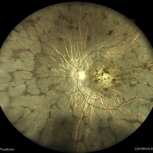 Atypical retinitis pigmentosa
May 27 2024 by Akansha Sharma
Color fundus photograph of a 47 year old male with atypical retinitis pigmentosa.
Photographer: Dr. Akansha Sharma, Bharati Eye Hospital
Condition/keywords: ATYPICAL RETINITIS PIGMENTOSA
-
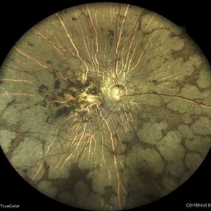 Atypical Retinitis Pigmentosa
May 27 2024 by Akansha Sharma
Color fundus photograph of a 47 year old male with atypical retinitis pigmentosa.
Photographer: Dr. Akansha Sharma, Bharati Eye Hospital
Condition/keywords: ATYPICAL RETINITIS PIGMENTOSA
-
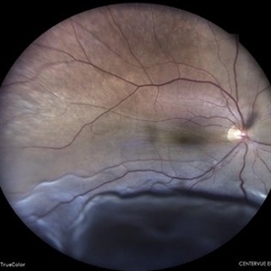 Retinal Detachment
May 27 2024 by Akansha Sharma
Color fundus photograph of a 65 year old female with retinal detachment.
Photographer: Dr. Akansha Sharma, Bharati Eye Hospital
Condition/keywords: RD, Retinal Detachment
-
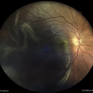 Retinal Detachment
May 27 2024 by Akansha Sharma
Color fundus photograph of a 64 year old male with retinal detachment.
Photographer: Dr. Akansha Sharma, Bharati Eye Hospital
Condition/keywords: RD, Retinal Detachment
-
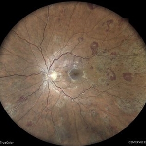 Chronic Myelogenous Leukemia
May 27 2024 by Akansha Sharma
Color fundus photograph of a 41 year old male presenting with ocular manifestations of chronic myelogenous leukemia.
Photographer: Dr. Akansha Sharma, Bharati Eye Hospital
Condition/keywords: CML, Roth Spots
-
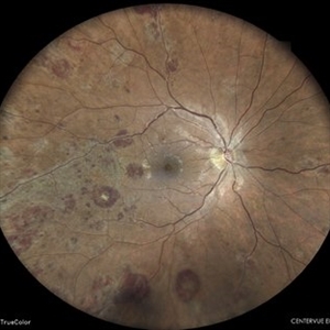 Chronic Myelogenous Leukemia
May 27 2024 by Akansha Sharma
Color fundus photograph of a 41 year old male presenting with ocular manifestations of chronic myelogenous leukemia.
Photographer: Dr. Akansha Sharma, Bharati Eye Hospital
Condition/keywords: CML, Roth Spots
-
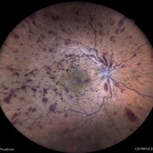 Ischaemic Central Retinal Vein Occlusion
Jun 5 2024 by Akansha Sharma
Color fundus photograph of a 64 year old hypertensive male with ischaemic central retinal vein occlusion.
Photographer: Dr. Akansha Sharma, Bharati Eye Hospital
Condition/keywords: ischemic CRVO
-
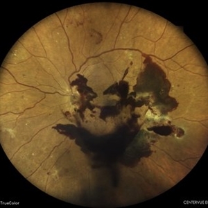 Subhyaloid Hemorrhage in a Case of Proliferative Diabetic Retinopathy
Jun 5 2024 by Akansha Sharma
Color fundus photograph of a 55 year old diabetic female with subhyaloid hemorrhage in a case of proliferative diabetic retinopathy.
Photographer: Dr. Akansha Sharma, Bharati Eye Hospital
Condition/keywords: PDR, proliferative diabetic retinopathy (PDR), SHH, subhyaloid hemorrhage
-
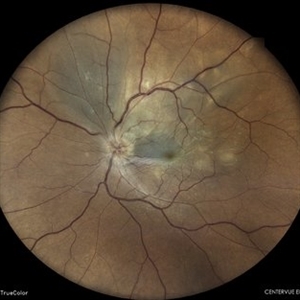 Multifocal Chorioditis
Jun 5 2024 by Akansha Sharma
Color fundus photograph of a 42 year old male with multifocal choroiditis.
Photographer: Dr. Akansha Sharma, Bharati Eye Hospital
Condition/keywords: choroiditis
-
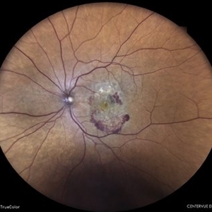 Subretinal Neovascular Membrane
Jun 5 2024 by Akansha Sharma
Color fundus photograph of a 94 year old female with subretinal bleed suggestive of subretinal neovascular membrane.
Photographer: Dr. Akansha Sharma, Bharati Eye Hospital
Condition/keywords: choroidal neovascular membrane (CNVM), CNVM, SRNVM, subretinal neovascularization (SRNV), wet age-related macular degeneration (wet AMD)
-
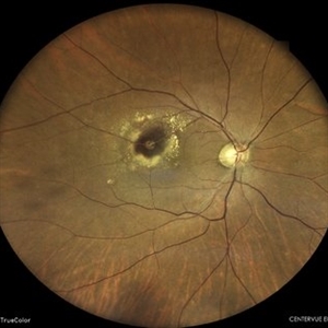 Retinal Arterial Macroaneurysm
Jun 5 2024 by Akansha Sharma
Color fundus photograph of a 61 year old hypertensive male with retinal arterial macroaneurysm.
Photographer: Dr. Akansha Sharma, Bharati Eye Hospital
Condition/keywords: optic disc pallor, RAM
-
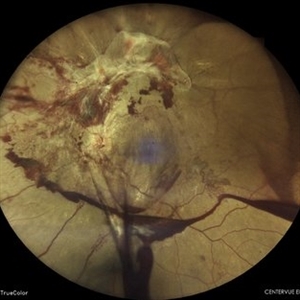 Subhyaloid Hemorrhage With Vascular Proliferations With Vitreous Hemorrhage in a Case of Proliferative Diabetic Retinopathy
Jun 5 2024 by Akansha Sharma
Color fundus photograph of a 24 year old type-1 diabetic and hypertensive female with subhyaloid hemorrhage with vascular proliferations with vitreous hemorrhage in a case of proliferative diabetic retinopathy.
Photographer: Dr. Akansha Sharma, Bharati Eye Hospital
Condition/keywords: fibrovascular proliferation, FVPs, SHH, subhyaloid hemorrhage, tractional retinal detachment, TRD, VH, vitreous hemorrhage
-
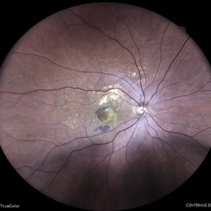 Subretinal Neovascular Membrane
Jun 5 2024 by Akansha Sharma
Color fundus photograph of a 49 year old female with subretinal bleed suggestive of subretinal neovascular membrane.
Photographer: Dr. Akansha Sharma, Bharati Eye Hospital
Condition/keywords: choroidal neovascular membrane (CNVM), CNVM, SRNVM, subretinal neovascularization (SRNV), wet age-related macular degeneration (wet AMD)
-
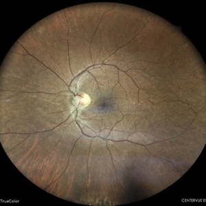 Status Post Pars Plana Vitrectomy With Silicone Oil Infusion Followed by Silicone Oil Removal
Jun 17 2024 by Akansha Sharma
Color fundus photograph of a 47 year old male with retina on status post pars plana vitrectomy with silicone oil infusion followed by silicone oil removal.
Photographer: Dr. Akansha Sharma, Bharati Eye Hospital
Condition/keywords: RD SURGERY, silicone oil
-
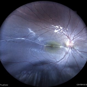 Old Retinal Detachment
Jun 17 2024 by Akansha Sharma
Color fundus photograph of a 10 year old male with old retinal detachment with subretinal band.
Photographer: Dr. Akansha Sharma, Bharati Eye Hospital
Condition/keywords: demarcation line, OLD RD, subretinal bands
-
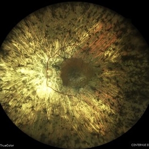 Retinitis Pigmentosa
Jun 17 2024 by Akansha Sharma
Color fundus photograph of a 59 year old male with retinitis pigmentosa with retinal thinning.
Photographer: Dr. Akansha Sharma, Bharati Eye Hospital
Condition/keywords: retinitis pigmentosa, RP
-
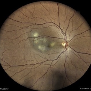 TB Choroiditis
Jun 17 2024 by Akansha Sharma
Color fundus photograph of a 40 year old male with TB choroiditis.
Photographer: Dr. Akansha Sharma, Bharati Eye Hospital
Condition/keywords: TB CHOROIDITIS
-
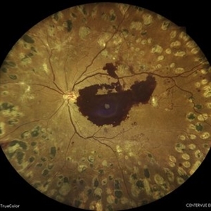 Subhyaloid Hemorrhage in a Case of Lasered Proliferative Diabetic Retinopathy
Jun 24 2024 by Akansha Sharma
Color fundus photograph of a 47 year old female with subhyaloid hemorrhage in a case of lasered proliferative diabetic retinopathy.
Photographer: Dr. Akansha Sharma, Bharati Eye Hospital
Condition/keywords: PDR, proliferative diabetic retinopathy (PDR), SHH
-
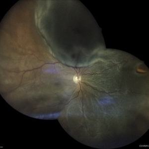 Retinal Detachment
Jun 24 2024 by Akansha Sharma
Color fundus photograph of a 56 year old male with retinal detachment with visible horse shoe tear.
Photographer: Dr. Akansha Sharma, Bharati Eye Hospital
Condition/keywords: RD, Retinal Detachment
-
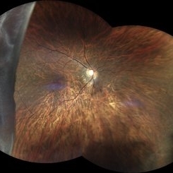 Giant Retinal Tear
Jun 24 2024 by Akansha Sharma
Color fundus photograph of a 30 year old female with a giant retinal tear.
Photographer: Dr. Akansha Sharma, Bharati Eye Hospital
Condition/keywords: GIANT RETINAL TEAR, GRT
-
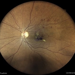 Old Vein Occlusion With Macular Edema
Jun 24 2024 by Akansha Sharma
Color fundus photograph of a hypertensive 53 year old female with vein occlusion with macular edema.
Photographer: Dr. Akansha Sharma, Bharati Eye Hospital
Condition/keywords: macular edema, Vein Occlusion
-
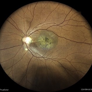 Optic Disc Pit With Macular Scar
Jun 24 2024 by Akansha Sharma
Color fundus photograph of a 42 year old male with optic disc pit with macular scar.
Photographer: Dr. Akansha Sharma, Bharati Eye Hospital
Condition/keywords: macular scar, Optic disc pit
-
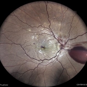 Venous Stasis Retinopathy
Jun 27 2024 by Akansha Sharma
Color fundus photograph of a 31 year old male patient with venous stasis retinopathy.
Photographer: Dr. Akansha Sharma, Bharati Eye Hospital
Condition/keywords: cystoid macular edema (CME), Disc Edema, SHH
-
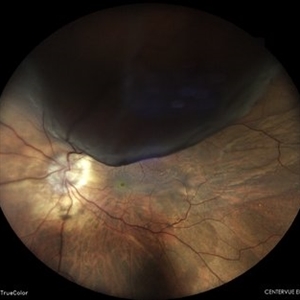 Retinal Detachment
Jun 27 2024 by Akansha Sharma
Color fundus photograph of a 65 year old male with retinal detachment with horse shoe tear at 12 o'clock.
Photographer: Dr. Akansha Sharma, Bharati Eye Hospital
Condition/keywords: RD, Retinal Detachment
-
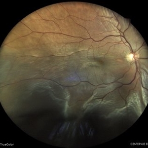 Retinal Detachment
Aug 6 2024 by Akansha Sharma
Color fundus photograph of a 39 year old male with retinal detachment status post trauma.
Photographer: Dr. Akansha Sharma, Bharati Eye Hospital
Condition/keywords: RD, Retinal Detachment
-
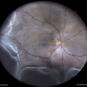 Retinal Detachment With Choroidal Detachment
Aug 6 2024 by Akansha Sharma
Color fundus photograph of a 49 year old male with retinal detachment and choroidal detachment.
Photographer: Dr. Akansha Sharma, Bharati Eye Hospital
Condition/keywords: CD, choroidal detachment, RD, Retinal Detachment
-
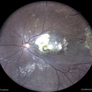 Coats' Disease
Aug 6 2024 by Akansha Sharma
Color fundus photograph of a 15 year old male with coats' disease.
Photographer: Dr. Akansha Sharma, Bharati Eye Hospital
Condition/keywords: Coats' disease
-
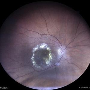 Circinate Retinopathy in a Case of Old Vein Occlusion
Aug 6 2024 by Akansha Sharma
Color fundus photograph of a 65 year old female with circinate retinopathy in a case of old vein occlusion.
Photographer: Dr. Akansha Sharma, Bharati Eye Hospital
Condition/keywords: circinate retinopathy, Vein Occlusion
-
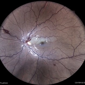 Venous Stasis Retinopathy
Aug 6 2024 by Akansha Sharma
Color fundus photograph of a 18 year old male with venous stasis retinopathy.
Photographer: Dr. Akansha Sharma, Bharati Eye Hospital
Condition/keywords: arterial occlusion, venous stasis retinopathy
-
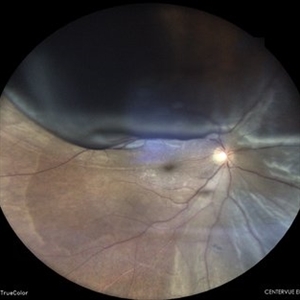 Retinal Detachment
Aug 6 2024 by Akansha Sharma
Color fundus photograph of a 36 year old male with retinal detachment.
Photographer: Dr. Akansha Sharma, Bharati Eye Hospital
Condition/keywords: RD, Retinal Detachment
-
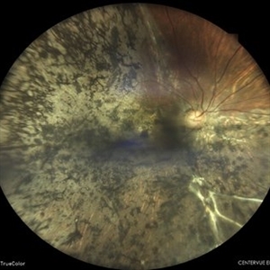 Spontaneously Settled Retinal Detachment
Aug 6 2024 by Akansha Sharma
Color fundus photograph of a 36 year old female with spontaneously settled retinal detachment.
Photographer: Dr. Akansha Sharma, Bharati Eye Hospital
Condition/keywords: OLD SETTLED RETINAL DETACHMENT, SPONTANEOUSLY SETTLED RETINAL DETACHMENT
-
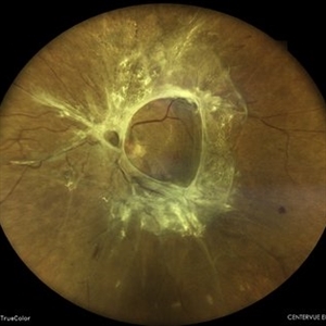 Tractional Retinal Detachment in a Case of Proliferative Diabetic Retinopathy
Aug 6 2024 by Akansha Sharma
Color fundus photograph of a 60 year old female with tractional retinal detachment in a case of proliferative diabetic retinopathy.
Photographer: Dr. Akansha Sharma, Bharati Eye Hospital
Condition/keywords: fibrovascular proliferation, PDR, Proliferative Diabetic retinopathy, tractional retinal detachment, TRD
-
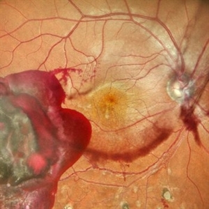 Subhyaloid Hemorrhage With Dispersed Vitreous Hemorrhage in a Case of Old Lasered Branch Retinal Vein Occlusion
Jul 12 2025 by Akansha Sharma
Color fundus photograph of a 32 year old hypertensive and diabetic male with subhyaloid hemorrhage with dispersed vitreous hemorrhage in a case of old lasered branch retinal vein occlusion.
Photographer: DR. AKANSHA SHARMA
Condition/keywords: branch retinal vein occlusion (BRVO), laser photocoagulation, SHH, subhyaloid hemorrhage, VH, vitreous hemorrhage
-
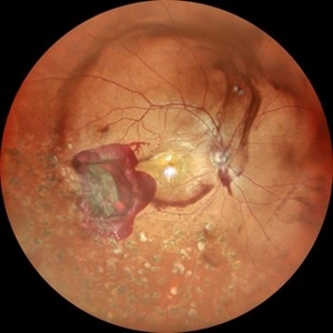 Subhyaloid Hemorrhage With Dispersed Vitreous Hemorrhage in a Case of Old Lasered Branch Retinal Vein Occlusion
Jul 12 2025 by Akansha Sharma
Color fundus photograph of a 32 year old hypertensive and diabetic male with subhyaloid hemorrhage with dispersed vitreous hemorrhage in a case of old lasered branch retinal vein occlusion.
Photographer: DR. AKANSHA SHARMA
Condition/keywords: branch retinal vein occlusion (BRVO), laser photocoagulation, SHH, subhyaloid hemorrhage, VH, vitreous hemorrhage
-
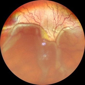 Exudative Retinal Detachment
Jul 12 2025 by Akansha Sharma
Color fundus photograph of a 33 year old male with exudative retinal detachment with a small axial length.
Photographer: DR. AKANSHA SHARMA
Condition/keywords: exudative detachment
-
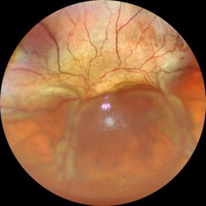 Exudative Retinal Detachment
Jul 12 2025 by Akansha Sharma
Color fundus photograph of a 33 year old male with exudative retinal detachment with a small axial length.
Photographer: DR. AKANSHA SHARMA
Condition/keywords: exudative detachment
-
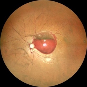 Valsalva Retinopathy
Jul 30 2025 by Akansha Sharma
Color fundus photograph of a 33 year old male with subhyaloid hemorrhage suggestive of valsalva retinopathy.
Photographer: DR. AKANSHA SHARMA
Condition/keywords: subhyaloid hemorrhage, valsalva retinopathy
-
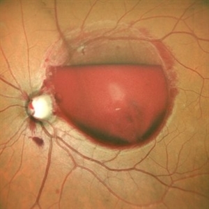 Valsalva Retinopathy
Jul 30 2025 by Akansha Sharma
Color fundus photograph of a 33 year old male with subhyaloid hemorrhage suggestive of valsalva retinopathy.
Photographer: DR. AKANSHA SHARMA
Condition/keywords: subhyaloid hemorrhage, valsalva retinopathy
-
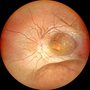 Retinoschisis
Jul 30 2025 by Akansha Sharma
Color fundus photograph of a 2 year old male with both eyes retinoschisis.
Photographer: DR. AKANSHA SHARMA
Condition/keywords: juvenile retinoschisis, RETINOSCHISIS
-
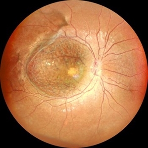 Retinoschisis
Jul 30 2025 by Akansha Sharma
Color fundus photograph of a 2 year old male with both eyes retinoschisis.
Photographer: DR. AKANSHA SHARMA
Condition/keywords: juvenile retinoschisis, RETINOSCHISIS
-
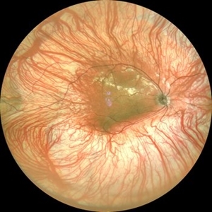 Oculo-cutaneous Albinisim
Aug 10 2025 by Akansha Sharma
Color fundus photograph of a 18 year old male with oculo-cutaneous albinisim.
Photographer: DR. AKANSHA SHARMA
Condition/keywords: albinism, foveal hypoplasia, oculocutaneous albinism
-
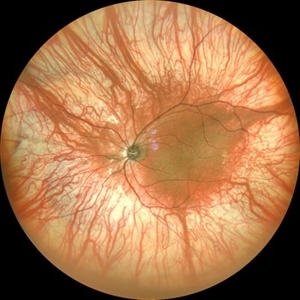 Oculo-cutaneous Albinisim
Aug 10 2025 by Akansha Sharma
Color fundus photograph of a 18 year old male with oculo-cutaneous albinisim.
Photographer: DR. AKANSHA SHARMA
Condition/keywords: albinism, foveal hypoplasia, oculocutaneous albinism
-
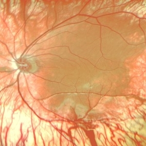 Oculo-cutaneous Albinisim
Aug 10 2025 by Akansha Sharma
Color fundus photograph of a 18 year old male with oculo-cutaneous albinisim.
Photographer: DR. AKANSHA SHARMA
Condition/keywords: albinism, foveal hypoplasia, oculocutaneous albinism
-
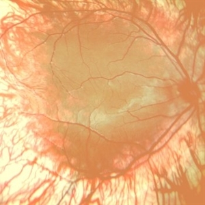 Oculo-cutaneous Albinisim
Aug 10 2025 by Akansha Sharma
Color fundus photograph of a 18 year old male with oculo-cutaneous albinisim.
Photographer: DR. AKANSHA SHARMA
Condition/keywords: albinism, foveal hypoplasia, oculocutaneous albinism
-
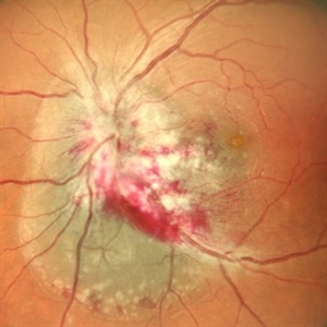 CMV Retinitis
Aug 14 2025 by Akansha Sharma
Color fundus photograph of a 42 year old seropositive female with cmv retinitis.
Photographer: DR. AKANSHA SHARMA
Condition/keywords: CMV retinitis, cytomegalovirus (CMV)
-
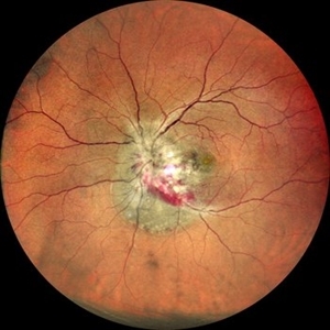 CMV Retinitis
Aug 14 2025 by Akansha Sharma
Color fundus photograph of a 42 year old seropositive female with cmv retinitis.
Photographer: DR. AKANSHA SHARMA
Condition/keywords: CMV retinitis, cytomegalovirus (CMV)
-
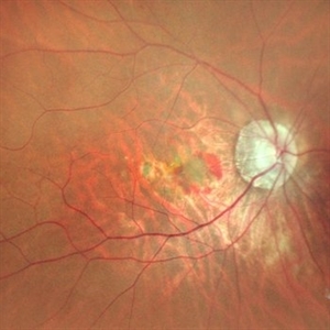 Subretinal Neovascular Membrane
Aug 15 2025 by Akansha Sharma
Color fundus photograph of a 40 year old male with subretinal neovascular membrane.
Photographer: DR. AKANSHA SHARMA
Condition/keywords: choroidal neovascular membrane (CNVM), CNVM, Myopic CNVM, SRNVM, subretinal neovascularization (SRNV), Wet age related macular degeneration
-
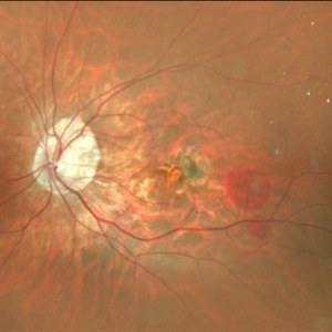 Subretinal Neovascular Membrane
Aug 15 2025 by Akansha Sharma
Color fundus photograph of a 40 year old male with subretinal neovascular membrane.
Photographer: DR. AKANSHA SHARMA
Condition/keywords: choroidal neovascular membrane (CNVM), CNVM, SRNVM, subretinal neovascularization (SRNV), wet age-related macular degeneration (wet AMD)
-
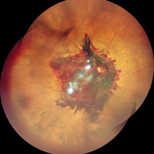 Subhyaloid Hemorrhage With Vitreous Hemorrhage
Sep 12 2025 by Akansha Sharma
Color fundus photograph of a 56 year old hypertensive and diabetic female who presented with subhyaloid hemorrhage along with vitreous hemorrhage after being administered high dose anti-platelet therapy pre- and post a cardiac procedure.
Photographer: DR. AKANSHA SHARMA
Condition/keywords: SHH, sub ILM hemorrhage, subhyaloid hemorrhage, VH, vitreous hemorrhage
-
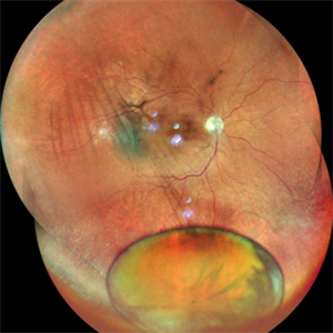 Dropped Nucleus With Disc Pallor With Posterior Pole Retinal Detachment
Sep 12 2025 by Akansha Sharma
Color fundus photograph of a 60 year old male with a dropped nucleus with disc pallor with posterior pole retinal detachment.
Photographer: DR. AKANSHA SHARMA
Condition/keywords: dropped nucleus, fragmatome, nucleus drop, optic disc pallor, PALE DISC, POSTERIOR POLE RETINAL DETACHMENT, RD, retinal detachment
-
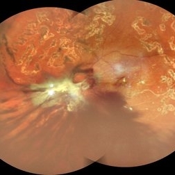 Table Top Tractional Retinal Detachment With Vitreous Hemorrhage in a Case of Proliferative Diabetic Retinopathy
Sep 12 2025 by Akansha Sharma
Color fundus photograph of a 56 year old male with table top tractional retinal detachment with vitreous hemorrhage in a case of proliferative diabetic retinopathy.
Photographer: DR. AKANSHA SHARMA
Condition/keywords: pan-retinal photocoagulation (PRP), PDR, proliferative diabetic retinopathy (PDR), PRP, TABLE TOP TRD, tractional retinal detachment, TRD, VH, vitreous hemorrhage
A project from the American Society of Retina Specialists