 Initializing download.
-
 By Joshua Friedman
Co-author(s): J. Sebag - Uploaded on May 31, 2022.
- Last modified by Joshua Friedman on May 31, 2022.
- Rating
- Appears in
- Miscellaneous
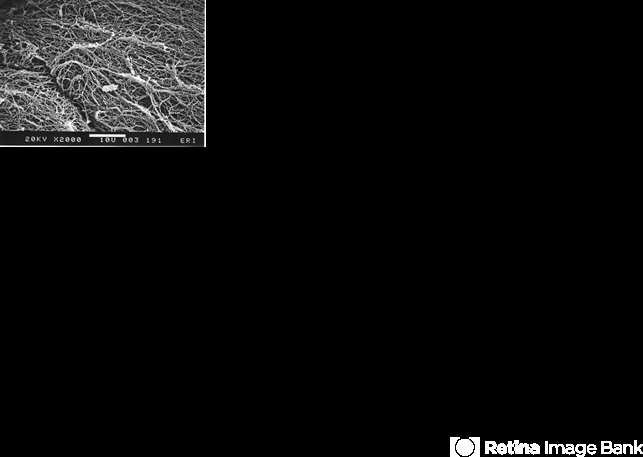
- Condition/keywords
- scanning EM, posterior vitreous cortex, collagen fibrils, vision degrading myodesopsia
- Photographer
- EM lab, Eye Research Institute of Retina Foundation, Boston, MA
- Description
- Scanning electron microscopy demonstrates the dense packing of collagen fibrils in the posterior vitreous cortex. To some extent this arrangement is exaggerated by the dehydration that occurs during specimen preparation for scanning electron microscopy (bar = 10 µm).


