-
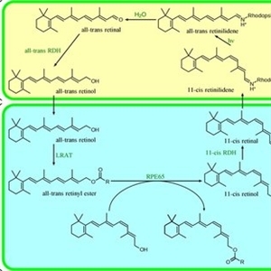 Diagram of the visual cycle
Mar 23 2022 by Joshua Friedman
This diagram is associated with the Retina Atlas chapter on Visual Cycle Disease, which can be found here: https://atlas.asrs.org/article/visual-cycle-disease-118#
Condition/keywords: visual cycle
-
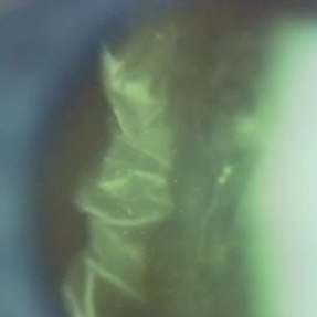 Folds in Detached Posterior Vitreous Cortex
May 31 2022 by Joshua Friedman
Slit lamp (video) image showing folds in the posterior vitreous cortex in an eye with PVD.
Photographer: Martin Snead, MD, Cambridge, England
Condition/keywords: folds, posterior vitreous cortex, PVD, vision degrading myodesopsia, vitreous
-
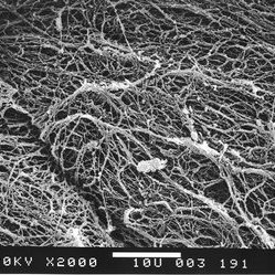 Scanning electron microscopy of the posterior aspect of the human posterior vitreous cortex
May 31 2022 by Joshua Friedman
Scanning electron microscopy demonstrates the dense packing of collagen fibrils in the posterior vitreous cortex. To some extent this arrangement is exaggerated by the dehydration that occurs during specimen preparation for scanning electron microscopy (bar = 10 µm).
Photographer: EM lab, Eye Research Institute of Retina Foundation, Boston, MA
Condition/keywords: collagen fibrils, posterior vitreous cortex, scanning EM, vision degrading myodesopsia
-
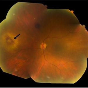 Sarcoidosis
Mar 27 2023 by Joshua Friedman
Fundus photograph of a 70-year-old woman with presumed sarcoidosis and sarcoid-associated choroidal granuloma.
Photographer: Christopher D Conrady, MD, PhD
Condition/keywords: sarcoid granuloma
-
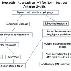 Posterior Manifestations of Sarcoidosis – Management of noninfectious anterior uveitis
Mar 29 2023 by Joshua Friedman
Management of noninfectious anterior uveitis.
Condition/keywords: uveitis
-
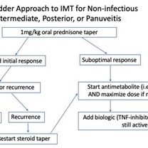 Posterior Manifestations of Sarcoidosis – Management of noninfectious intermediate, posterior, or panuveitis
Mar 29 2023 by Joshua Friedman
Management of noninfectious intermediate, posterior, or panuveitis.
Condition/keywords: panuveitis, sarcoidosis
-
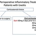 Posterior Manifestations of Sarcoidosis – Perioperative inflammatory supplementation
Mar 29 2023 by Joshua Friedman
Perioperative inflammatory supplementation.
Condition/keywords: sarcoidosis
-
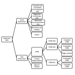 Figure 1: Classification of stationary inherited retinal disease
Dec 15 2023 by Joshua Friedman
Abbreviations: AD, autosomal dominant; AR, autosomal recessive; CSNB, congenital stationary night blindness; IRD, inherited retinal disease; XL, X-linked.
Condition/keywords: stationary IRD
-
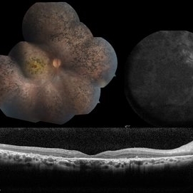
 LCA type 10
Apr 10 2025 by Joshua Friedman
LCA type 10 due to mutations in CEP290. 36-year-old male with best corrected visual acuity of light perception in both eyes since childhood. On color fundus imaging, there is a mix of polymorphous white flecks and pigmentary changes. On autofluorescence imaging, there is almost complete loss of macular RPE. On OCT, there is complete loss of inner and outer retinal layers, the greatest losses occurring centrally.
Photographer: Stephen Tsang, MD, PhD
Condition/keywords: Leber Congenital Amaurosis
-
 LCA Type 8
Apr 10 2025 by Joshua Friedman
LCA Type 8 due to a pathogenic mutations in CRB1. 5-year-old male with a visual acuity of count fingers at 3 feet. Note the pseudopapilledema, para-arteriolar sparing, and nummular intraretinal pigment migration.
Photographer: Stephen Tsang, MD, PhD
Condition/keywords: Leber Congenital Amaurosis
-
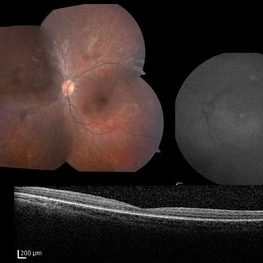 LCA Type 2
Apr 10 2025 by Joshua Friedman
LCA Type 2 (RPE65) showing characteristic hypoautofluorescence and retinal thinning. 8F with best corrected visual acuity of 20/400 (OD) and 20/150 (OS). Small white intraretinal spots and RPE mottling are visible on color fundus photography. Blue light autofluorescence reveals near-complete loss of signal, while OCT demonstrates widespread outer retinal thinning.
Photographer: Stephen Tsang, MD, PhD
Condition/keywords: Leber Congenital Amaurosis
-
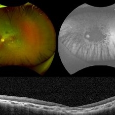
 LCA type 12
Apr 10 2025 by Joshua Friedman
LCA type 12 due to pathogenic mutations in RDH12. 13-year-old male with a visual acuity of 20/80 and 20/300 in the right and left eye, respectively. There is extensive pigment migration in the peripheral retina and macula. Like RPE65, there is widespread hypoautofluorescent signal, however, the peripapillary retina is uniquely spared in this form of LCA. On OCT, there is almost complete loss of the retina centrally.
Photographer: Stephen Tsang, MD, PhD
Condition/keywords: Leber Congenital Amaurosis
-
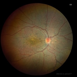
 LCA Type 1
Apr 10 2025 by Joshua Friedman
LCA Type 1 (GUCY2D) demonstrating the hallmark discordance between severe vision loss (light perception) in a 10-year-old female and a near-normal fundus appearance in childhood, typical of guanylate cyclase dysfunction.
Photographer: Stephen Tsang, MD, PhD
Condition/keywords: Leber Congenital Amaurosis (LCA)
A project from the American Society of Retina Specialists