 Initializing download.
-
 By Olivia Rainey
Retina Specialists of Michigan
Co-author(s): Greg Bever, MD - Uploaded on Oct 7, 2020.
- Last modified by Caroline Bozell on Oct 8, 2020.
- Rating
- Appears in
- Miscellaneous
- Condition/keywords
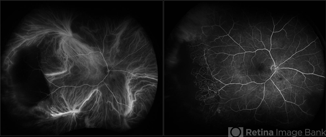
- subretinal hemorrhage, periphery, peripheral exudative hemorrhagic chorioretinopathy (PEHCR), indocyanine green (ICG) angiography, ultra-wide field imaging, Optos, monocular
- Photographer
- Olivia Rainey, OCT-C, COA
- Imaging device
-
Fundus camera
Optos California - Description
- Fluorecein and ICG angiography of a 80-year-old male with peripheral exudative hemorrhagic chorioretinopathy affecting his right eye. Patient noted only mild floaters for a couple weeks OD on 10/5/2020. The physician strongly suspects that the lesion is subretinal blood (likely from PEHCR) rather than choroidal melanoma. There is blocking on FA and ICG with a definite lack of intrinsic vessels within the mass lesion. He will monitor closely, as the patient is monocular with a history of multiple surgeries (which the family believes PPV for "scar tissue") OS mostly in 2013. His family also reports remembering possibly being told there was a "small mass" in the left eye at one point in their surgical course. The physician believes that it's possible this was a bleed related to PEHCR as it typically exists as a bilateral condition.
")


