 Initializing download.
-
 By David L Kilpatrick, MD
Co-author(s): John O. Mason, Retina Consultants of Alabama, P. C. - Uploaded on Apr 12, 2019.
- Last modified by Caroline Bozell on Jul 2, 2019.
- Image of the week
-
Jul 7, 2019
View all images of the week - Rating
- Appears in
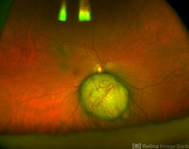
- Amelanotic choroidal melanoma
- Condition/keywords
- amelanotic melanoma
- Photographer
- Retina Consultants of Alabama, P. C.
- Imaging device
-
Fundus camera
Optos - Description
- Fundus photograph of a 69-year-old male with an amelanotic choroidal melanoma and corresponding exudative retinal detachment. Transvitreal biopsy was performed at the time of radioactive I-125 plaque placement. The genetic expression profile revealed a Class 1A, PRAME negative tumor.


